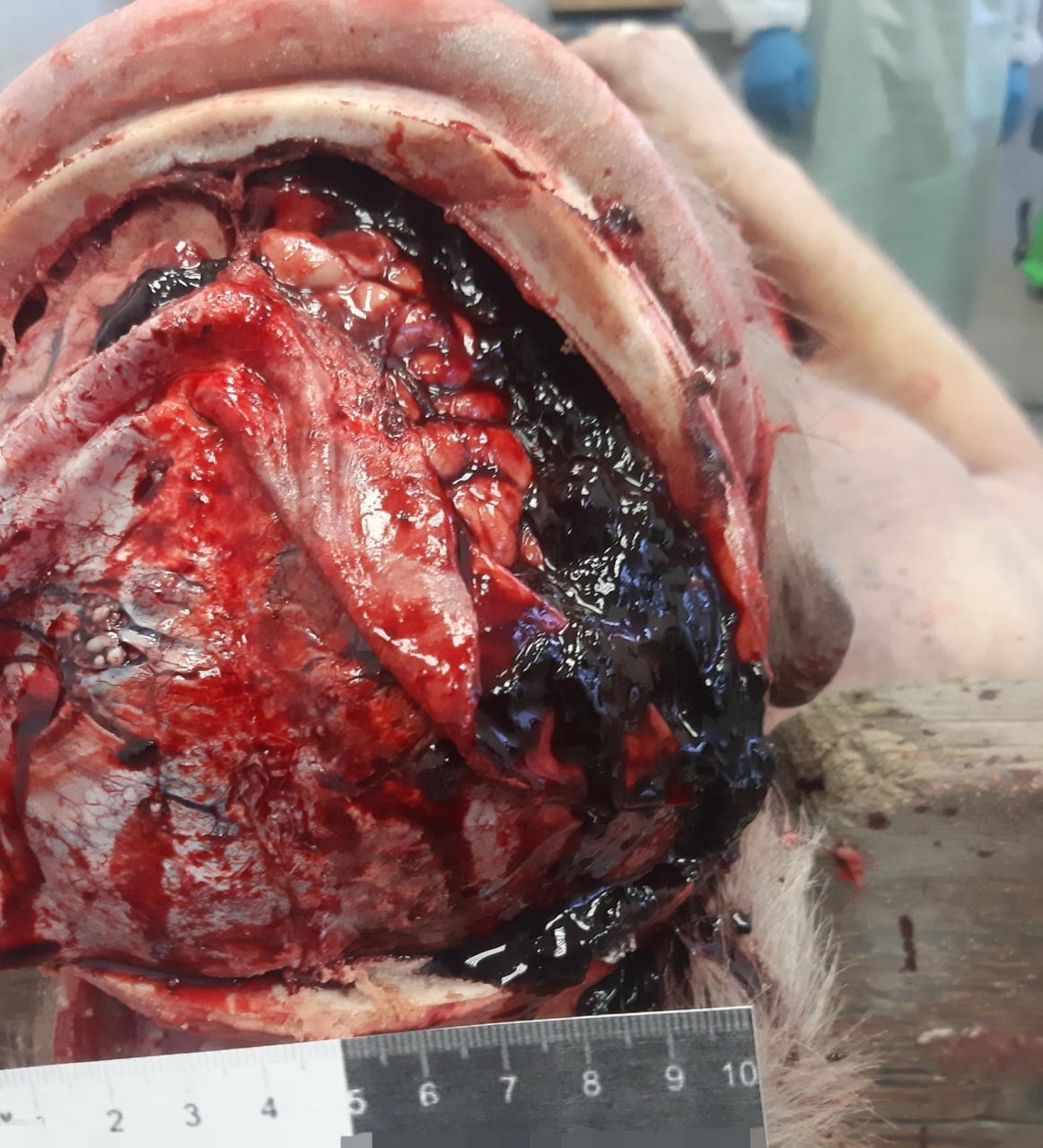
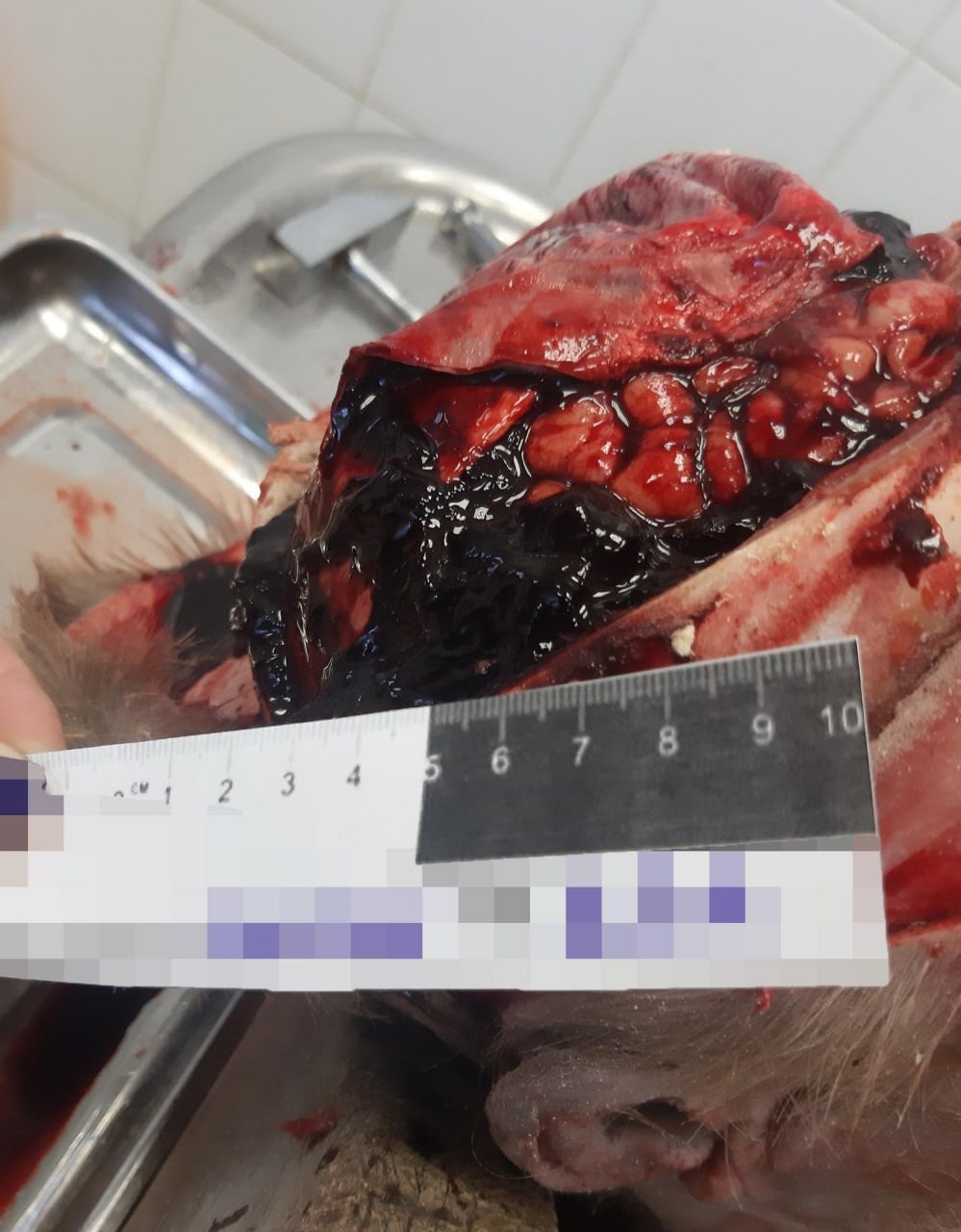
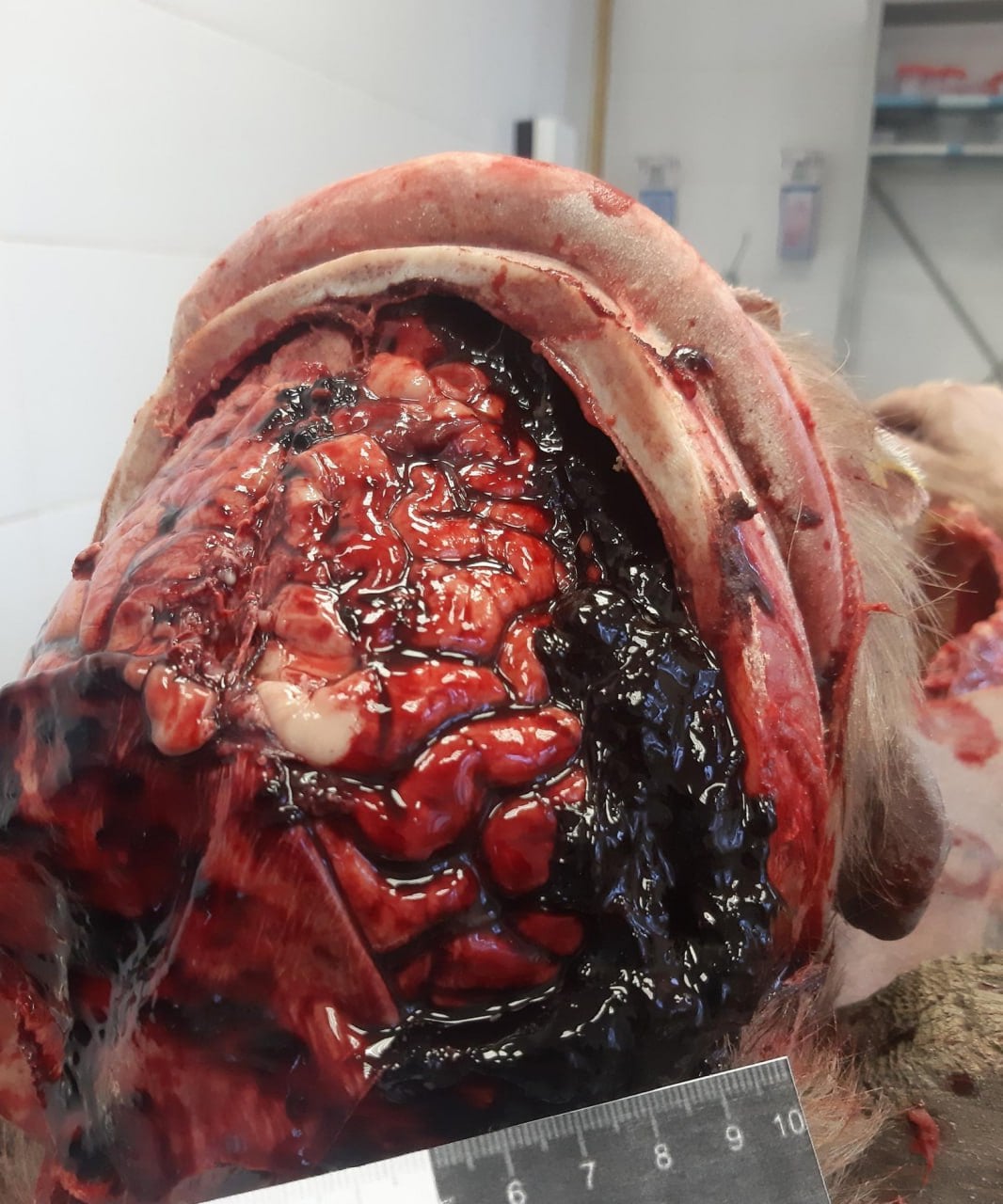
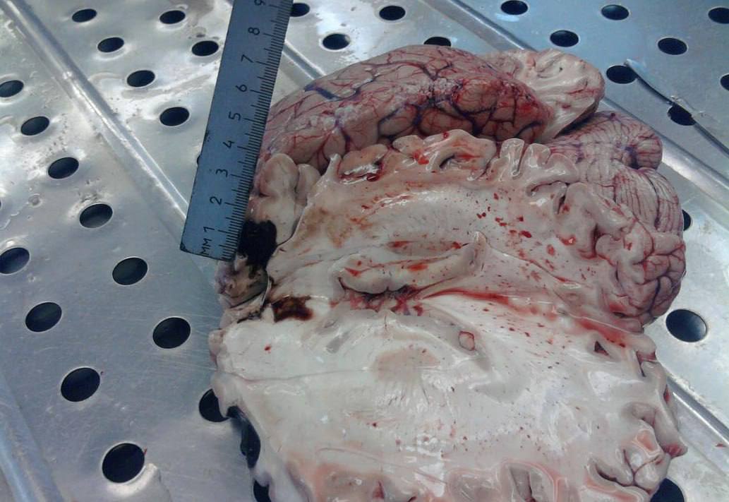
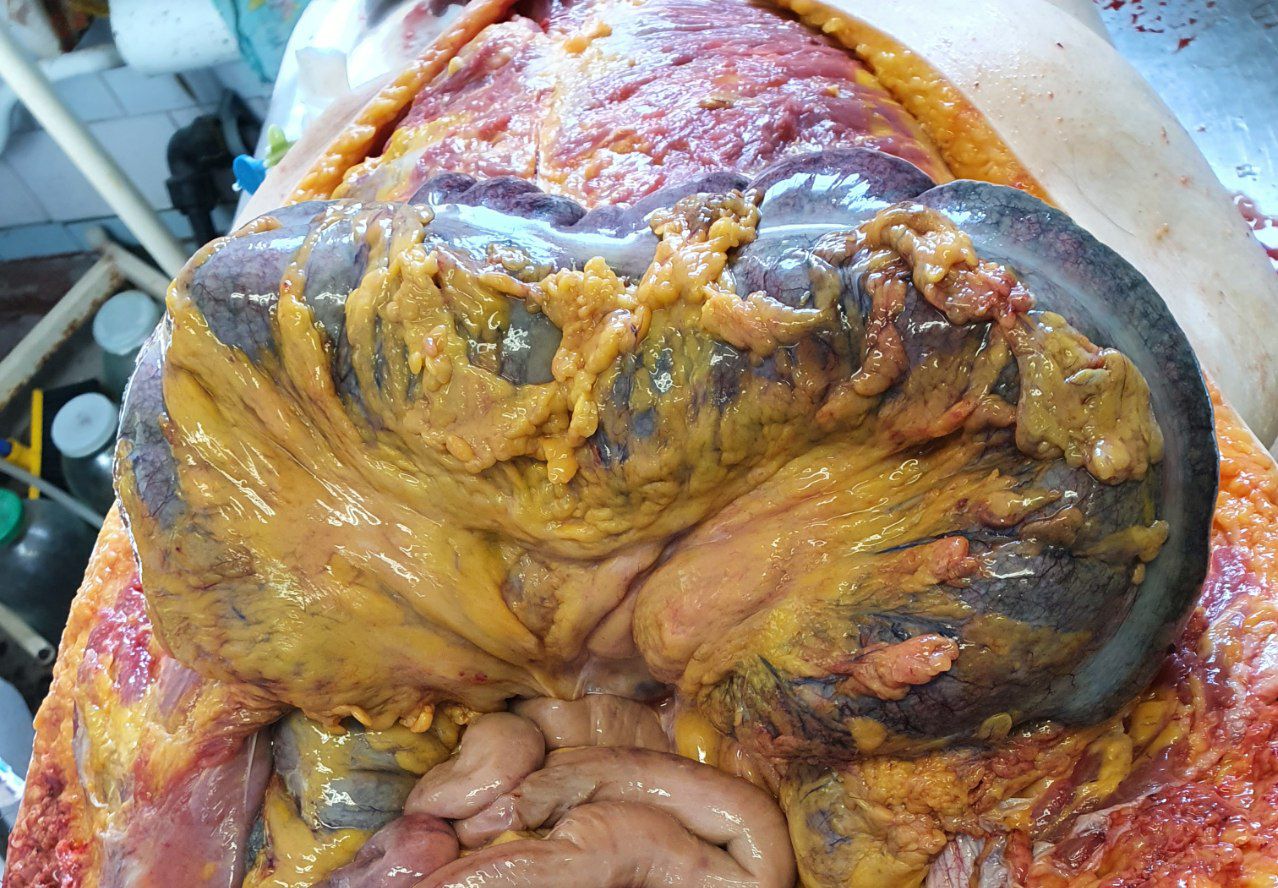
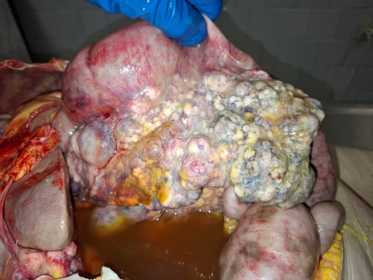
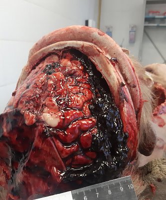
Note: the text accompanying these images are translations from the posts on the Telegram channels. I am not a medical expert, and thus cannot comment on said info's reliability (as well as any possible translation defects). 1. An elderly man was admitted [to the morgue] without signs of external injuries. At autopsy, pronounced liver cirrhosis was found, characteristic of chronic alcoholism, as well as coagulopathy - a blood clotting disorder typical in liver failure. Inside the cranial cavity there was a non-traumatic subdural haematoma.
Such haemorrhages can occur spontaneously: weakened cerebral vessels rupture even without a blow, especially if the blood clotting system is disrupted due to cirrhosis.

 2.
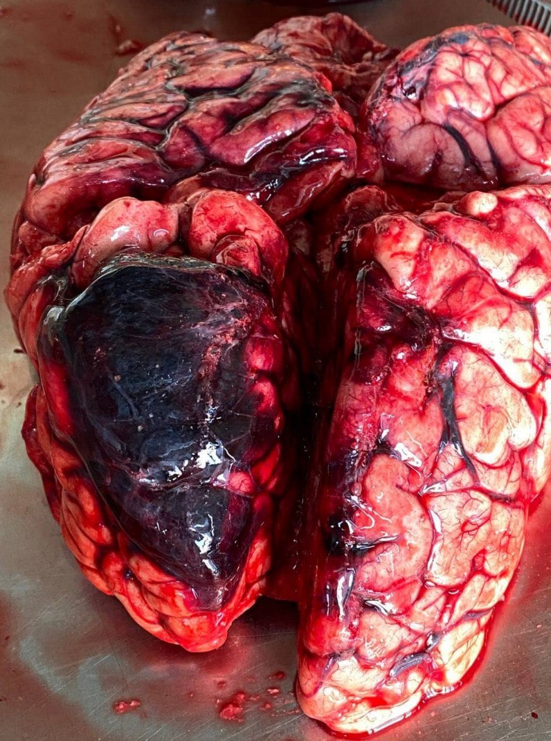
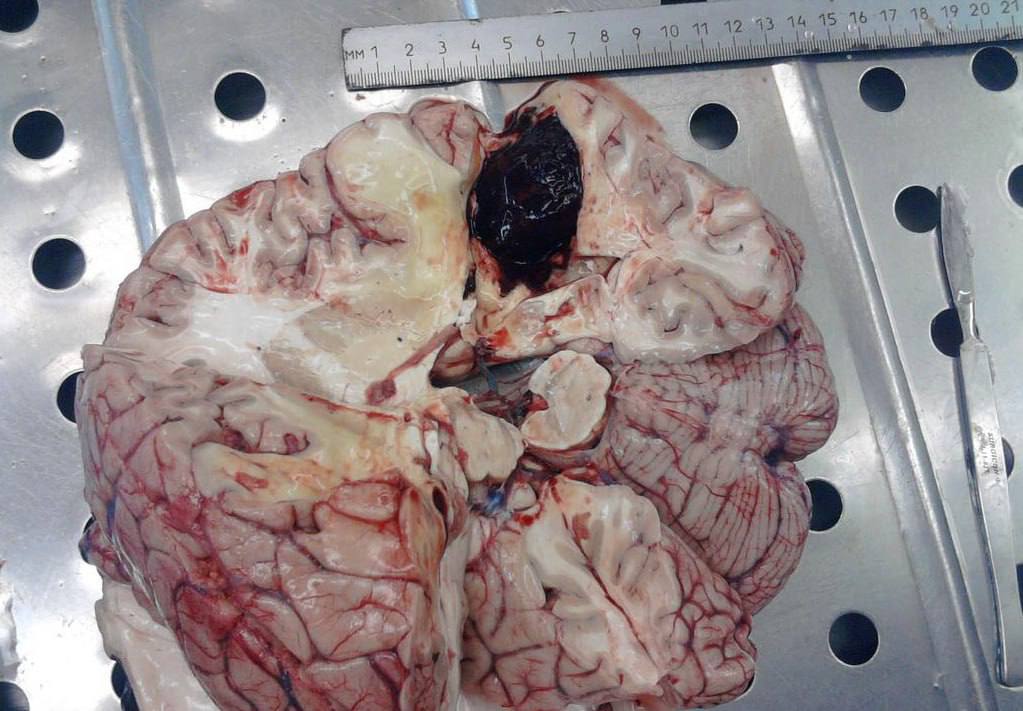
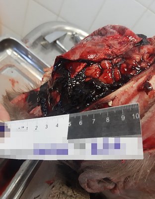
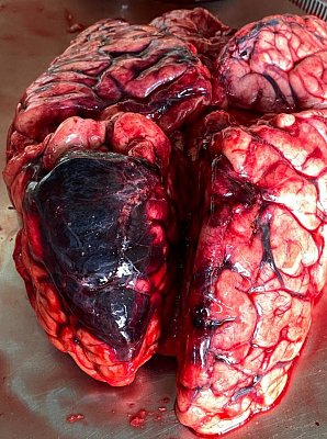
2. Haemorrhagic stroke with massive bleeding under the pia mater.
Because of the haematoma, the brain increased in volume and came under pressure. As a result, part of the brainstem shifted and became wedged into the occipital foramen of the skull. It is precisely in this area that the centres responsible for breathing and cardiac activity are located. As soon as such a displacement occurs, breathing stops - this is what causes death in such strokes.
 3.
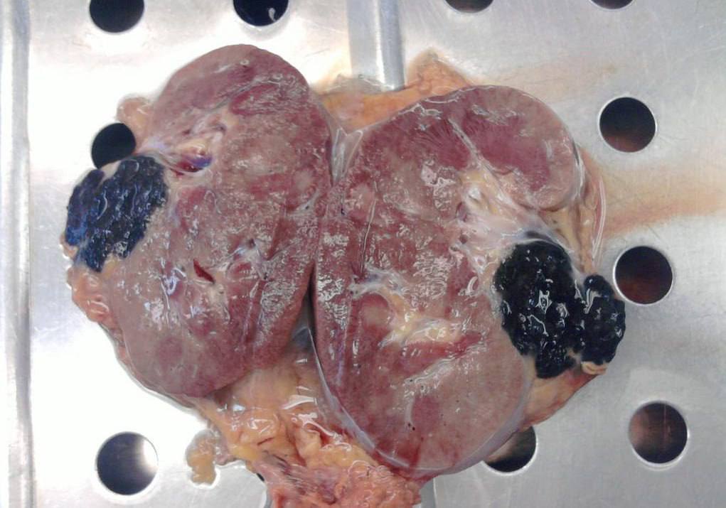
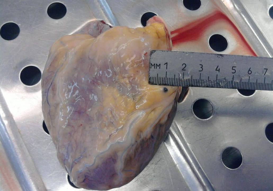
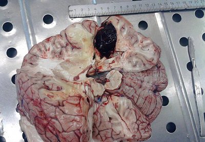
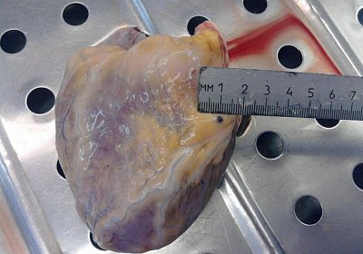
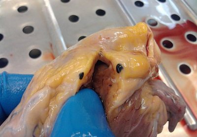
3. The melanoma spread to the brain, kidneys, and heart, taking control of these vital organs. The tumour left no chance, rapidly worsening the body’s functions and leading to a tragic end. When cancer attacks so aggressively, the consequences become irreversible.


 4.
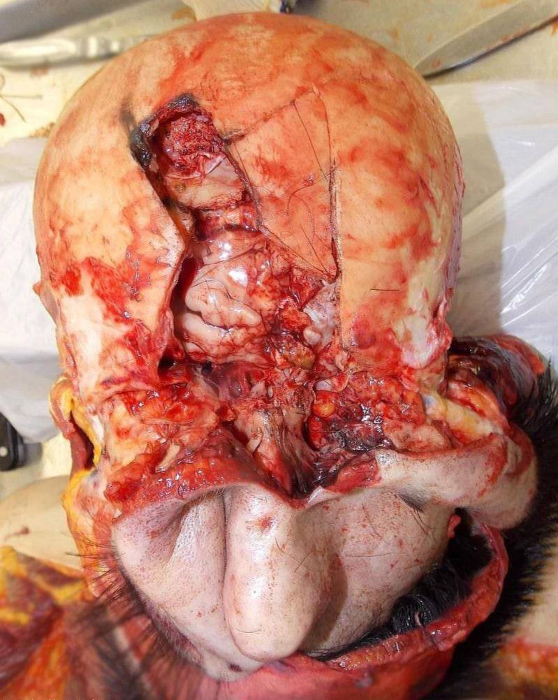
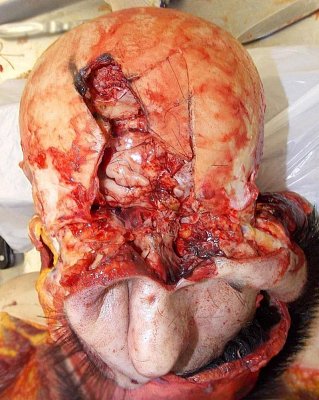
4. A fatal fracture of the frontal bone after a blow from a heavy blunt object. Massive bleeding, crushed tissue, an open defect in the skull area. The brain was left practically unprotected.
Death came quickly. The combination of severe traumatic brain injury and heavy blood loss left the victim with minimal chances of survival. Such injuries, especially without immediate medical assistance, almost always result in a fatal outcome.
 5.
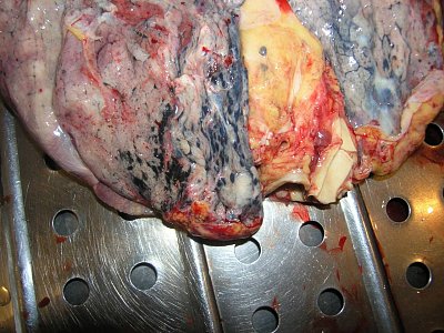
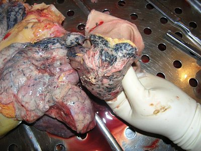
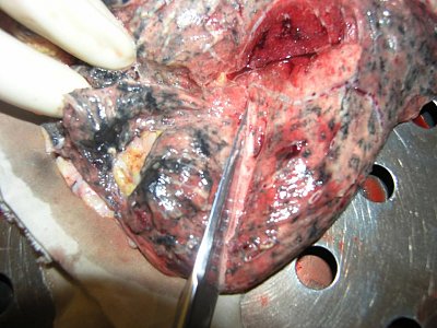
5. This is what the lungs affected by tuberculosis look like at autopsy: dense (with multiple whitish-grey lesions), cavities (areas of tissue breakdown), and scars. Sometimes the entire upper portion of the lung turns into one continuous area of necrosis.
On cross-section, we can observe a cheese-like mass, characteristic of caseous necrosis. To the touch, such areas are denser than normal lung tissue and do not compress easily. All of this is the result of chronic inflammation, tissue destruction, and the body’s attempts to contain the infection with a dense capsule.
 6.
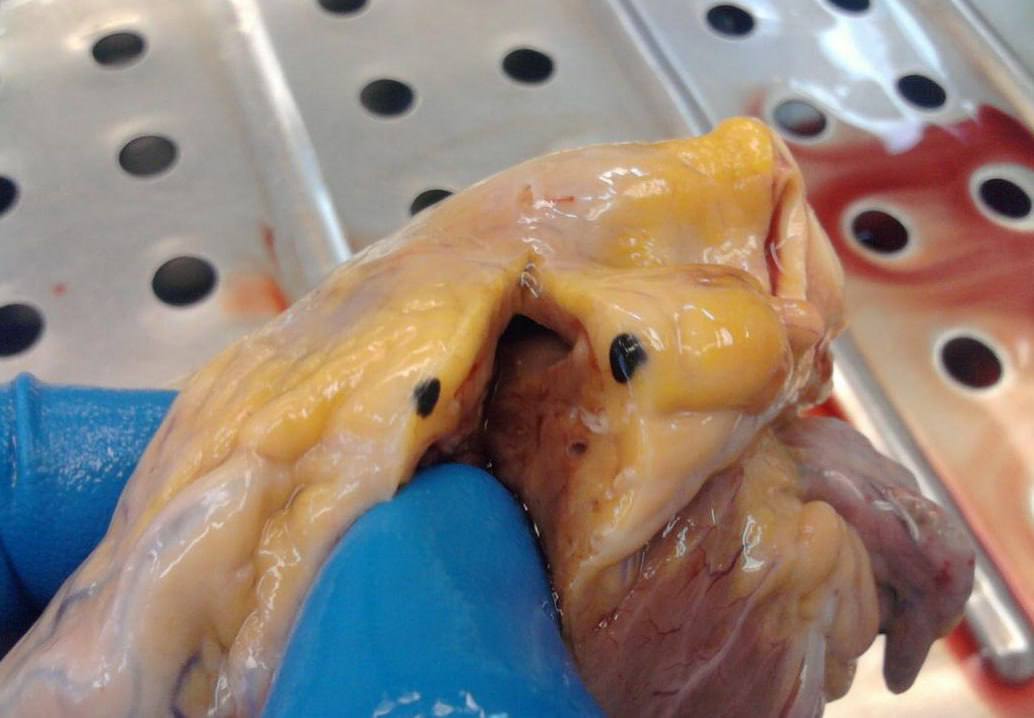
6. Sometimes bodies arrive with not a single complaint in their medical history. At autopsy - a torn carotid artery, an aneurysm in the carotid region. It all happened quietly: internal bleeding, and no one realized anything.
The carotid sinus is the area where baroreceptors monitor blood pressure. And nearby, there may be an aneurysm that ruptures suddenly.
This thing can be detected in 15 minutes with a routine ultrasound. But many never make it to the doctor’s office - they end up with us.
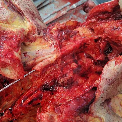 7.
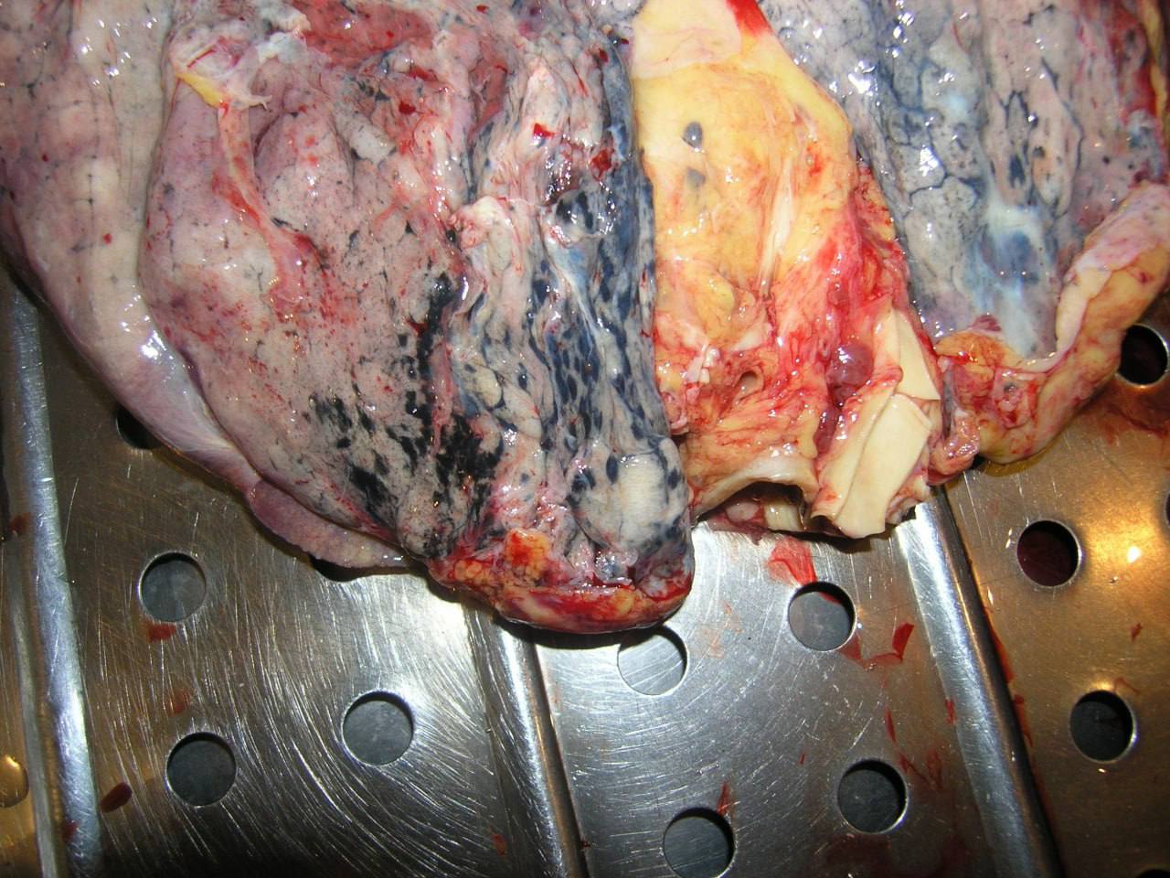
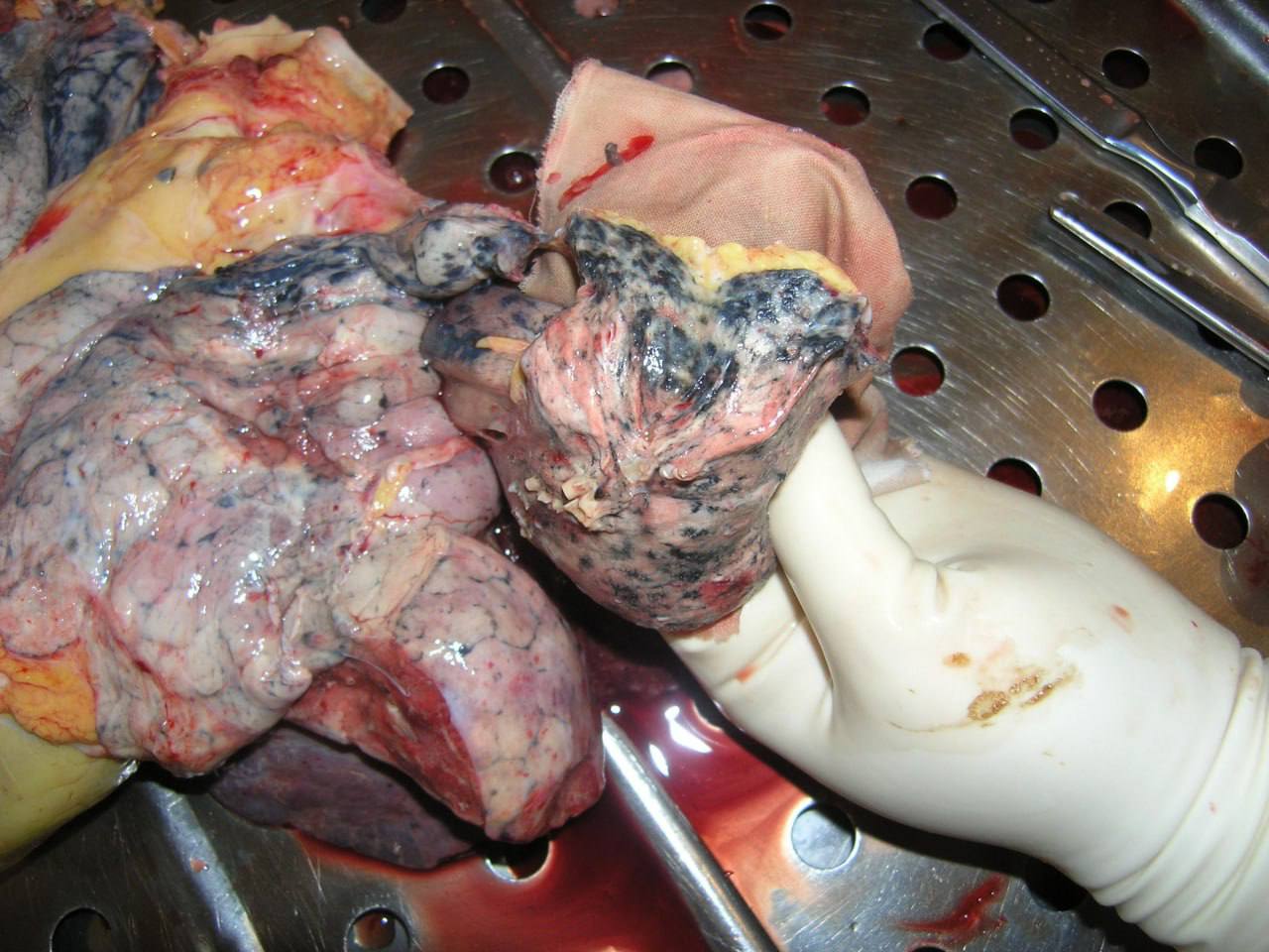
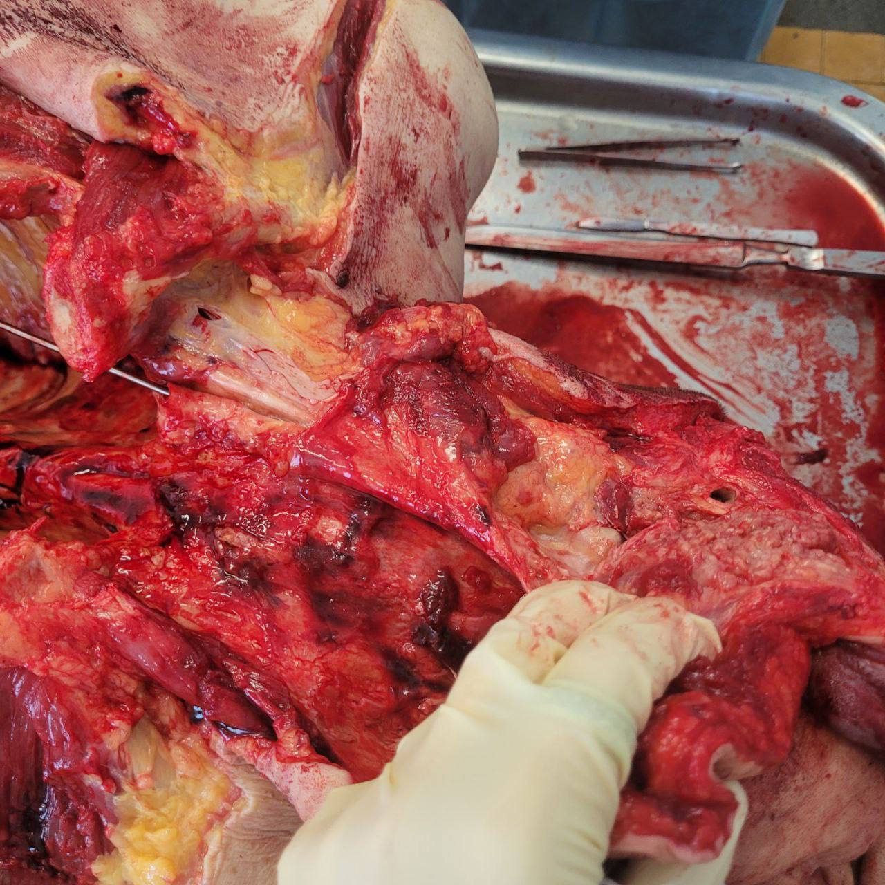
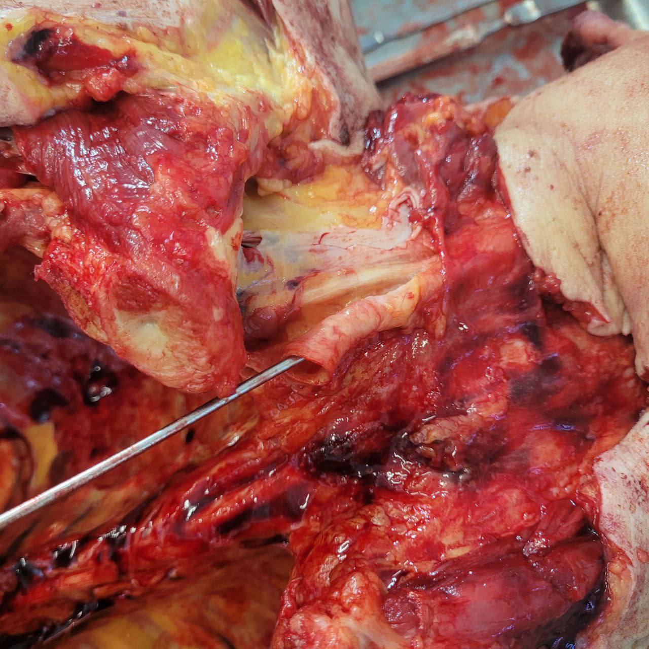
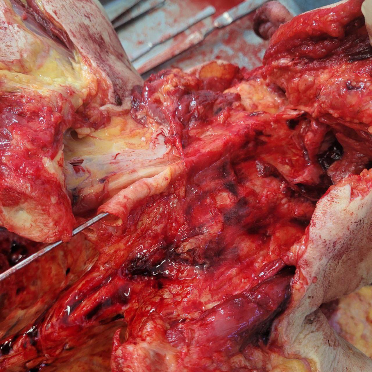
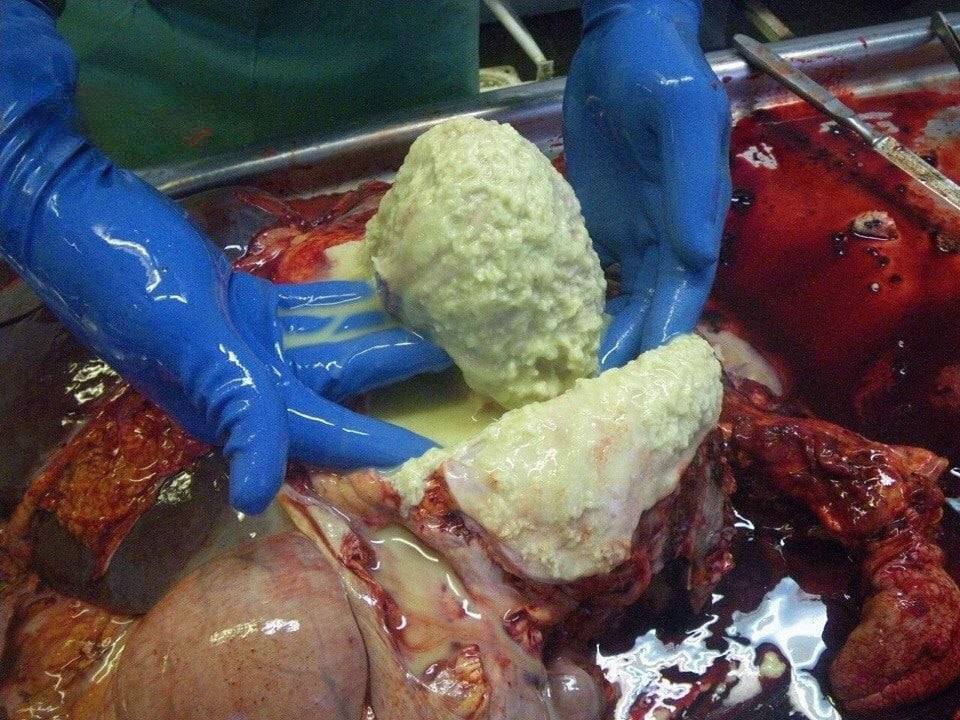
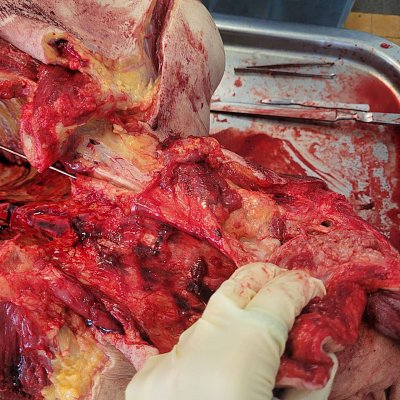
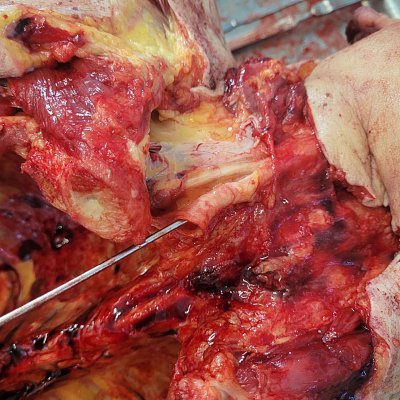
7. Clinical case of postmortem examination: the body of a patient with total cancer of the left lung, complicated by purulent pericarditis.
Lung cancer in its late stages often affects nearby tissues, which worsens the course of the disease and causes severe complications such as pericarditis.
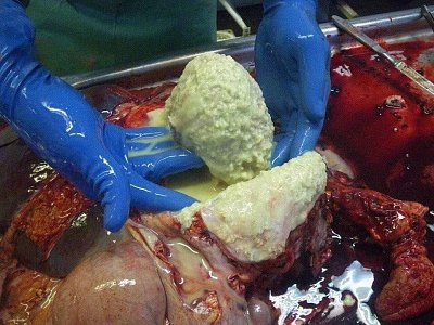 8.
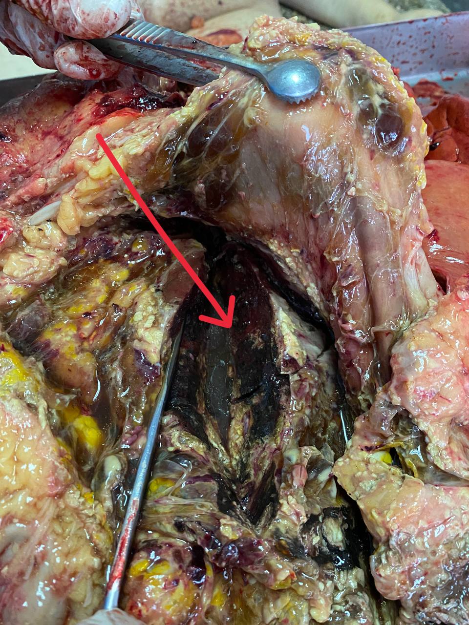
8. This green-black mash is what’s left of the pancreas.
When it undergoes necrosis, enzymes are released into the surrounding tissues and “digest” them. In other words, nothing living remains. A very frightening and deadly consequence.
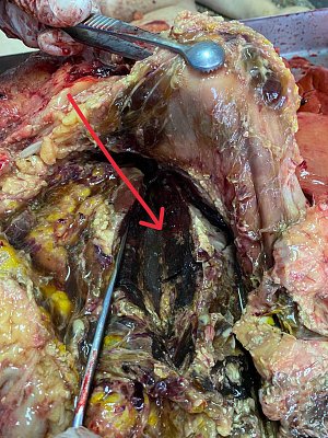 9.
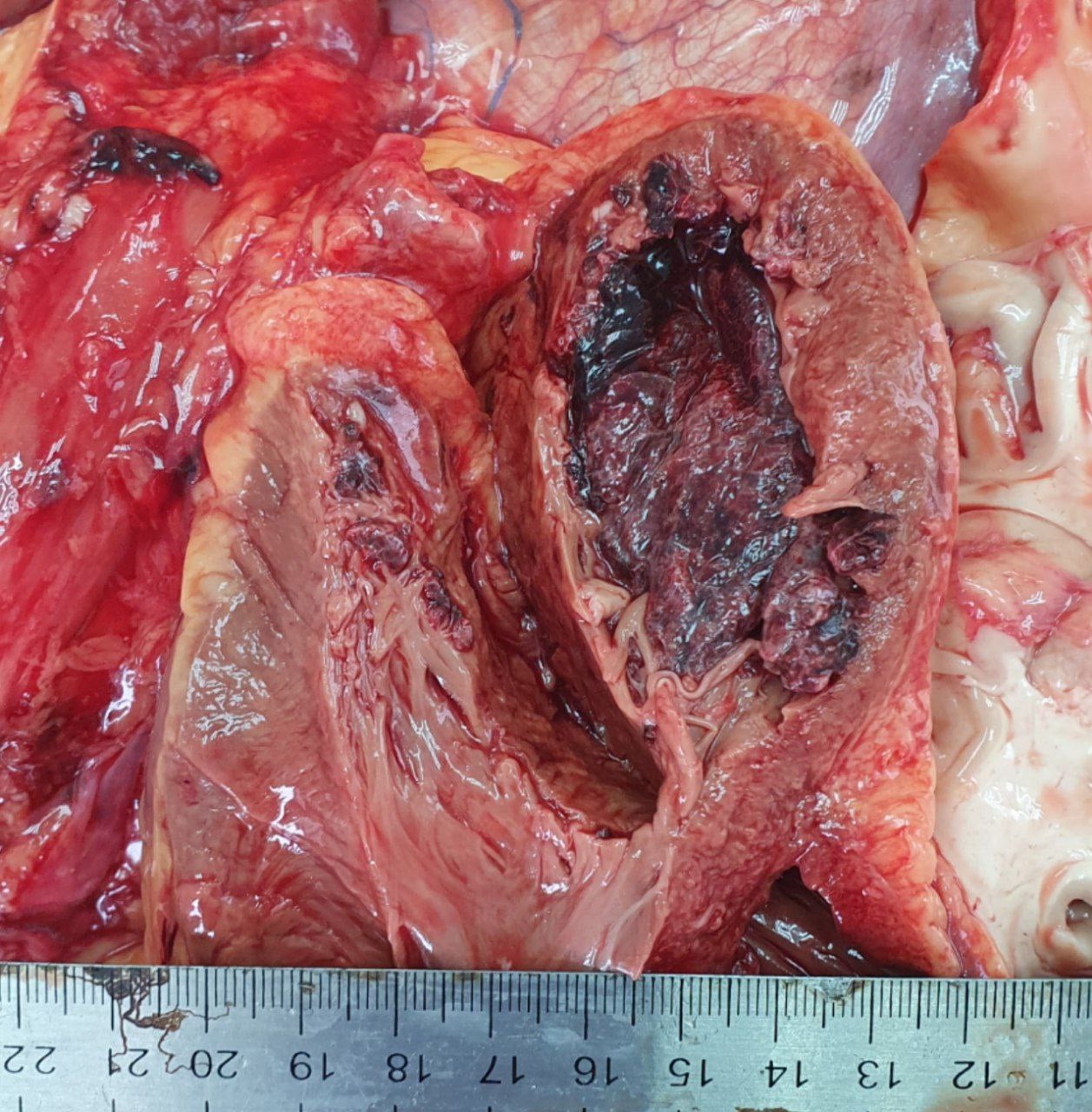
9. Autopsy of a man admitted with a diagnosis of myocardial infarction.
A massive rupture of the left ventricular wall was found, the heart surrounded by blood clots, and thrombi within the cavity itself.
A classic fatal scenario for myocardial rupture: first, a section of the muscle dies due to acute oxygen deprivation, the tissue softens, and the pressure of the blood literally tears through it. The result is almost instantaneous death from cardiac tamponade and blood loss.


 10.
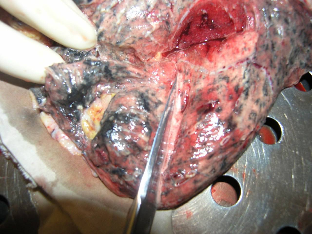
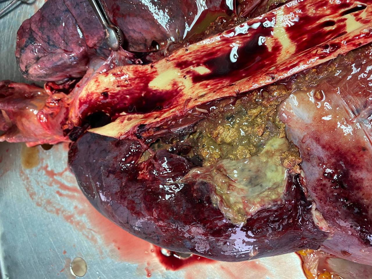
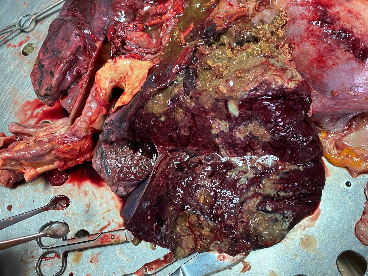
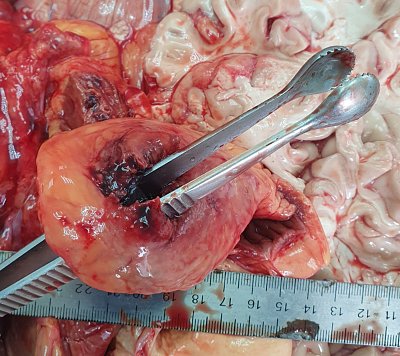
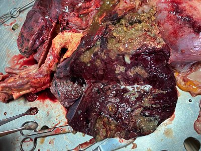
10. Death occurred against the background of lung gangrene in a patient with a tracheoesophageal fistula — a defect connecting the trachea and esophagus, causing food and liquids to enter directly into the airways. Through this ‘hole,’ food was regularly aspirated into the lung, which provoked chronic inflammation and, ultimately, purulent destruction of lung tissue.
The picture was compounded by an advanced oncological process. At autopsy, multiple metastases were found in all groups of lymph nodes and in the liver.
 11.
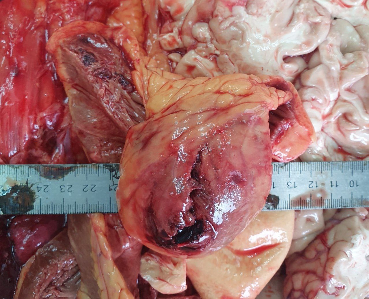
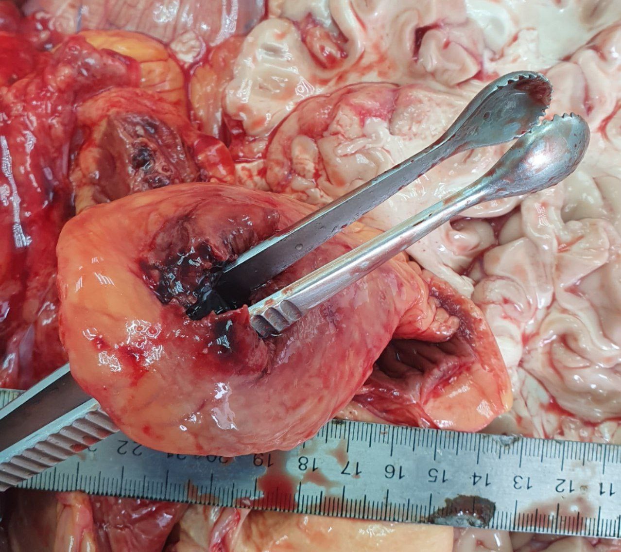
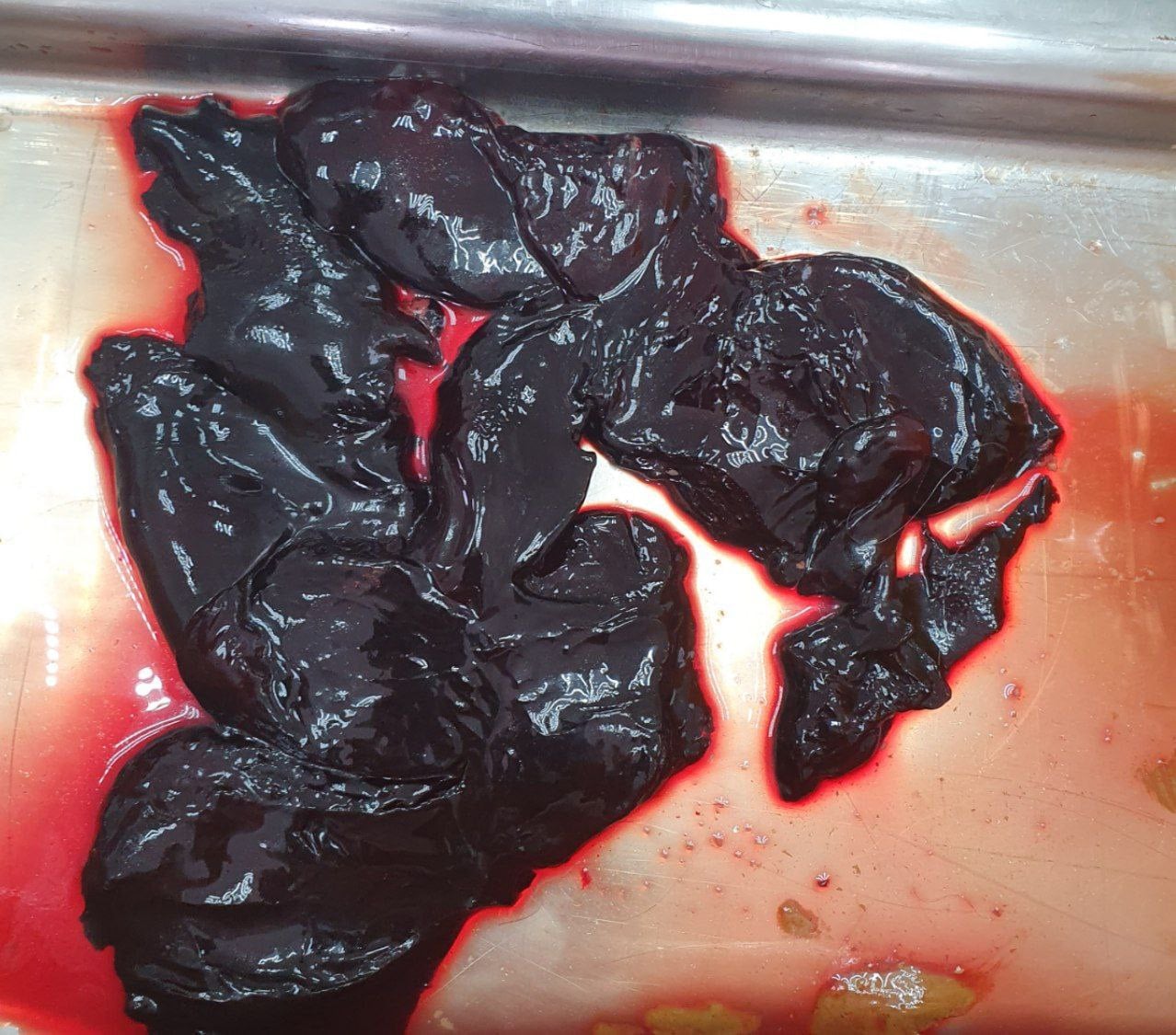
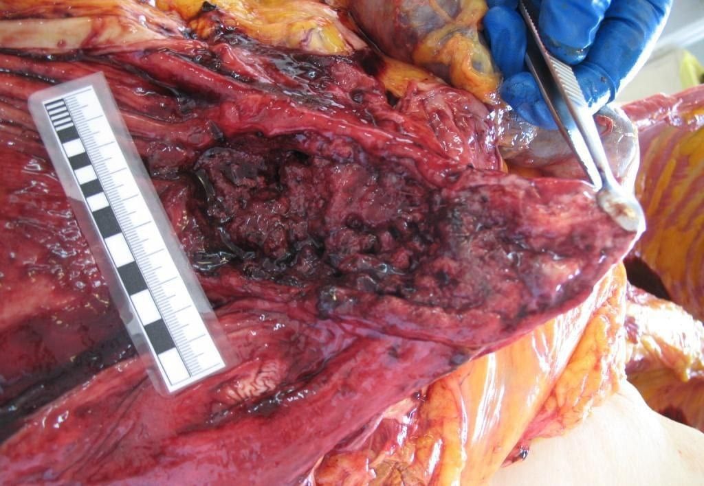
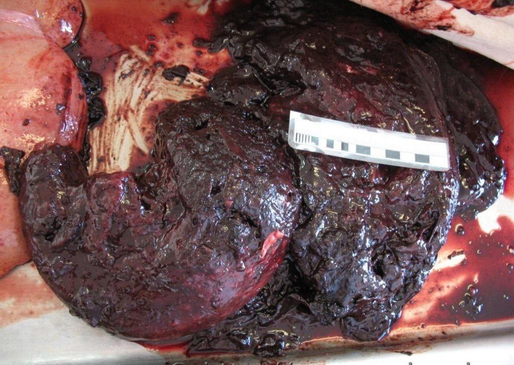
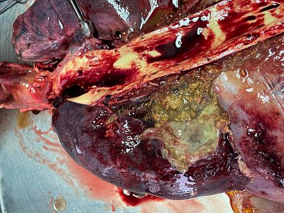
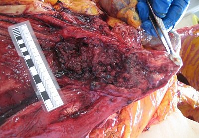
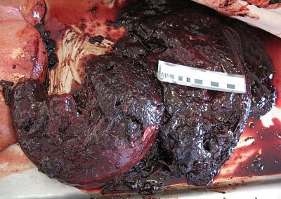
11. An elderly man passed away without ever knowing his diagnosis, as stomach cancer had gone unnoticed during his lifetime. At autopsy, an extensive gastric ulcer with signs of malignant growth was found (photo 1), along with a large amount of clotted blood that was removed from the stomach cavity (photo 2).
The cause of death was gastrointestinal bleeding due to tumour breakdown. Early symptoms are often vague, and the disease is sometimes only discovered after death.
 12.
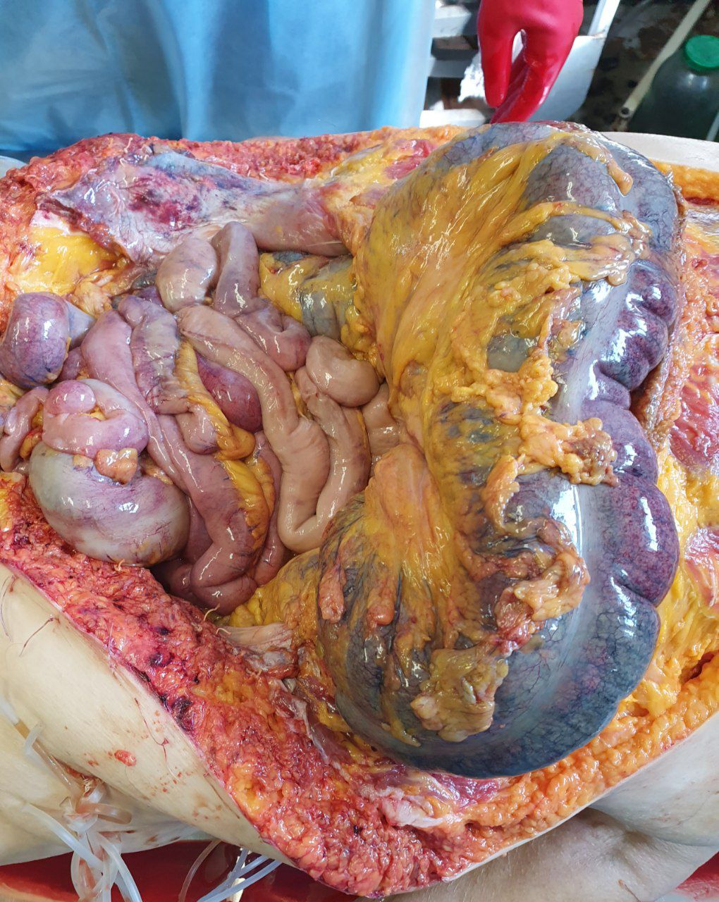
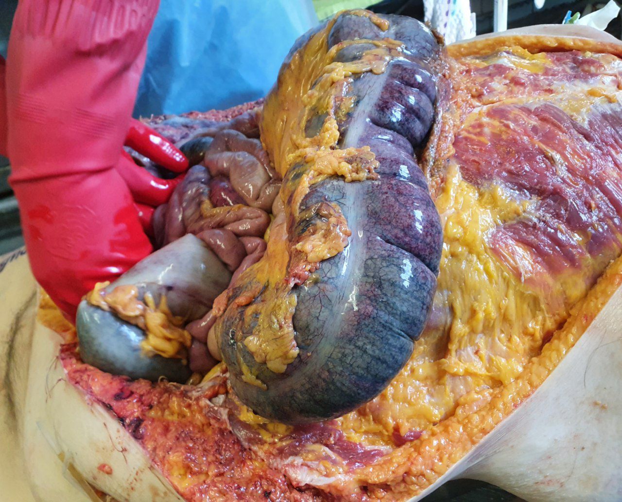
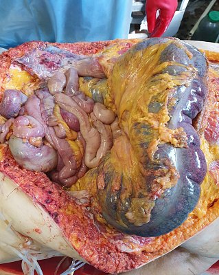
12. Cause of death – acute intestinal obstruction.
The patient was admitted to the hospital with complaints of sudden abdominal pain, severe bloating, and absence of stool and gas. His condition rapidly deteriorated: signs of intoxication, tachycardia, and a drop in blood pressure were observed.
Examination revealed symptoms of peritonitis. Despite emergency surgical intervention, cardiac arrest occurred.

 13.
13. Final stage of malignant colon tumour.
Clinically, such patients often present with symptoms of intestinal obstruction, severe intoxication, rapid weight loss, and signs of metastatic involvement. Even with palliative therapy, the prognosis is unfavourable. The tumour progresses rapidly and leads to death from complications: perforation, peritonitis, cachexia.
Colon cancer remains one of the leading causes of death in oncology, especially when medical care is sought at a late stage.