|
#1
●
06-15-2024, 05:59 PM
| ||||||||
 These are the rooms Poster Rank:26 of ruin. Join Date: Sep 2014 Posts: 54,011 Mentioned: 145 Post(s) Quoted: 30403 Post(s)
| ||||||||
|
Oral Myiasis and Squamous Cell Carcinoma
A 62-year-old woman presented to the ENT clinic of our hospital, with a history of a non-healing ulcer on the floor of mouth for the past six months. On external examination, there was a hard, fixed, and non-tender swelling approximately 6 x 4 cm in size, involving the bilateral submental and the submandibular region. On examination of the oral cavity, there was an ulcerative region on the left lower alveolus. It was approximately 4 x 2 cm in size, extending from the first to the last molar, sparing the retro-molar trigone. The rest of the floor of mouth was normal and mouth opening was adequate. A biopsy was planned which resulted in a diagnosis of well-differentiated squamous cell carcinoma of the left lower alveolus. There were no other comorbidities. She was subsequently referred to the oncology department of our hospital for palliative chemotherapy which was initiated with paclitaxel and carboplatin in April 2023 with 21-day cycles. According to the patient, after receiving the third dose of chemotherapy, a discharging sinus developed between the ulcer and her neck. The emergency department dressed the wound and urgently directed her to follow up at our hospital's ENT clinic. However, she presented to the clinic, three weeks later, with an extensive lesion involving the submental and submandibular regions of the neck. The lesion was necrotic with elevated edges, there was a loss of overlying skin substance, and there were live larvae inside the lesion (Figure 1). The patient admitted to self-dressing her wounds for three weeks, avoiding medical attention due to embarrassment. The malodor associated with the patient's advanced maggot infestation ultimately resulted in the patient’s family taking her to the hospital. Initially, turpentine-soaked dressings were applied to the wound. This was followed by the manual removal of larvae with forceps. After removal, the wound was washed with saline and dressed with 10% povidone-iodine. The larvae were removed on every alternate day, in three sessions. The patient was sent home after each session, with instructions about wound care. She was also instructed to keep her house free of flies. The patient was given 6 mg ivermectin, once daily for three days along with 400mg Ibuprofen on an as-needed basis. No antibiotics were initiated. The number of live larvae was reduced after the first visit and on the third follow-up, only dead maggots remained. On each follow-up visit, the degree of odor and exudation from the wound was less than the preceding one. Following her last visit, when it was ensured that all larvae were removed, she was referred to the plastic surgery department for consultation regarding reconstruction as the infestation had caused enormous tissue loss. Unfortunately, the patient's prognosis is unknown as she did not come for a follow-up after the four initial treatment sessions. 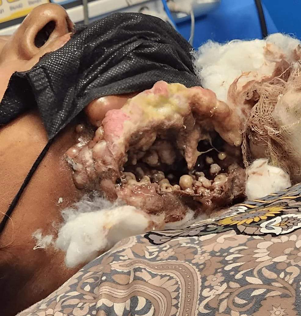 · |
|
#2
●
06-15-2024, 11:52 PM
| ||||||||
 ★ Legacy Member ★ Poster Rank:252 So many choices now Join Date: Jul 2015 Posts: 5,548 Mentioned: 15 Post(s) Quoted: 2120 Post(s)
| ||||||||
|
Re: Oral Myiasis and Squamous Cell Carcinoma
So the smell is what finally got her? Really? The gaping face wound and her ability to whistle without opening her mouth were not enough? I get the embarrassment thing, but sheesh...
|
|
#5
●
06-18-2024, 07:35 PM
| ||||||||
 My Rank: CORPORAL Poster Rank:1272 female Join Date: May 2009 Posts: 488 Mentioned: 0 Post(s) Quoted: 35 Post(s)
| ||||||||
|
Re: Oral Myiasis and Squamous Cell Carcinoma
She's still out there doing nothing about it. Seeing she didn't return for the follow up.  |
|
#6
●
06-18-2024, 09:41 PM
| ||||||||
 ♚ Legacy Gold Member ♚ Poster Rank:1292 Femme fatale Join Date: May 2024 Posts: 477 Mentioned: 1 Post(s) Quoted: 191 Post(s)
| ||||||||
|
Re: Oral Myiasis and Squamous Cell Carcinoma
She must have been getting hungry after all the food falling through her chin. That's what happens when people crap in the street, then vendors sell food from the streets. |