|
#1
●
01-14-2023, 04:09 AM
| ||||||||
| These are the rooms Poster Rank:25 of ruin. Join Date: Sep 2014 Posts: 54,011 Mentioned: 145 Post(s) Quoted: 30403 Post(s)
| ||||||||
|
Knife in the Skull
A 34-year-old male presented to the trauma bay neurologically intact after sustaining a stabbing to the left temporal region (Figure 1). A CT of the head showed that the knife had penetrated the left temporal bone, left sphenoid bone, left pterygopalatine fossa, and left maxillary sinus (Figure 2). A computed tomography angiogram (CTA) showed no obvious vascular injury, but the process was limited due to streak artifact. Since the knife penetrated the maxillary sinus and there was a concern for a vascular injury, the patient was planned for multidisciplinary treatment with neurosurgery, otolaryngology, and neuroendovascular surgery. The patient was taken to the operating room for the planned removal of the knife under endoscopic visualization and wound debridement. A small incision was made around the knife and the endoscope was advanced into the maxillary sinus with immediate visualization of the knife. Under direct visualization, the knife was removed with minimal bleeding. The wound was then debrided of bone fragments and copiously irrigated. The patient was taken immediately to the endovascular suite where digital subtraction angiography (DSA) showed a left distal internal maxillary artery and proximal middle meningeal artery injury . The left distal internal maxillary artery and proximal middle meningeal artery were embolized with Onyx (ev3, Irvine, CA) due to active bleeding. The postoperative head CT showed a very small left temporal lobe hemorrhage with a small amount of pneumocephalus without any hydrocephalus . The patient was extubated on post-injury day one without any cranial nerve palsies or other neurologic deficits and was discharged on postoperative day two. 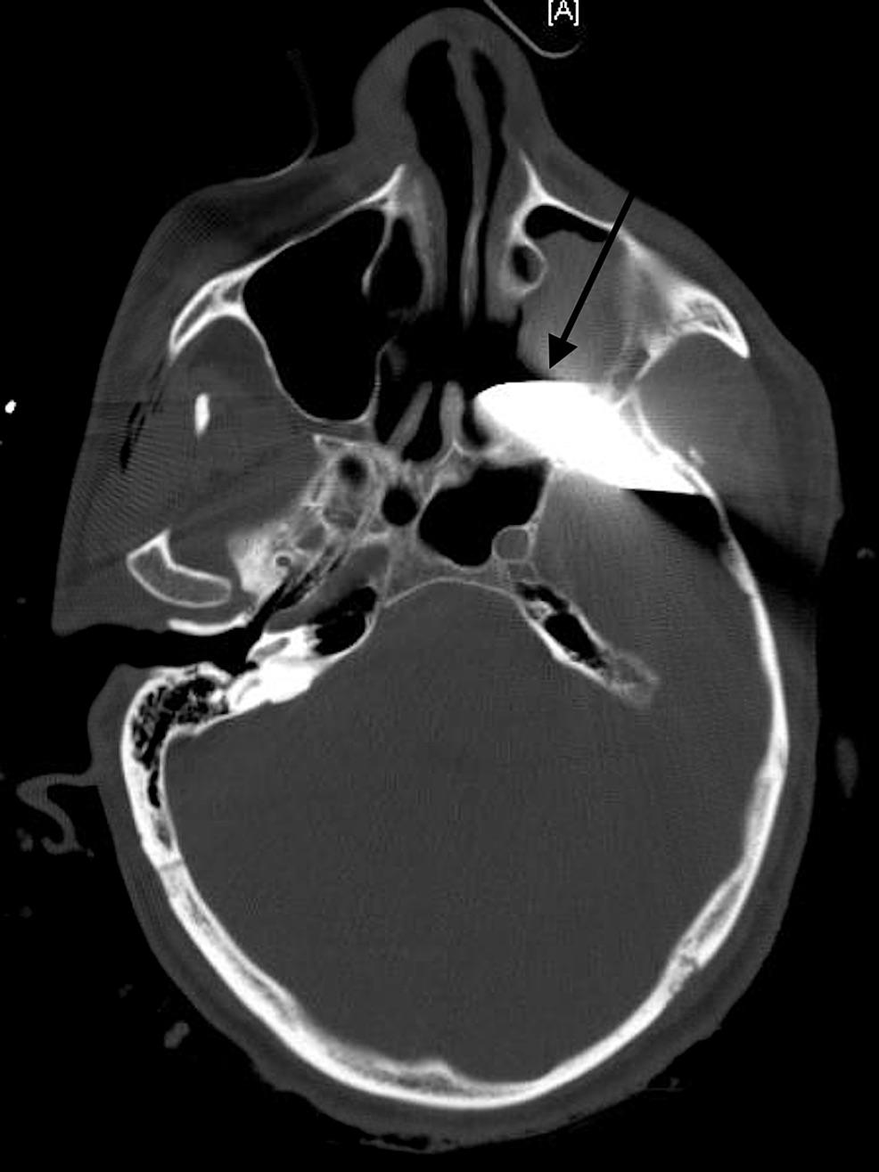 · 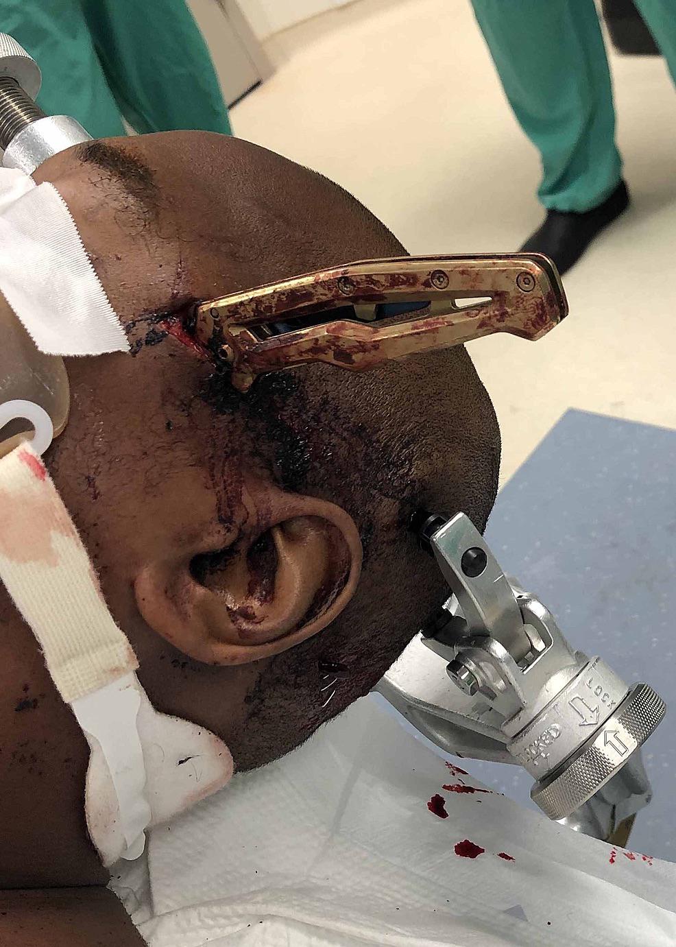 · |
|
#2
●
01-14-2023, 04:16 AM
| ||||||||
| My Rank: FIRST SERGEANT Poster Rank:393 Join Date: Apr 2011 Posts: 3,054 Mentioned: 2 Post(s) Quoted: 276 Post(s)
| ||||||||
|
Re: Knife in the Skull
This is how you crack a coconut |