|
#1
●
08-06-2024, 07:14 PM
| ||||||||
| These are the rooms Poster Rank:25 of ruin. Join Date: Sep 2014 Posts: 54,011 Mentioned: 145 Post(s) Quoted: 30403 Post(s)
| ||||||||
|
IV Drug Use Complications
A 54-year-old man, who was an intravenous drug abuser (IVDA), presented to a hospital nearby with left hand and forearm swelling for a week. There was a necrotic area at the antecubital fossa with surrounding bullae formation as well. Upon initial assessment, there was reduced functionality of the elbow joint due to pain. An ultrasound showed features of left brachial arteriovenous fistula (at the medial antebrachial region) complicated with brachial artery pseudoaneurysm (outpouching with narrow neck seen arising from the left brachial artery measuring 2.4 cm × 1.5 cm) and surrounding hematoma. Subsequently, he started bleeding from his wound site at the antecubital fossa. The patient was then referred to our center. Upon initial assessment, he was noted to be septic with tachycardia up to 120 beats per minute and a temperature of 38.7°C. He also had generalized swelling over his left upper limb from the elbow joint to his fingers with a large necrotic patch over his proximal forearm involving his antecubital fossa. There was bluish discoloration over the fingertip of his fifth digit. There was also a deep wound over the lateral aspect of the necrotic patch with an active arterial bleed (Figure 1). A bedside Doppler ultrasound showed a monophasic signal over the distal radial artery and no blood flow over the distal ulnar artery. A biphasic Doppler signal was obtained over the proximal brachial artery above the necrotic patch. He underwent left brachial artery exploration, ligation, and proximal brachial artery to distal radial artery bypass using the right long saphenous vein graft. Intraoperatively, wound exploration showed grossly infected tissue with some necrotic muscle tissue and a hematoma of about 200 ml. The brachial artery had ruptured just above the cubital fossa with no backflow distally. The brachial artery was then ligated proximally at the proximal edge of the wound, then distal dissection was done until brachial artery bifurcation where the proximal radial and ulnar arteries were ligated separately (Figure 2). A full-length great saphenous vein (GSV) was harvested. The vein was reversed and tunneled subcutaneously along normal (non-infected) tissue lateral to the wound. Anastomosis was done at the proximal brachial artery and distal radial artery (Figures 3, 4). Postoperative examination showed biphasic signal over graft, and distal radius and ulnar arteries. Blood culture, pus culture, and tissue culture grew mixed organisms with the Staphylococcus group predominating in all cultures. The patient was then given antibiotics, wound care, and physiotherapy post surgery. He was discharged home well with outpatient follow-up for his wound (Figure 5). 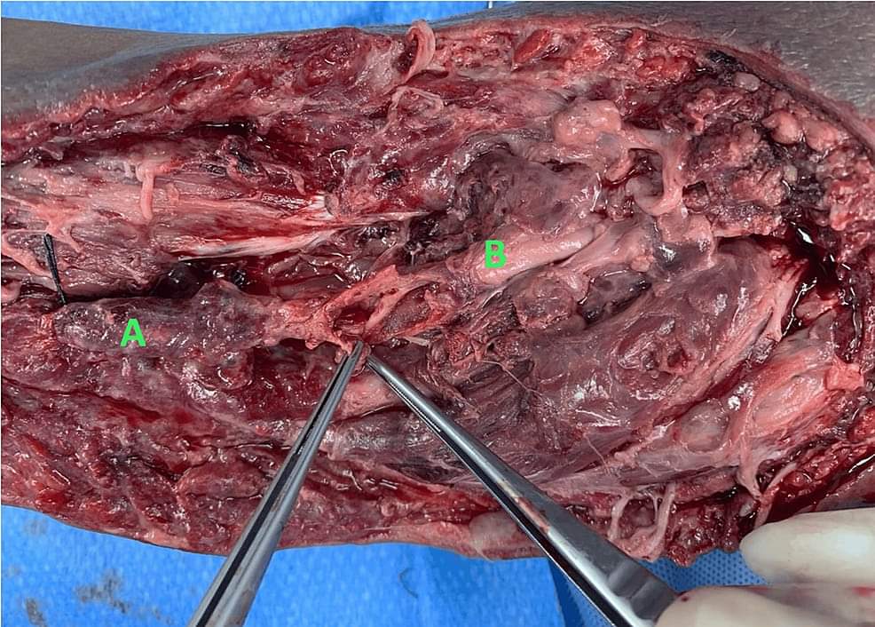 · 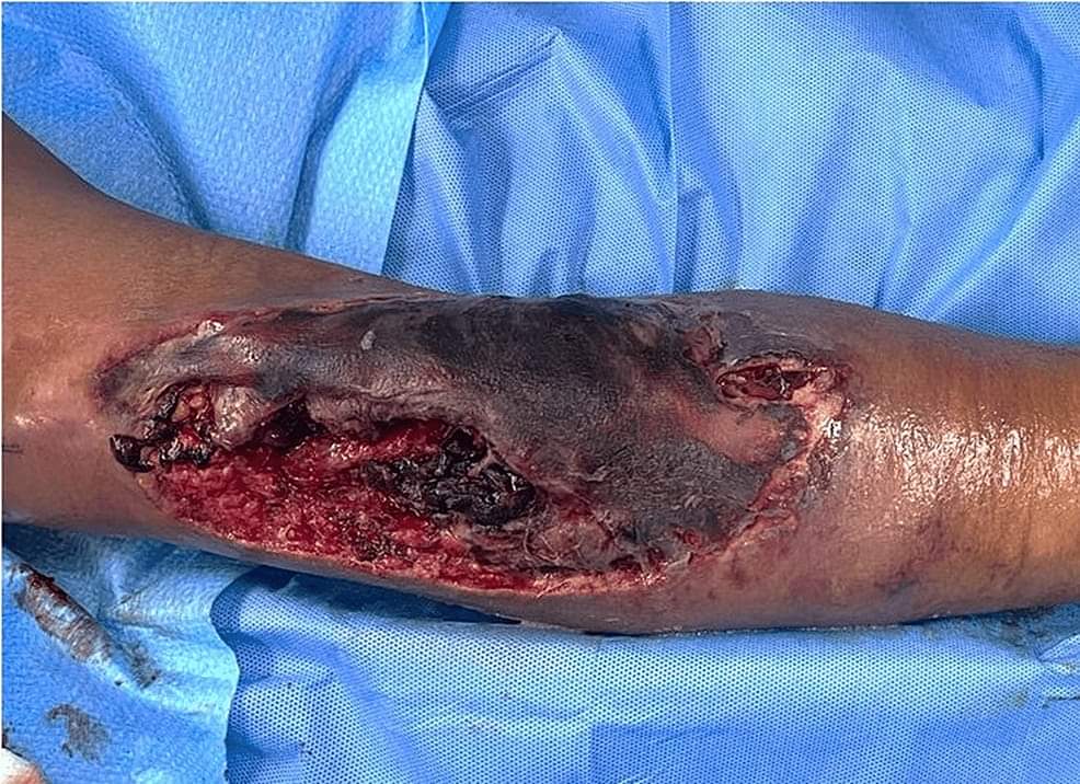 · 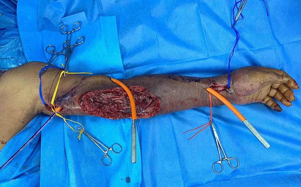 · 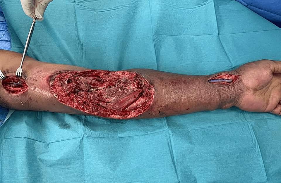 · 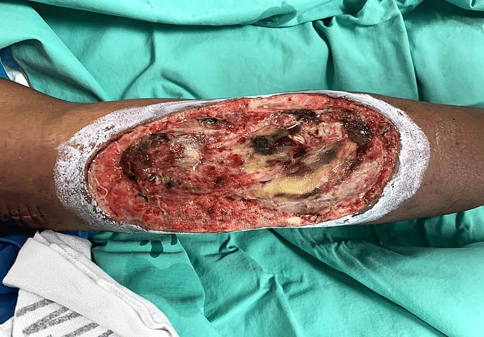 · |
|
#2
●
08-06-2024, 09:06 PM
| ||||||||
| My Rank: SERGEANT Poster Rank:968 Join Date: Jul 2023 Posts: 731 Mentioned: 0 Post(s) Quoted: 106 Post(s)
| ||||||||
|
Re: IV Drug Use Complications
gnarlah. fig 1's looking somewhat similar to the shredded pork sandwich i just ate. |
|
#3
●
08-06-2024, 10:14 PM
| ||||||||
 ♚ Legacy Gold Member ♚ Poster Rank:1280 Femme fatale Join Date: May 2024 Posts: 477 Mentioned: 1 Post(s) Quoted: 191 Post(s)
| ||||||||
|
Re: IV Drug Use Complications
Doctors have so much patience, at that point I'd be like just cut it off. And looking at that first picture I wouldn't even know what is what, I guess that's why I'm not a dr. |
|
#6
●
08-07-2024, 03:23 AM
| ||||||||
| ✝Mudderator from Hell✝ Poster Rank:10 e-mail Join Date: Oct 2006 Posts: 94,986
Contributions: 817
Mentioned: 472 Post(s) Quoted: 10081 Post(s)
| ||||||||
|
Re: IV Drug Use Complications
i wonder if he's still a drug abuser after all this.
|
|
#7
●
08-07-2024, 11:48 AM
| ||||||||
| ★ Legacy Member ★ Poster Rank:119 Secret Agent Join Date: Dec 2009 Posts: 13,216 Mentioned: 6 Post(s) Quoted: 2789 Post(s)
| ||||||||
|
Re: IV Drug Use Complications
I think it would be better if they cut that arm off at his neck. |
|
#8
●
08-07-2024, 02:06 PM
| ||||||||
| My Rank: MAJOR Poster Rank:118 Male Join Date: Feb 2010 Posts: 13,428 Mentioned: 15 Post(s) Quoted: 3534 Post(s)
| ||||||||
|
Re: IV Drug Use Complications
This should have been a wake up call but I think the urge made him start shooting that shit up again as soon as he left the hospital. It’s tragic .
|