|
#1
●
08-30-2024, 07:05 PM
| ||||||||
| These are the rooms Poster Rank:25 of ruin. Join Date: Sep 2014 Posts: 54,011 Mentioned: 145 Post(s) Quoted: 30403 Post(s)
| ||||||||
|
Building a Face from Scratch
A 15-year-old child was admitted to our department with severe scars in the craniofacial region in December 2015 (Fig. 1). When he was 4 months old, the child’s head and face were severely burned due to an accidental fire at home, resulting in extensive disfiguring scars. Due to family financial difficulties, the child did not receive timely treatment. Over the years, the scars on the top of the head have repeatedly broken. He was afraid to go out because of his ugly appearance. Volunteers found him, encouraged him to contact a foundation for financial support, and sent him to our department for head and face reconstruction. On admission, his eyelids failed to close due to severe cicatricial valgus, resulting in corneal leukoplakia and visual impairment. He underwent eight plastic surgery procedures in 10 months. The first operation was performed to implant an 800-ml dilator in his right chest and to release the scars on the eyelids and apply an autologous skin graft, which temporarily solved the problem of him not being able to close his eyes. After an inflating period of 4 months, the flap was delayed in the second operation. One week later, the third operation was performed. A Doppler flowmeter was used to determine the course of the cervical cutaneous branch of the transverse cervical artery, which was then marked on the surface of the skin. After removing the expander, the perforator skin flap was turned upward to repair the facial scar above the bilateral oral horn and perform autologous skin grafting around the pedicle (Fig. 2a, b, c, d). Perforations and sutures were applied where the eyes and nostrils were to be located on the skin flaps. At approximately 2 weeks after the third operation, the pedicle of the supraclavicular perforator flap was cut off and transferred to resurface the face below the mouth corners (Fig. 3). After half a month, the fifth operation was performed to further open the eyelids and corners of the mouth. A sixth operation was performed after 1 week. A prosthesis was implanted in the nose, and a skin graft was placed on the forehead wound. After the facial plastic surgery was completed, in May 2016, we placed two dilators on the top of the head and the occipital part of the patient (Fig. 4a and b). After 4 and a half months of inflation, the eighth operation was performed in collaboration with a neurosurgeon (Fig. 5). According to the preoperative CT reconstruction of the skull, a titanium mesh for repairing the skull defect was printed. The dilator was removed during the operation, and the neurosurgeon used a titanium nail to fix the titanium mesh. We covered the wound with a well-expanded scalp flap. Skin necrosis occurred at the margin of the reconstructed prefrontal region at the distal end of the flap, and all the other flaps and skin grafts survived. After 3 years of follow-up, the child’s head and face have achieved satisfactory aesthetic remodeling and functional recovery (Fig. 6). 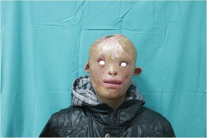 · 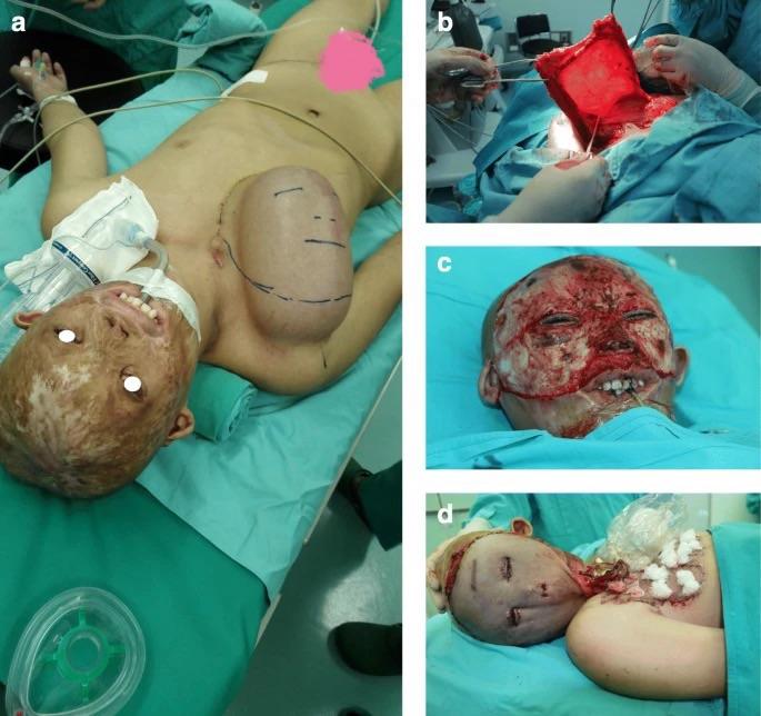 · 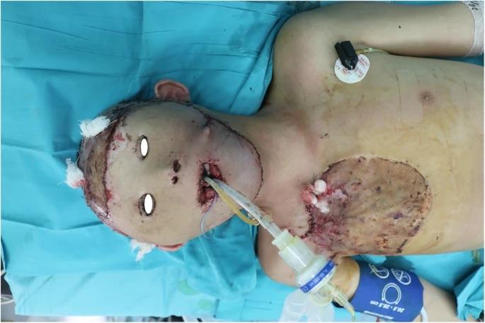 · 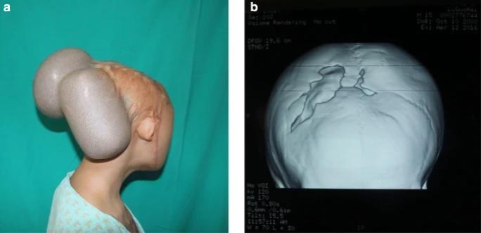 · 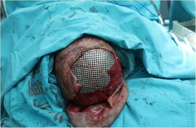 · 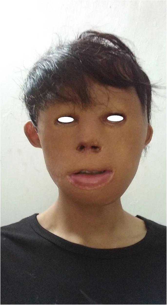 · |
|
#2
●
08-30-2024, 09:37 PM
| ||||||||
| ★ Legacy Member ★ Poster Rank:119 Secret Agent Join Date: Dec 2009 Posts: 13,185 Mentioned: 6 Post(s) Quoted: 2787 Post(s)
| ||||||||
|
Re: Building a Face from Scratch
Great improvement! Amazing surgeons! |
|
#3
●
08-30-2024, 10:51 PM
| ||||||||
| My Rank: CORPORAL Poster Rank:1285 female Join Date: May 2009 Posts: 474 Mentioned: 0 Post(s) Quoted: 35 Post(s)
| ||||||||
|
Re: Building a Face from Scratch
Almost new - ish. Amazing what medical science is capable of. Now cure cancer.
|
|
#4
●
08-31-2024, 02:07 AM
| ||||||||
| ✝Mudderator from Hell✝ Poster Rank:10 e-mail Join Date: Oct 2006 Posts: 94,965
Contributions: 817
Mentioned: 472 Post(s) Quoted: 10077 Post(s)
| ||||||||
|
Re: Building a Face from Scratch
Background: The reconstruction of large head and face missing structures in the craniofacial region in children is very challenging for plastic surgeons. Expanded local and expanded axial-pattern flaps are widely used for the reconstruction of large-area scars. Free flaps are used very cautiously in children. 3D printing technology is a new technology with great development potential. 3D printing technology is used to assist in individualizing titanium alloy restorations for prefabricated skull defect repair. This application has great advantages in the repair of large skull loss. However, it is crucial to choose appropriate techniques and treat deformities of the head and face with integrated approaches and collaboration among multiple departments. In this study, through the perfect combination of the expanded flap and 3D printing technology, a large-area missing structures of a child’s head and face was successfully repaired. Case presentation: This study proposes a method to combine the expanded flap method and 3D printing technology to achieve natural remodeling of the craniofacial region in a child. Conclusion: Large area of head and face missing structures can be reconstructed by using expanded skin flaps combined with 3D printing, and patients can get better new faces. |