JavaScript and Cookies are required to view this site. Please enable both in your browser settings.
Scanning Electron Microscope Images

Scanning Electron Microscope Images 

Current Rating:

Unlimited Views No Ads No Algorithms Lifetime Account

Documenting Reality

Community Forum · Est. 2006

Join Now
Thread Tools
  #1  
06-14-2012, 04:43 AM
dean mclean's Avatar
dean mclean
Offline:
My Rank: MASTER SERGEANT
Poster Rank:497
male
Join Date: Apr 2012
Posts: 2,080
Contributions: 1
 
Mentioned: 2 Post(s)
Quoted: 24 Post(s)
Activity Longevity
0/20 15/20
Today Posts
0/11 sssss2080
Scanning Electron Microscope Images

A scanning electron microscope (SEM) is a type of electron microscope that images a sample by scanning it with a beam of electrons in a raster scan pattern. The electrons interact with the atoms that make up the sample producing signals that contain information about the sample's surface topography, composition, and other properties such as electrical conductivity.

Magnification in a SEM can be controlled over a range of up to 6 orders of magnitude from about 10 to 500,000 times. Unlike optical and transmission electron microscopes, image magnification in the SEM is not a function of the power of the objective lens. SEMs may have condenser and objective lenses, but their function is to focus the beam to a spot, and not to image the specimen. Provided the electron gun can generate a beam with sufficiently small diameter, a SEM could in principle work entirely without condenser or objective lenses, although it might not be very versatile or achieve very high resolution. In a SEM, as in scanning probe microscopy, magnification results from the ratio of the dimensions of the raster on the specimen and the raster on the display device. Assuming that the display screen has a fixed size, higher magnification results from reducing the size of the raster on the specimen, and vice versa. Magnification is therefore controlled by the current supplied to the x, y scanning coils, or the voltage supplied to the x, y deflector plates, and not by objective lens power.
in order-

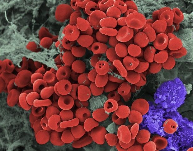
blood clot on gauze strip removed from a wound
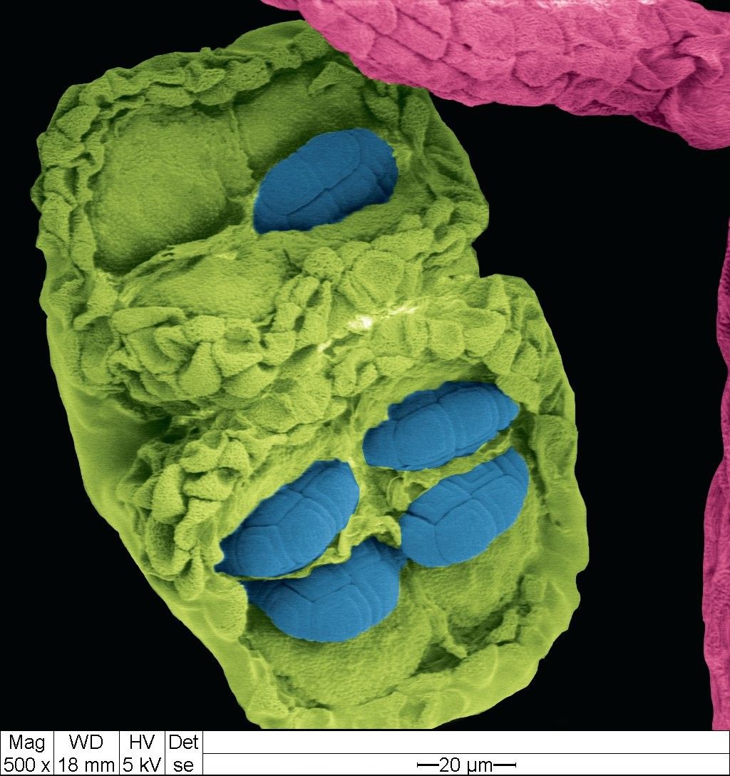
acacia dealbata flower- pollen (blue), anther(green), and filament(pink)
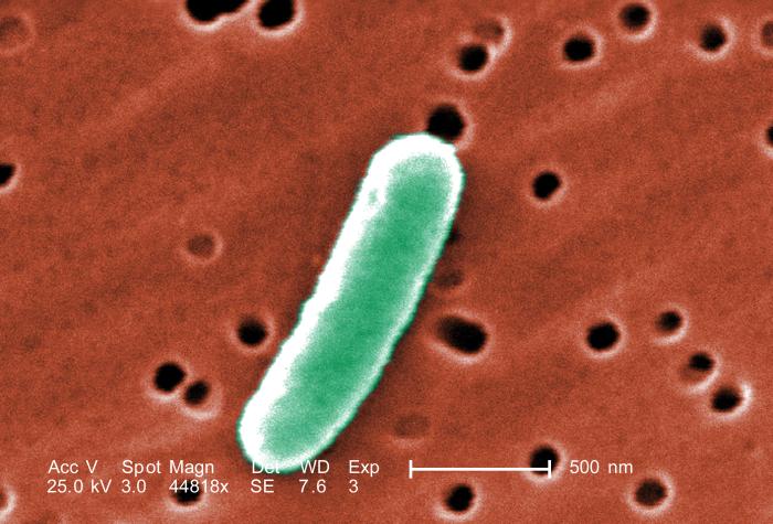
e. coli cells
blood cell growing into a melanoma
hamster oocyte-cumulus complex (fertilization area in the uterus)
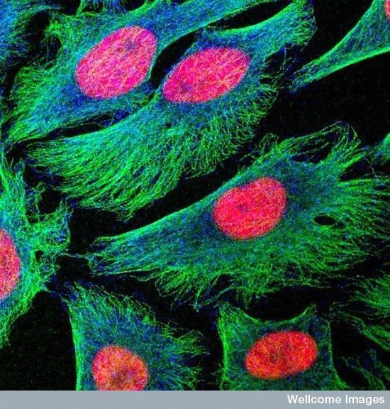
HeLa cancer cells
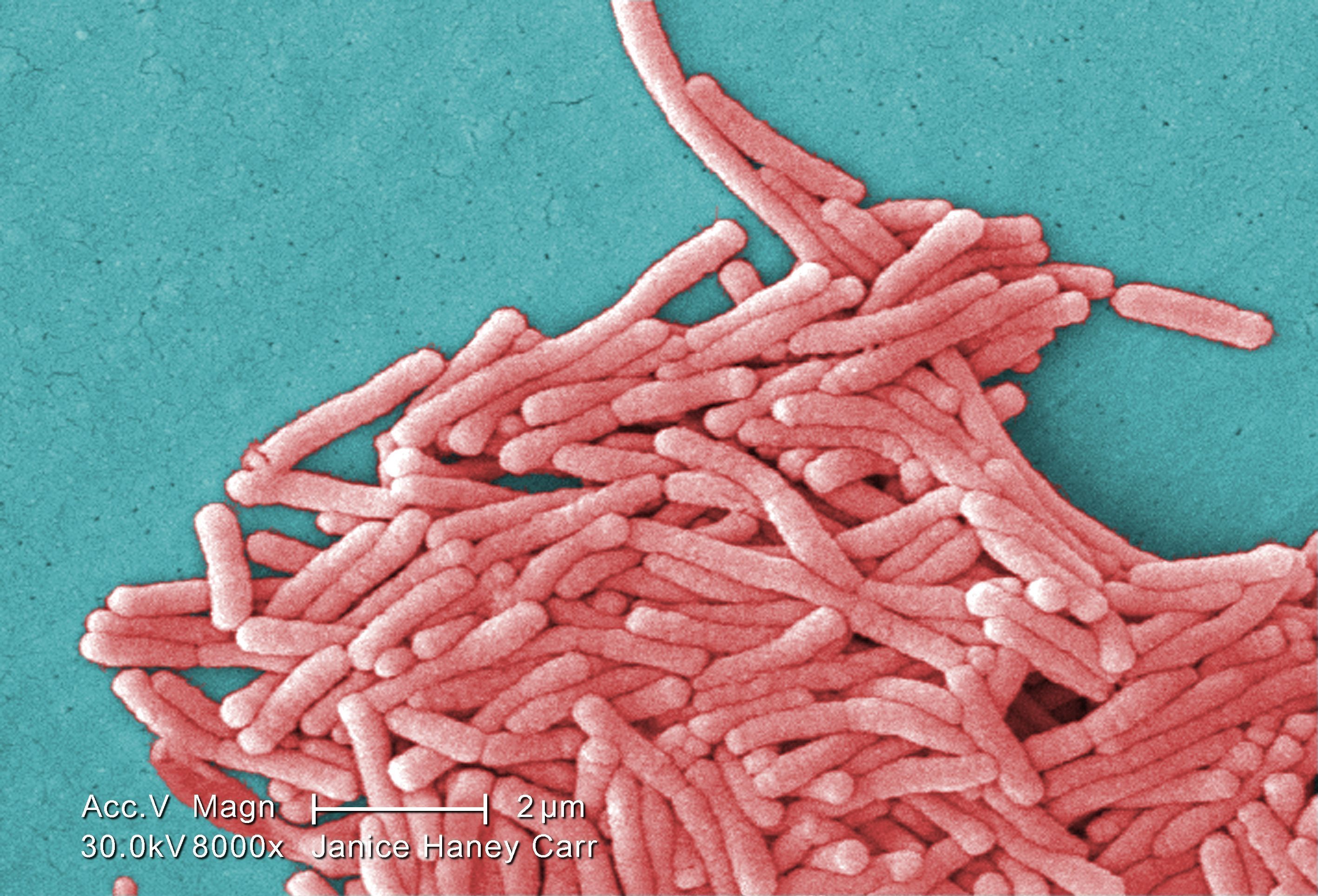
legionella bacteria
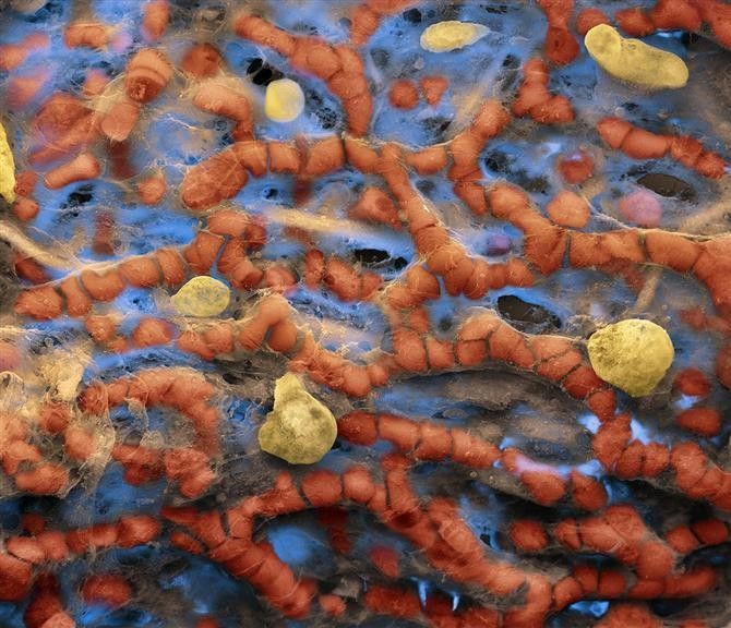
liver sinusoid of a brown rat
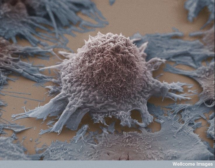
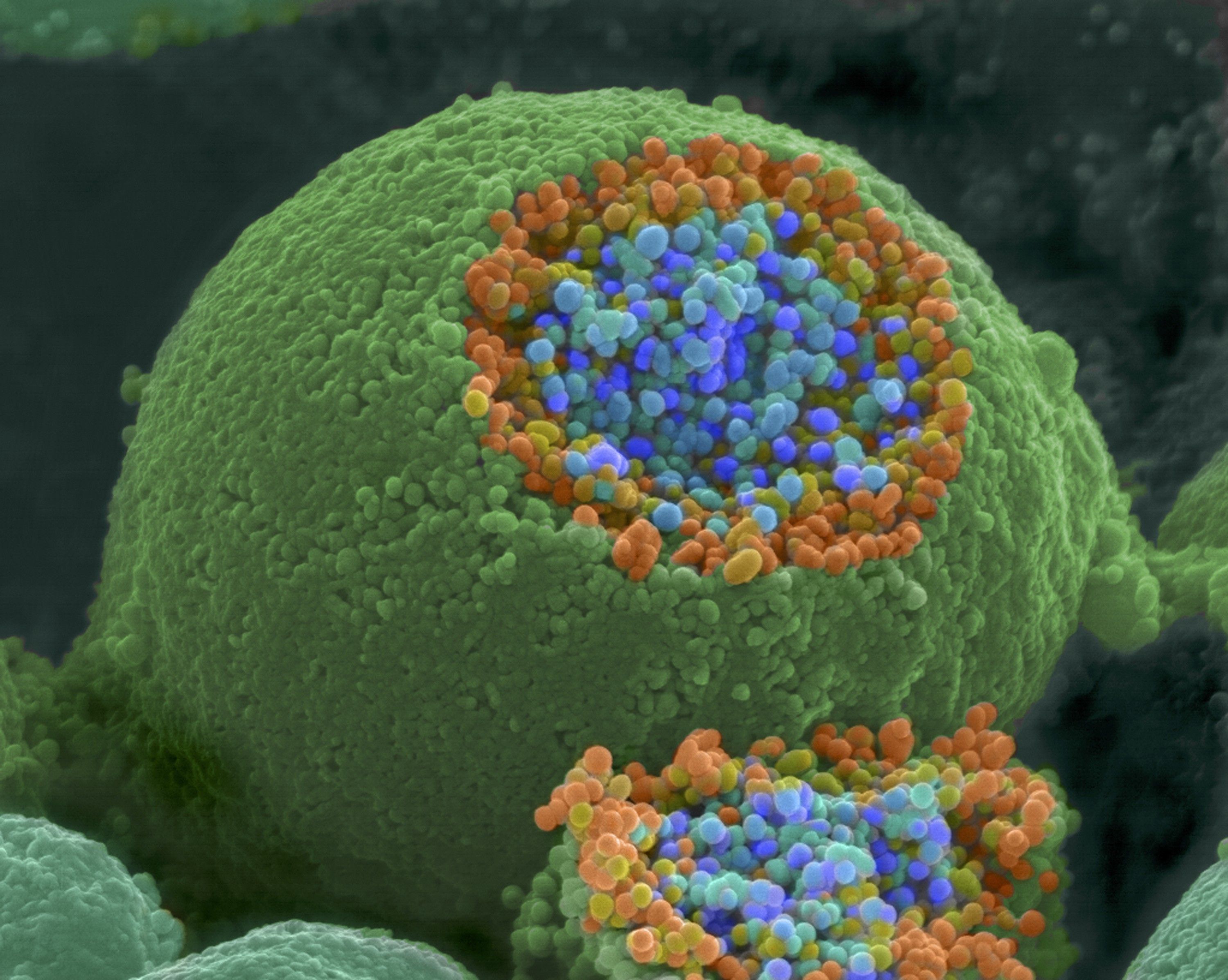
lung cancer cell
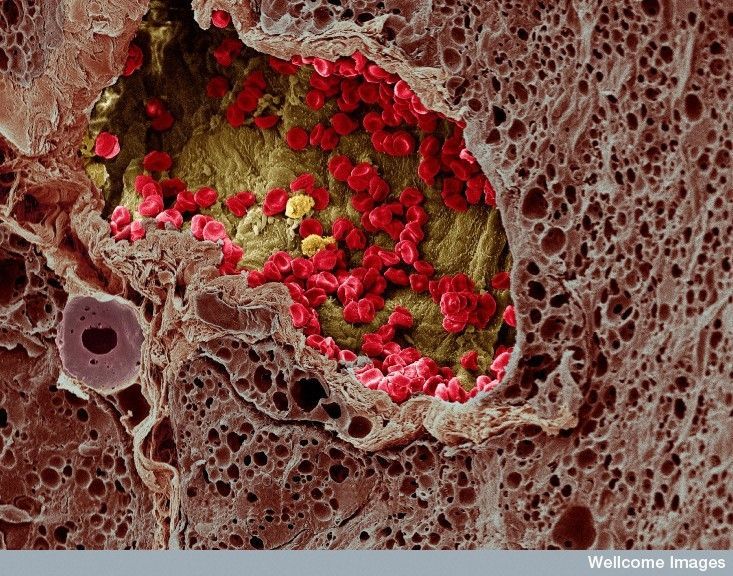
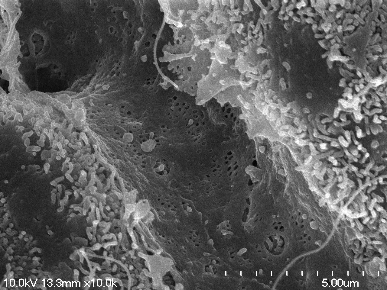
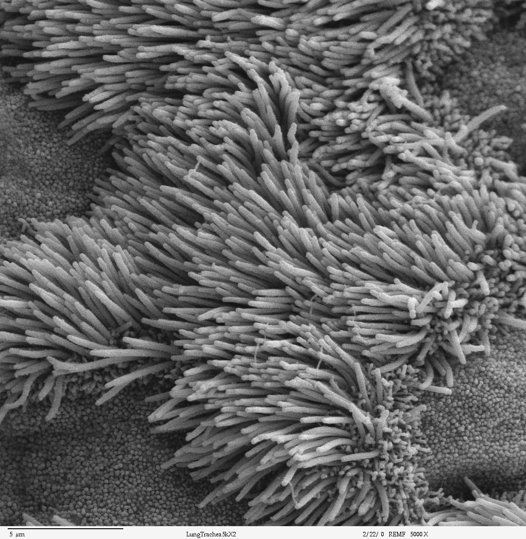
inner wall of a lung
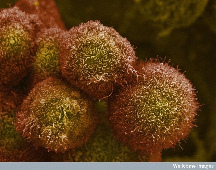
pancreatic cancer cell
nerve ending broken to show synaptic vessels
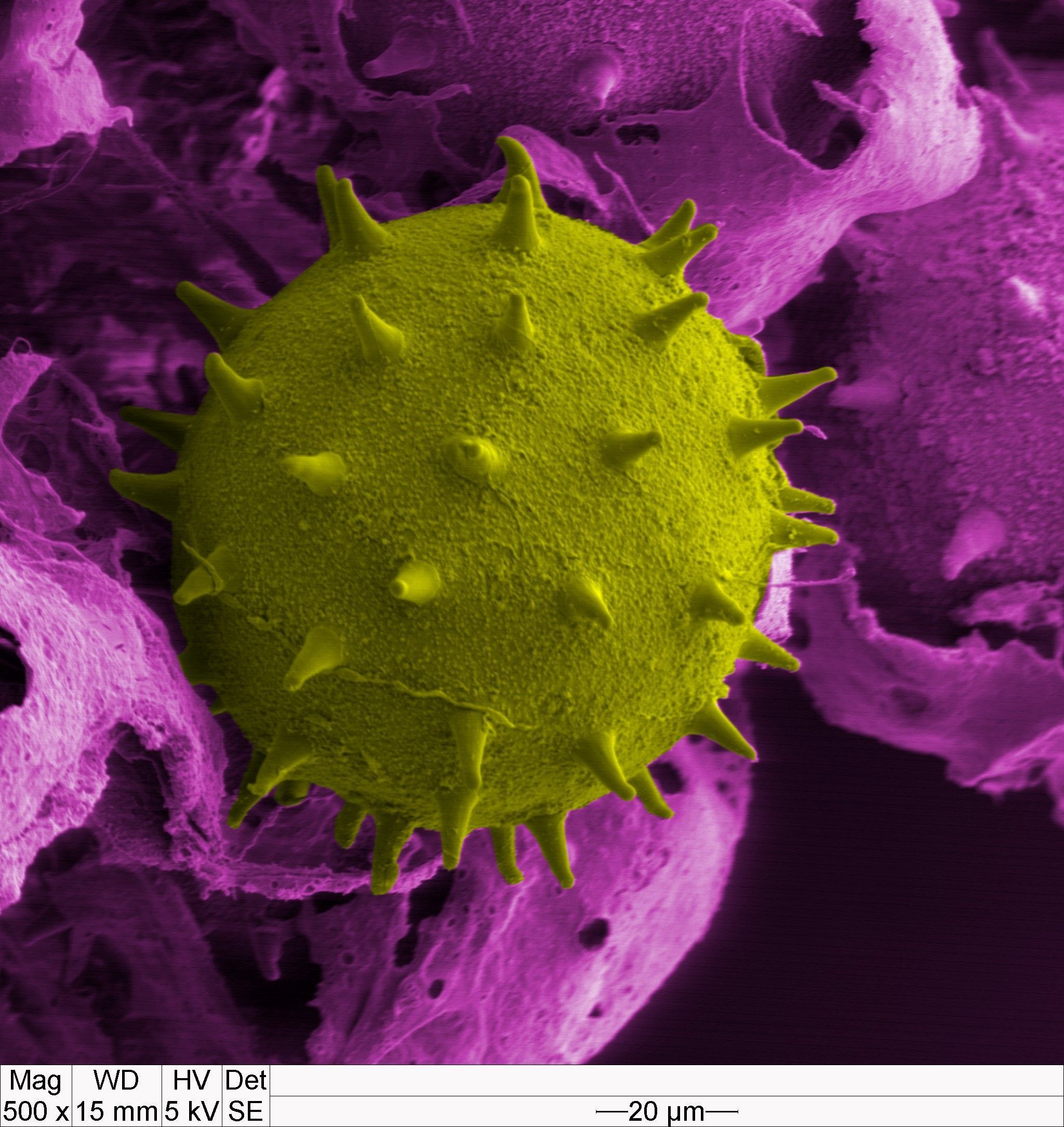
pollen
red blood cells within alveolar capillary network
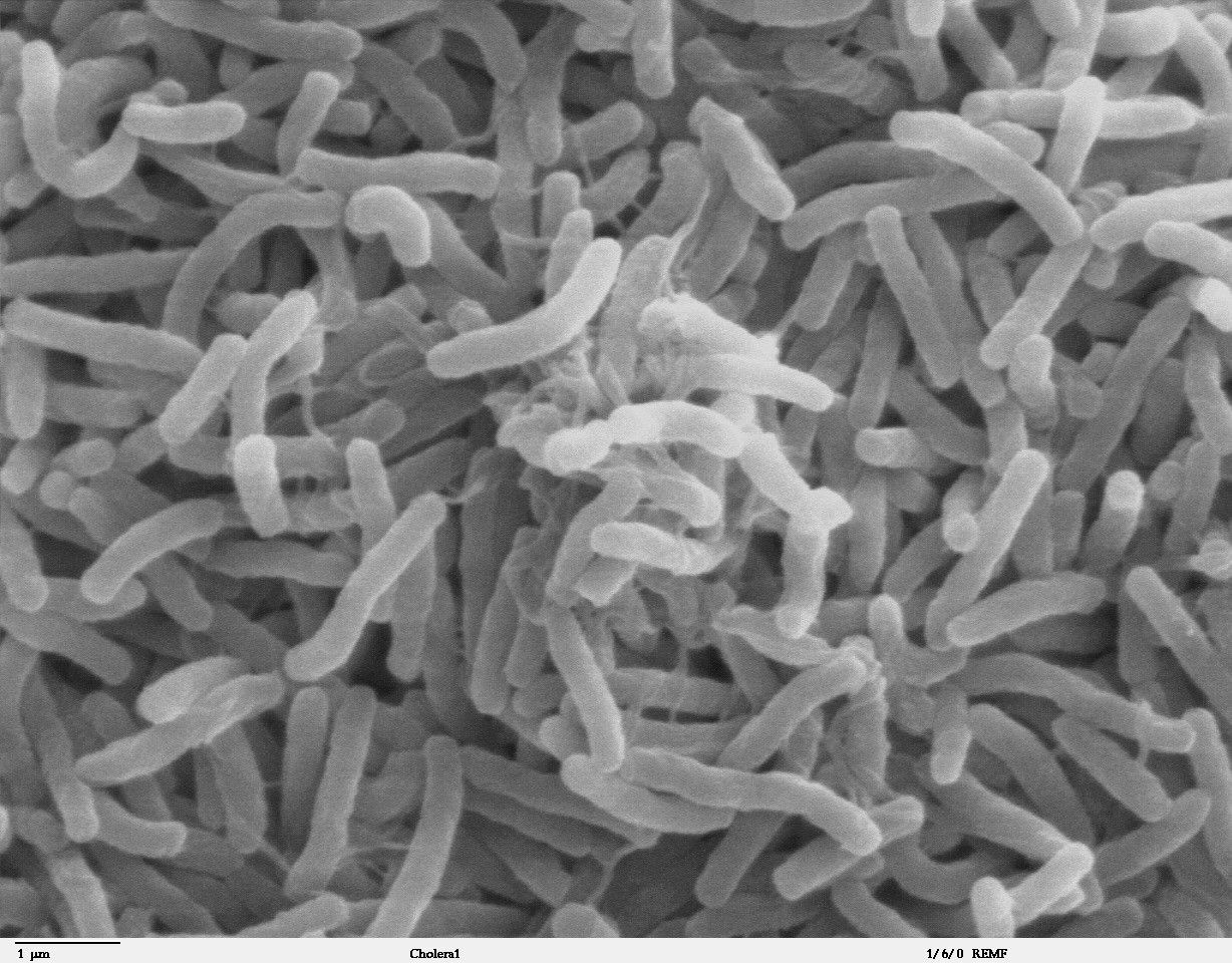
cholera
soybean cyst nematode and egg
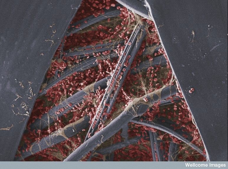
blood_clot_on_gauze.jpg
158.2 KB ·1501 views
acacia_dealbata_pollen_anther_filament.jpg
221.7 KB ·1073 views
ecoli.jpg
56.5 KB ·676 views
blood_vessel_grown_into_melanoma.jpg
215.4 KB ·2242 views
hamster_oocyte_cumulus_complex.jpg
97.3 KB ·859 views
HeLa_cancer_cells.jpg
151.0 KB ·1500 views
legionella_pneumophila__sem_.jpg
744.9 KB ·867 views
liver_sinusoid_of_brown_rat.jpg
413.0 KB ·3088 views
lung_cancer_cell.jpg
107.7 KB ·3534 views
lungtrachea2a.jpg
459.9 KB ·4703 views
pancreatic_cancer_cell.jpg
122.3 KB ·1718 views
nerve_ending_with_synaptic_vessels.jpg
950.1 KB ·4877 views
pollen.jpg
702.1 KB ·1684 views
red_blood_cells_within_alveola_capillary_network.jpg
117.5 KB ·815 views
Vibrio_cholerae.jpg
328.6 KB ·583 views
Soybean_cyst_nematode_and_egg_SEM.jpg
970.0 KB ·1448 views
4 Users Say Thank You For This Post:
Arsedestroyer, Ashen, Fulcanelli, MCBR
▼ PROMO FROM DOCUMENTING REALITY
No surprises here
Join Now
Hidden for upgraded members.
  #2  
06-14-2012, 07:30 AM
Fulcanelli's Avatar
Fulcanelli
Offline:
My Rank: MASTER SERGEANT
Poster Rank:507
Join Date: Mar 2008
Posts: 2,047
 
Mentioned: 3 Post(s)
Quoted: 163 Post(s)
Activity Longevity
1/20 19/20
Today Posts
0/11 sssss2047
Re: Scanning Electron Microscope Images

Amazing, thanks for sharing
  #3  
06-14-2012, 08:16 AM
MCBR's Avatar
MCBR
Offline:
So Fucking Banned
Poster Rank:124
Join Date: Sep 2009
Posts: 12,829
 
Mentioned: 17 Post(s)
Quoted: 2350 Post(s)
Activity Longevity
0/20 17/20
Today Posts
0/11 ssss12829
Re: Scanning Electron Microscope Images

  #4  
06-14-2012, 05:20 PM
kasapin's Avatar
kasapin
Offline:
My Rank: STAFF SERGEANT
Poster Rank:815
Join Date: May 2010
Posts: 955
 
Mentioned: 0 Post(s)
Quoted: 282 Post(s)
Activity Longevity
0/20 17/20
Today Posts
0/11 ssssss955
Re: Scanning Electron Microscope Images

thanks for sharing .. i am always amazed by that kind of pictures.


Powered by vBulletin Copyright 2000-2010 Jelsoft Enterprises Limited.

Search Engine Friendly URLs by vBSEO