A scanning electron microscope (SEM) is a type of electron microscope that images a sample by scanning it with a beam of electrons in a raster scan pattern. The electrons interact with the atoms that make up the sample producing signals that contain information about the sample's surface topography, composition, and other properties such as electrical conductivity.
Magnification in a SEM can be controlled over a range of up to 6 orders of magnitude from about 10 to 500,000 times. Unlike optical and transmission electron microscopes, image magnification in the SEM is not a function of the power of the objective lens. SEMs may have condenser and objective lenses, but their function is to focus the beam to a spot, and not to image the specimen. Provided the electron gun can generate a beam with sufficiently small diameter, a SEM could in principle work entirely without condenser or objective lenses, although it might not be very versatile or achieve very high resolution. In a SEM, as in scanning probe microscopy, magnification results from the ratio of the dimensions of the raster on the specimen and the raster on the display device. Assuming that the display screen has a fixed size, higher magnification results from reducing the size of the raster on the specimen, and vice versa. Magnification is therefore controlled by the current supplied to the x, y scanning coils, or the voltage supplied to the x, y deflector plates, and not by objective lens power.
in order-
blood clot on gauze strip removed from a wound
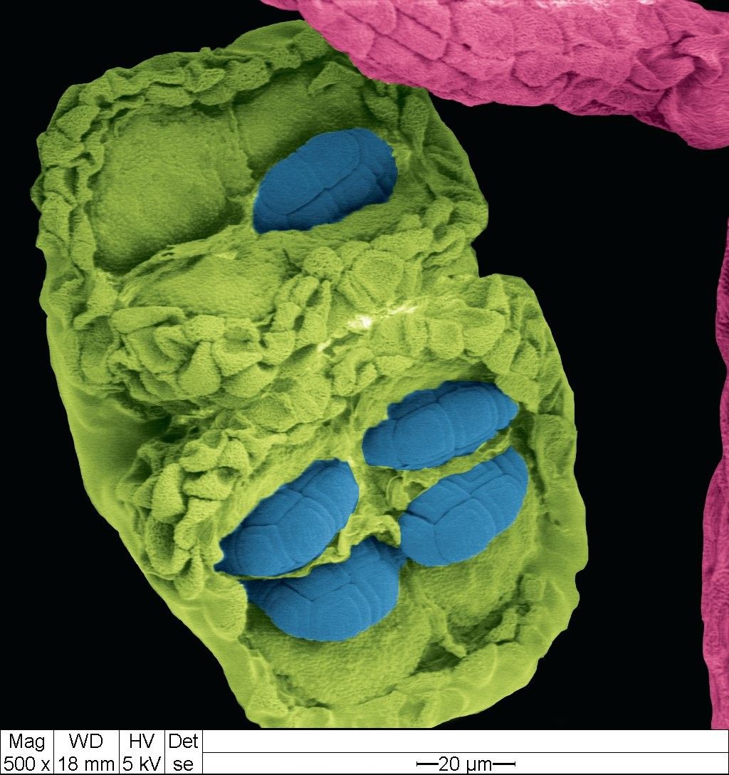
acacia dealbata flower- pollen (blue), anther(green), and filament(pink)
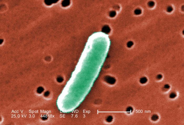
e. coli cells
blood cell growing into a melanoma
hamster oocyte-cumulus complex (fertilization area in the uterus)
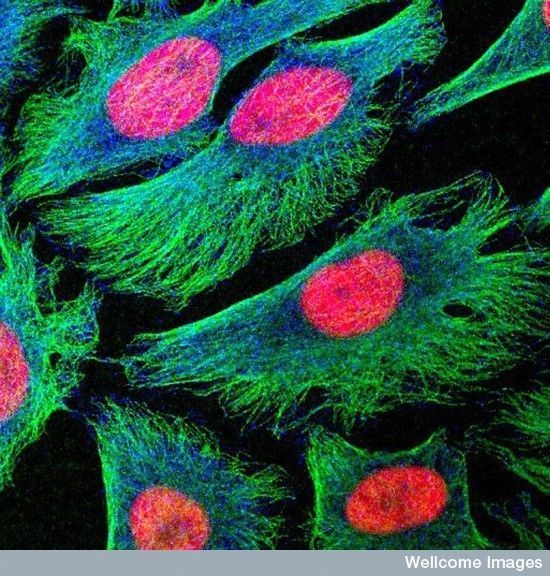
HeLa cancer cells
legionella bacteria
liver sinusoid of a brown rat
lung cancer cell
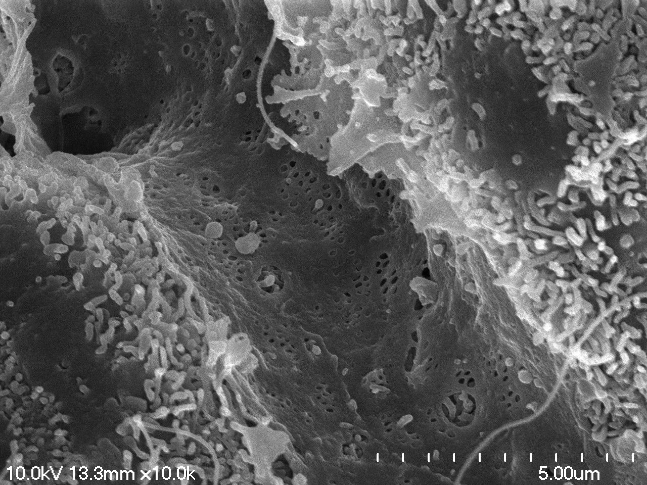
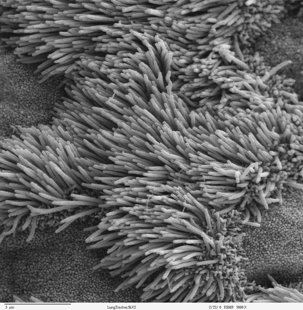
inner wall of a lung
pancreatic cancer cell
nerve ending broken to show synaptic vessels
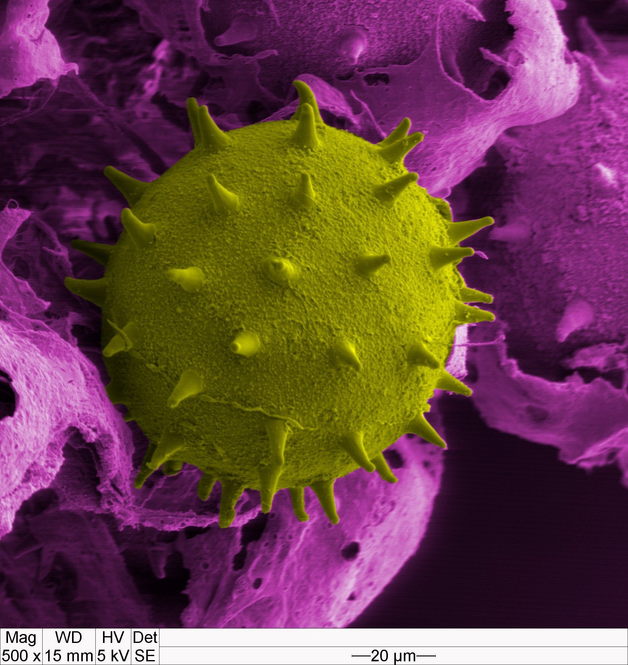
pollen
red blood cells within alveolar capillary network
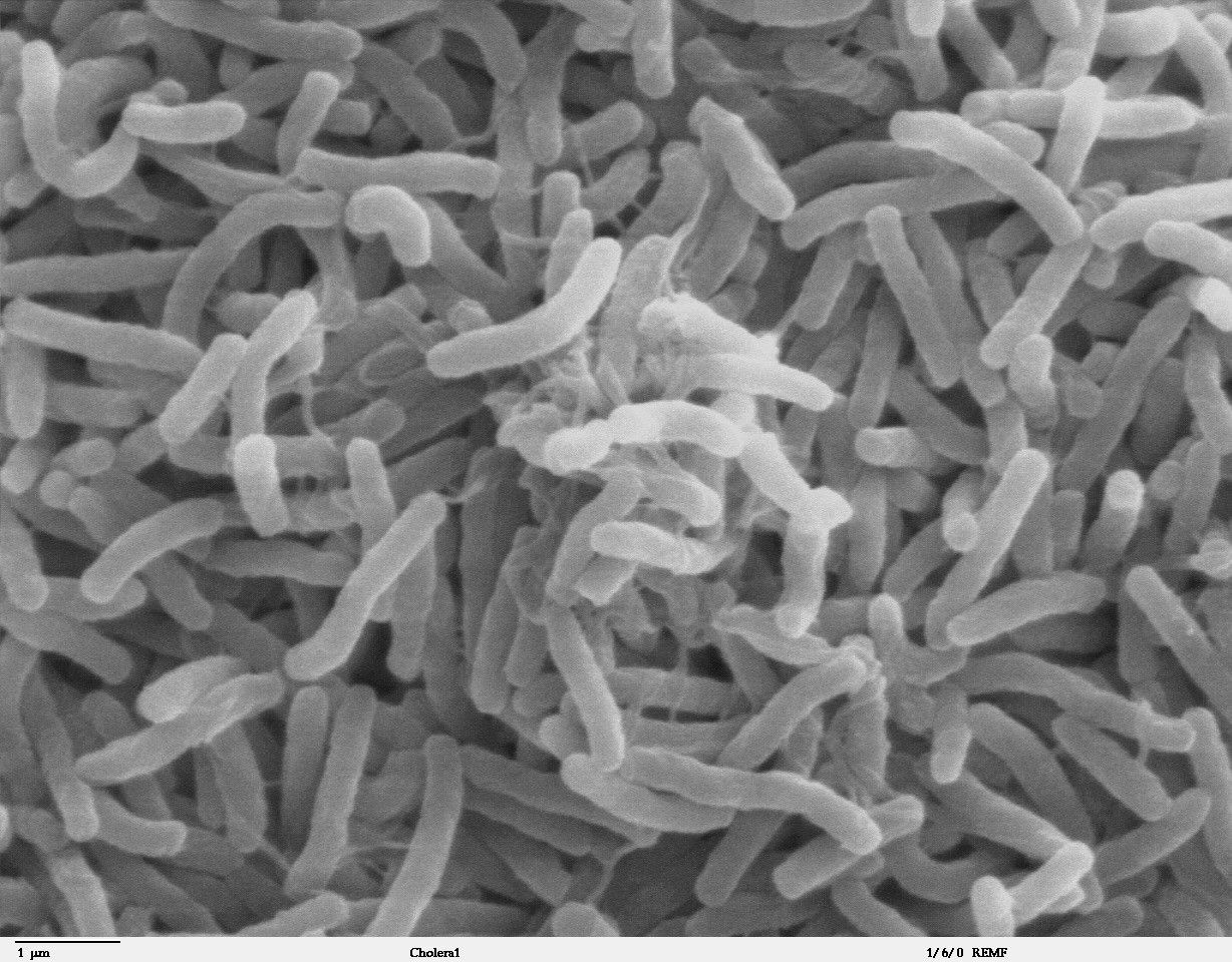
cholera
soybean cyst nematode and egg