|
#1
●
11-20-2011, 09:03 AM
| ||||||||
 ♚ Legacy Gold Member ♚ Poster Rank:35 Female Join Date: Nov 2008 Posts: 43,394
Contributions: 204
Mentioned: 95 Post(s) Quoted: 2282 Post(s)
| ||||||||
|
Amazing Colour Insect Close-Ups
Oliver Meckes and Nicole Ottawa form a two-person team: Eye of Science who produce images of the world beyond human vision.  Hornet head. Coloured scanning electron micrograph (SEM) of the head of a European hornet (Vespa crabro). The compound eyes are on either side of its head, with two antennae branching off to left and right from between the eyes. The biting jaws (mandibles) are at lower centre. Part of the rest of the body, the wings, and two legs, make up the rest of the image. This hornet is the largest of the European wasps, and has a reputation for being aggressive, though it only stings if provoked.  Dust mite. Coloured Scanning Electron Micrograph (SEM) of the dust mite Dermatophagoides pteronyssinus. This mite is eight legged of which six legs are visible (at lower frame). The mite has an unsegmented, round body. It's mouthparts (at lower centre) are highly adapted to feeding on the dead scales of human skin found in household dust. Dust mites are relatives of spiders and scorpions. They are smaller than full stops and therefore invisible to the naked eye. Millions of dust mites live inside furniture and fabric in the average home. The dead bodies and excrement of dust mites can cause allergic reactions to household dust.  Annelid worm. Coloured scanning electron micrograph (SEM) of an annelid worm, belonging to the subclass Oligochaeta. This subclass encompasses terrestrial earthworms and species that live in freshwater environments.  Coloured Scanning Electron Micrograph (SEM) of a malaria causing mosquito, Anopheles sp. The blue compound eyes of this insect are seen at upper centre of image. Emerging from the head (towards lower right) is a characteristic three- pronged fork made up of 2 mandibular palps and the piercing proboscis. The proboscis is used by the females of the species to draw blood. Anopheles are one of several species of mosquito which transmit malaria. The parasite that causes malaria is present in the saliva of female mosquitos. Malaria is a major health problem in many countries, causing a potentially fatal fever.  Coloured scanning electron micrograph (SEM) of the head of a silkworm moth caterpillar (Bombyx mori). The silkworm uses its chewing mouthparts (upper centre) to feed on mulberry leaves. On either side of the mouthparts is an antenna and several simple eyes (ocelli, black round structures, upper left and right). The caterpillar has three pairs of jointed legs behind the head (lower centre). Most caterpillars also have prolegs further along the body (not seen). The silkworm produces threads of silk with which it constructs a cocoon. People farm silkworms for their silk, which is woven into cloth.  Cat flea (Ctenocephalides felis) amongst cat hairs, coloured scanning electron micrograph (SEM). Looks like a walrus!  Sheep tick nymph. Coloured scanning electron micrograph (SEM) of a nymph of the sheep tick (Ixodes ricinus). A nymph is a sexually immature stage before reaching adult size. This tick is a blood-sucking parasite of animals and humans. At centre is its long, barbed hypostome used to suck blood from the host; sensory pedipalps occur on either side of the hypostome. The nymph has four pairs of walking legs. It is only the adult female tick that sucks blood from humans. Lyme disease occurs when the biting tick transmits a bacterium to humans. Lyme disease causes fever, muscle pain, and inflamed joints.  Coloured scanning electron micrograph (SEM) of the head of a fruit fly (Drosophila sp.). Its two compound eyes (red) are seen on either side of its head. At lower centre is its proboscis, used for sucking up food. This fly is widely used in genetic experiments as it breeds rapidly and has a small, well-understood genome (genetic code).  Coloured scanning electron micrograph (SEM) of a velvet mite (Family Trombidiidae). Velvet mites are small predatory arachnids that live in the upper layers of soil. They are covered in fine hairs give them an appearance of velvet.  Coloured scanning electron micrograph (SEM) of the head of a maggot (larva) of a bluebottle fly (Protophormia sp.). The maggots of this fly are used medicinally to clean wounds. Its mouthparts are seen at centre right (grey). The maggots are sterilised and placed in the wound, where they feed on dead tissue and leave healthy tissue untouched. Their saliva contains anti- bacterial chemicals which maintain sterility in the area. Maggots are used on ulcers and deep wounds away from organs or body cavities, most often being used to treat diabetic ulcers on the feet.  Blood-filled tick  The tick is swollen in size after feeding on the blood of its mammal host. The legs are protruding from its body either side of its mouthparts (centre) This is a common sheep tick (Ixodes ricinus), the principal vector of Lyme disease in Europe. It is common in the damp underbrush of European woods and attacks various domestic and wild animals, including dogs and humans. It carries the bacterium (Borrelia burgdorferi) that causes Lyme disease. The tick is swollen in size after feeding on the blood of its mammal host. The legs are protruding from its body either side of its mouthparts (centre) This is a common sheep tick (Ixodes ricinus), the principal vector of Lyme disease in Europe. It is common in the damp underbrush of European woods and attacks various domestic and wild animals, including dogs and humans. It carries the bacterium (Borrelia burgdorferi) that causes Lyme disease. Coloured scanning electron micrograph (SEM) of a tsetse fly Glossina sp., a blood- sucking parasitic fly of tropical Africa. The fly pierces the skin of its host with the sharp probo- scis at lower right. The tsetse fly transmits Trypanosoma protozoa, of which T. gambiense & T. rhodesiense cause sleeping sickness in humans. Both male & female flies are bloodsuckers. Their habitat is varied, ranging from forest to river banks to savanna. Unlike most flies the tsetse gives birth to fully developed larvae, which immediately pupate. The female nurtures one larva at a time, with a total of 8-10 per lifetime. 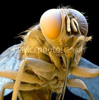 · |
|
#3
●
11-20-2011, 09:31 AM
| ||||||||
| ♚ Legacy Gold Member ♚ Poster Rank:35 Female Join Date: Nov 2008 Posts: 43,394
Contributions: 204
Mentioned: 95 Post(s) Quoted: 2282 Post(s)
| ||||||||
|
Re: Amazing Colour Insect Close-Ups
Made me love maggots now  Isn't he great? Looks like a weird wee critter in a big thick sheepskin coat!! Isn't he great? Looks like a weird wee critter in a big thick sheepskin coat!!All the ones we see swarming over corpses - they're cutey-pies |