|
#111
●
03-05-2011, 03:17 PM
| ||||||||
| My Rank: PRIVATE FIRST CLASS Poster Rank:3666 Male Join Date: Feb 2010 Posts: 91 Mentioned: 0 Post(s) Quoted: 0 Post(s)
| ||||||||
|
Re: JonBenét Ramsey Autopsy & Crime Scene Photos
Of course the parents did it. POS mom and her damned pageants.. |
|
#112
●
03-31-2011, 12:02 AM
| ||||||||
| My Rank: LANCE CORPORAL Poster Rank:3250 i have a PP Join Date: Feb 2010 Posts: 114 Mentioned: 0 Post(s) Quoted: 5 Post(s)
| ||||||||
|
Re: JonBenét Ramsey Autopsy & Crime Scene Photos
December 23, 1996: The Ramseys host a Christmas party, with approximately 30 guests attending, and with former journalism professor Bill McReynolds playing Santa Claus. At 6:47 p.m., someone attending the party placed a 911 call, which was answered by police dispatcher Therese Hilleary. The caller hung up without saying anything. Police call back only to get the Ramsey's anwering machine. Officer "B.O. 266" goes to the home at 6:54 p.m. and leaves at 7:09 p.m., after being assured that there was no emergency. December 24, 1996: The Ramseys attend twilight service at St. John's Episcopal Church in Boulder. At 9 p.m., John Ramsey retrieves a brand new silver girl's bike stored in neighbor Joe Barnhill's garage and places it under the Christmas tree for JonBenét. December 25, 1996: The Ramseys attend a Christmas dinner at 5:00 p.m at the Fleet White residence. After the family returns home, JonBenét is carried? to bed at about 9:30 p.m. The family had plans to fly to Michigan early the next morning. Sometime before dawn, JonBenét is killed; her skull is fractured, she is strangled with a cord, duct tape is put over her mouth, and her body is placed downstairs in a small windowless room in the basement. She is wrapped in a blanket, with the ligature still around her neck, head uncovered, and her arms above her head. December 26, 1996: Patsy Ramsey calls the police at 5:52 A.M., shouting "send help!" and saying that her daughter is missing and that a 2½ page ransom note demanding $118,000 had been left by the kidnapper on the steps of the back stairs leading to the kitchen. The note begins: "Dear Mr. Ramsey, We have your daughter..." and includes the words "behead" and "attaché." It was printed in block letters with a "Sharpie" felt tipped pen. Four misspellings in the note appear to be intentional. Patsy Ramsey screams for John and they check Burke's room, but JonBenét is not in there. Patsy begins to phone friends. Friends begin gathering at the home almost immediately. Police arrive at approximately 6:00 A.M and perform a cursory search of the premises. A window in the basement was found broken with a suitcase underneath. There is no other indication of forced entry. They contact the FBI and begin making plans to deal with the kidnapper. A detective does not arrive until two hours later. John Ramsey begins arranging to obtain cash for the $118,000 ransom. At approximately 8:00 AM Law Enforcement arrive and set up a wiretap and recording equipment. Around 1:00 p.m., Linda Arndt asked Fleet White, a friend of the Ramseys, to take John and search the house for "anything unusual." At approximately 1:30 p.m., John Ramsey and his friend Fleet White discover JonBenét's body in the basement. John Ramsey removes the tape from her mouth and carries her upstairs in outstretched arms, where he lay her on the floor at the top of the stairs and requests a blanket from the couch to cover her. Patsy flings herself onto JonBenét's body and shouts "help me Jesus!". Linda Arndt shortly thereafter, moves JonBenét's body over near the Christmas tree and places a Colorado Avalanche's sweatshirt over her. Twenty minutes later, John is overheard placing a phone call to his pilot to ready the plane to head for Atlanta. Police instructed them not to leave town, so they began staying at a friend's home in Boulder. At approximately 2:00 P.M., as the Ramseys are leaving their home after JonBenét's body is discovered and it is declared a crime scene, John Andrew, Melinda and Stewart Long (Melinda's fiancé) arrive in front of the house. Investigators begin a 10 day evidence gathering quest. The coroner arrived at approximately 8 PM and entered the house where the decedent's body was located at approximately 8:20 PM. The Boulder County coronor's staff removed JonBenét's body from the house at approximately 9:45 p.m. December 27, 1996: The Boulder County coroner reported that an autopsy revealed that the cause of death was asphyxia due to strangulation, and her death was ruled a homicide. December 28, 1996: Detectives inspect the body for evidence of semen, blood and saliva, and take blood, hair and handwriting samples from the Ramseys and their relatives and friends. December 29, 1996: A memorial service is held for JonBenét at St. John's Episcopal Church in Boulder. December 30, 1996: The family takes the body by private jet to Atlanta, which was their former place of residency before their move to Colorado. It is also reported that the Ramseys have retained Bryan Morgan and their attorney, and that JonBenét's two older half-siblings, John Andrew (who lived at the Ramsey home) and Melinda, were out of town on the night of the murder. A press conference is held by Leslie Aaholm, city of Boulder spokeperson. December 31, 1996: JonBenét is buried at a cemetary in Marietta, Georgia, her birthplace, next to her half-sister Elizabeth, who had been killed at the age of 22 in a car accident in 1992. 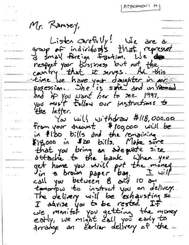 · 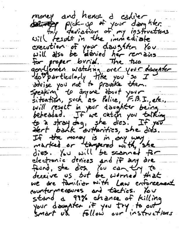 · 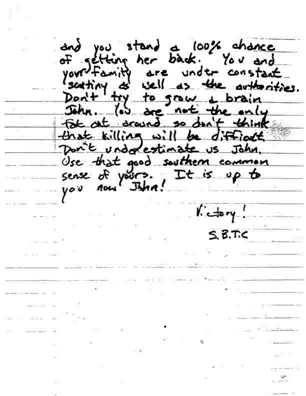 · 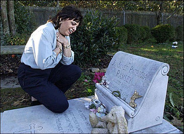 · 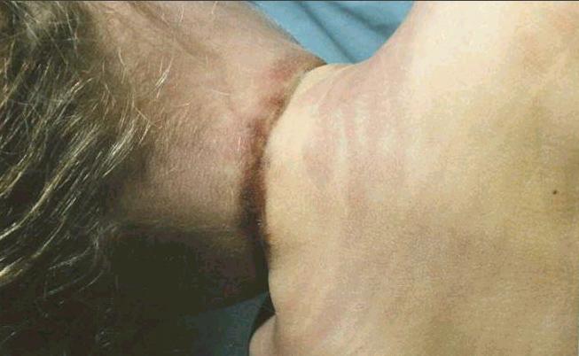 · 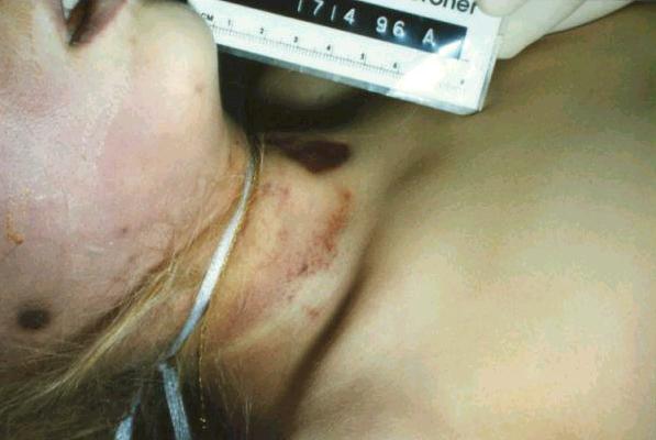 ·  · 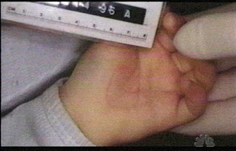 · 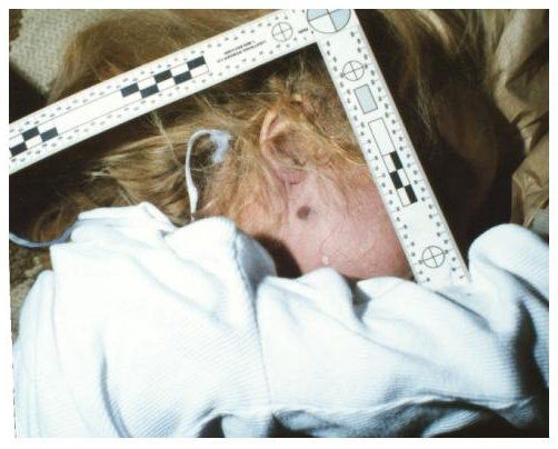 · 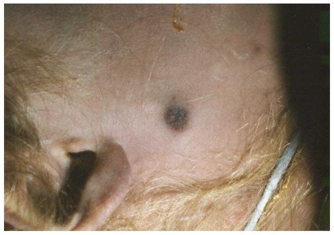 · 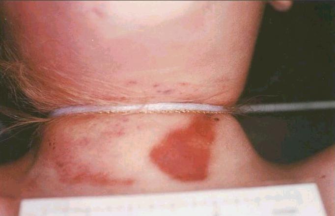 ·  ·  · 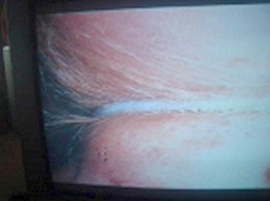 · 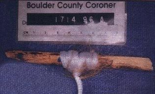 · 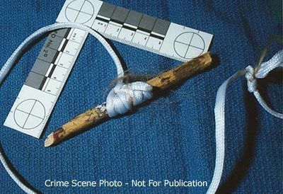 · 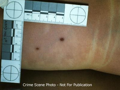 · 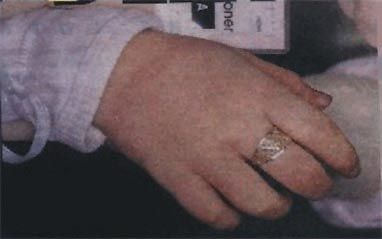 · 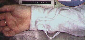 · 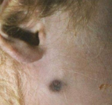 ·  · |
|
#113
●
03-31-2011, 12:03 AM
| ||||||||
| My Rank: LANCE CORPORAL Poster Rank:3250 i have a PP Join Date: Feb 2010 Posts: 114 Mentioned: 0 Post(s) Quoted: 5 Post(s)
| ||||||||
|
Re: JonBenét Ramsey Autopsy & Crime Scene Photos
NAME: RAMSEY, JONBENET AUTOPSY NO. 96A-155 DOB: 08/06/90 DEATH D/T: 12/26/96 @ 1323 AGE: 6Y SEX: F AUTOPSY D/T: 12/27/96 @ 0815 ID NO: 137712 PATH MD: MEYER COR/MEDREC#: 1714-96-A TYPE: COR FINAL DIAGNOSIS: I. Ligature strangulation A. Circumferential ligature with associated ligature furrow of neck B. Abrasions and petechial hemorrhages, neck C. Petechial hemorrhages, conjunctival surfaces of eyes and skin of face II. Craniocerebral injuries A. Scalp contusion B. Linear, comminuted fracture of right side of skull C. Linear pattern of contusions of right cerebral hemisphere D. subarachnoid and subdural hemorrhage E. Small contusions, tips of temporal lobes III. Abrasion of right cheek IV. Abrasion/contusion, posterior right shoulder V. Abrasions of left lower back and posterior left lower leg VI. Abrasion and vancular congestion of vaginal mucosa VII. Ligature of right wrist Toxicologic Studies blood ethanol - none detected blood drug screen - no drugs detected CLINOCOPATHOLIGICAL CORRELATION: Cause of death of this six year old female is asphyxia by strangulation associated with craniocerebral trauma. John E. Meyer M.D. Pathologist jn/12/27/96 The body of this six year old female was first seen by me after I was called to an address identified as 755 - 15th street in Boulder, Colorado, on 12/26/96. I arrived at the scene approximately 8 PM on 12/26 and entered the house where the decedent's body was located at approximately 8:20 PM. A brief examination of the body disclosed a ligature around the neck and a ligature around the right wrist. Also noted was a small area of abrasion or contusion below the right ear on the lateral aspect of the right cheek. A prominent dried abrasion was present on the lower left neck. After examining the body, I left the residence at approximately 8:30 PM. EXTERNAL EVIDENCE OF INJURY: Located just below the right ear at the right angle of the mandible, 1.5 inches below the right external auditory canal is a 3/8 x 1/4 inch area of rust colored abrasion. In the lateral aspect of the left lower eyelid on the inner conjunctival surface is a 1 mm in maximum dimension petechial hemorrhage. Very fine, less than 1 mm petechial hemorrhages are present on the skin of the upper eyelids bilaterally as well as on the lateral left cheek. On everything the left upper eyelid there are much smaller, less than 1 mm petechial hemorrhages located on the conjunctival surface. Possible petechial hemorrhages are also seen on the conjunctival surfaces of the right upper and lower eyelids, but liver mortis on this side of the face makes definite identification difficult. A deep ligature furrow encircles the entire neck. The width of the furrow varies from one- eight of an inch to five/sixteenths of an inch and is horizontal in orientation, with little upward deviation. The skin of the anterior neck above and below the ligature furrow contains areas of petechial hemorrhage and abrasion encompassing an area measuring approximately 3 x 2 inches. The ligature furrow crosses the anterior midline of the neck just below the laryngeal prominence, approximately at the level of the cricoid cartilage. It is almost completely horizontal with slight upward deviation from the horizontal towards the back of the neck. The midline of the furrow mark on the anterior neck is 8 inches below the top of the head. The midline of the furrow mark on the posterior neck is 6.75 inches below the top of the head. The area of abrasion and petechial hemorrhage of the skin of the anterior neck includes on the lower left neck, just to the left of the midline, a roughly triangular, parchment-like rust colored abrasion which measures 1.5 inches in length with a maximum width of 0.75 inches. This roughly triangular shaped abrasion is obliquely oriented with the apex superior and lateral. The remainder of the abrasions and petechial hemorrhages of the skin above and below the anterior projection of the ligature furrow are nonpatterned, purple to rust colored, and present in the midline, right, and left areas of the anterior neck. The skin just above the ligature furrow along the right side of the neck contains petechial hemorrhage composed of multiple confluent very small petechial hemorrhages as well as several larger petechial hemorrhages measuring up to one-sixteenth and one-eight of an inch in maximum dimension. Similar smaller petechial hemorrhages are present on the skin below the ligature furrow on the left lateral aspect of the neck. Located on the right side of the chin is a three-sixteenths by one-eight of an inch area of superficial abrasion. On the posterior aspect of the right shoulder is a poorly demarcated, very superficial focus of abrasion/contusion which is pale purple in color and measures up to three-quarters by one-half inch in maximum dimension. Several linear aggregates of petechial hemorrhages are present in the anterior left shoulder just above deltopectoral groove. These measure up to one inch in length by one-sixteenth to one-eight of an inch in width. On the left lateral aspect of the lower back, approximately sixteen and one-quarter inches and seventeen and one-half inches below the level of the top of the head are two dried rust colored to slightly purple abrasions. The more superior of the two measures one-eight by one-sixteenth of an inch and the more inferior measures three-sixteenths by one-eight of an inch. There is no surrounding contusion identified. On the posterior aspect of the left lower leg, almost in the midline, approximately 4 inches above the level of the heel are two small scratch-like abrasions which are dried and rust colored. They measure one-sixteenth by less than one- sixteenth of an inch and one-eight by less than one-sixteenth of an inch respectively. On the anterior aspect of the perineum, along the edges of closure of the labia majora, is a small amount of dried blood. A similar small amount of dried and semifluid blood is present on the skin of the fourchette and in the vestibule. Inside the vestibule of the vagina and along the distal vaginal wall is reddish hyperemia. This hyperemia is circumferential and perhaps more noticeable on the right side and posteriorly. The hyperemia also appears to extend just inside the vaginal orifice. A 1 cm red-purple area of abrasion is located on the right posterolateral area of the 1 x 1 cm hymeneal orifice. The hymen itself is represented by a rim of mucosal tissue extending clockwise between the 2 and 10:00 positions. The area of abrasion is present at approximately the 7:00 position and appears to involve the hymen and distal right lateral vaginal wall and possibly the area anterior to the hymen. On the right labia majora is a very faint area of violent discoloration measuring approximately one inch by three-eighths of an inch. Incision into the underlying subcutaneous tissue discloses no hemorrhage. A minimal amount of semiliquid thin watery red fluid is present in the vaginal vault. No recent or remote anal or other perineal trauma is identified. REMAINDER OF EXTERNAL EXAMINATION: The unembalmed, well developed and well nourished Caucasian female body measures 47 inches in length and weights n estimated 45 pounds. No scalp trauma is identified. The external auditory canals are patent and free of blood. The eyes are green and the pupils equally dilated. The sclerae are white. The nostrils are both patent and contain a small amount of tan mucous material. The teeth are native and in good repair. The tongue is smooth, pink-tan and granular. No buccal mucosal trauma is seen. The frenulum is intact. There is slight drying artifact of the tip of the tongue. On the right cheek is a pattern of dried saliva and mucous material which does not appear to be hemorrhagic. The neck contains no palpable adenopathy or masses and the trachea and larynx are midline. The chest is symmetrical. Breasts are prepubescent. The abdomen is flat and contains no scars. No palpable organomegaly or masses are identified. The external genitalia are that of a prepubescent female. No pubic hair is present. The anus is patent. Examination of the extremities is unremarkable. The fingernails of both hands are of sufficient length for clipping. Examination of the back is unremarkable. There is dorsal 3+ to 4+ livor mortis which is nonblanching. Livor mortis is also present on the right side of the face. At the time of the initiation of the autopsy there is mild 1 to 2+ rigor mortis of the elbows and shoulders with more advanced 2 to 3+ rigor mortis of the joints of the lower extremities. INTERNAL EXAM: The anterior chest musculature is well developed. No sternal or rib fractures are identified. Mediastinum: The mediastinal contents are normally distributed. The 21 gm thymus gland has a normal external appearance. The cut sections are finely lobular and pink-tan. No petechial hemorrhages are seen. The aorta and remainder of the mediastinal structures are unremarkable. Body Cavities: The right and left thoracic cavities contain approximately 5 cc of straw colored fluid. The pleural surfaces are smooth and glistening. The pericardial sac contains 3-4 cc of straw colored fluid and the epicardium and pericardium are unremarkable. The abdominal contents are normally distributed and covered by a smooth glistening serosa. No intra-abdominal accumulation of fluid or blood is seen. Lungs: The 200 gm right lung and 175 gm let lung have a normal lobar configuration. An occasional scattered subpleural petechial hemorrhage is seen on the surface of each lung. The cut sections of the lungs disclose an intact alveolar architecture with a small amount of watery fluid exuding from the cut surfaces with mild pressure. The intrapulmonary bronchi and vasculature are unremarkable. No evidence of consolidation is seen. Heart: The 100 gm heart has a normal external configuration. There are scattered subepicardial petechial hemorrhages over the anterior surface of the heart. The coronary arteries are normal in their distribution and contain no evidence of atherosclerosis. The tan- pink myocardium is homogeneous and contains no areas of fibrosis or infarction. The endocardium is unremarkable. The valve cusps are thin, delicate and pliable and contain no vegetation or thrombosis. The major vessels enter and leave the heart in the normal fashion. The foramen ovale is closed. Aorta and Vena Cava: The aorta is patent throughout its course as are its major branches. No atherosclerosis is seen. The Vena Cava is unremarkable. Spleen: The 61 gm spleen has a finely wrinkled purple capsule. Cut sections are homogeneous and disclose readily identifiable red and white pulp. No intrinsic abnormalities are identified. Adrenals: The adrenal glands are of normal size and shape. A golden yellow cortex surmounts a thin brown-tan medullary area. No intrinsic abnormalities are identified. Kidneys: The 40 gm right kidney and 40 gm left kidney have a normal external appearance. The surfaces are smooth and glistening. Cut sections disclose an intact corticomedullary architecture. The renal papilae are sharply demarcated. The pelvocaliceal system is lined by gray-white mucosa which is unremarkable. Both ureters are patent throughout their course to the bladder. Liver: The 625 gm liver has a normal external appearance. The capsule is smooth and glistening. Cut sections disclose an intact lobular architecture with no intrinsic abnormalities identified. Pancreas: The pancreas is of normal size and shape. Cut sections are finely lobular and tan. No intrinsic abnormalities are identified. Bladder: The bladder is contracted and contains no urine. The bladder mucosa is smooth and tan-gray. No intrinsic abnormalities are seen. Genitalia: The upper portions of the vaginal vault contain no abnormalities. The prepubescent uterus measures 3 x 1 x 0.8 cm and is unremarkable. The cervical os contains no abnormalities. Both fallopian tubes and ovaries are prepubescent and unremarkable by gross examination. Gallbladder: The gallbladder contains 2-3 cc of amber bile. No stones are identified and the mucosa is smooth and velvety. The cystic duct, right and left hepatic duct and common bile duct are patent throughout their course to the duodenum. G.I. Tract: The esophagus is empty. It is lined by gray-white mucosa. The stomach contains a small amount (8-10 cc) of viscous to green to tan colored thick mucous material without particulate matter identified. The gastric mucosa is autolyzed but contains no areas of hemorrhage or ulceration. The proximal portion of the small intestine contains fragmented pieces of yellow to light green-tan apparent vegetable or fruit material which may represent fragments of pineapple. No hemorrhage is identified. The remainder of the small intestine is unremarkable. The large intestine contains soft green fecal material. The appendix is present. Lymphatic System: Unremarkable. Musculoskeletal System: Unremarkable. Skull and Brain: Upon reflection of the scalp there is found to be an extensive area of scalp hemorrhage along the right temporoparietal area extending from the orbital ridge, posteriorly all the way to the occipital area. This encompasses an area measuring approximately 7 x 4 inches. This grossly appears to be fresh hemorrhage with no evidence of organization. At the superior extension of this area of hemorrhage is a linear to comminuted skull fracture which extends from the right occipital to posteroparietal area forward to the right frontal area across the parietal portion of the skull. the posteroparietal area of this fracture is a roughly rectangular shaped displaced fragment of skull measuring one and three-quarters by one-half inch. The hemorrhage and the fracture extend posteriorly just past the midline of the occipital area of the skull. This fracture measures approximately 8.5 inches in length. On removal of the skull cap there is found to be a thin film of subdural hemorrhage measuring approximately 7-8 cc over the surface of the right cerebral hemisphere and extending to the base of the cerebral hemisphere. The 1450 gm brain has a normal overall architecture. Mild narrowing of the sulci and flattening of the gyri are seen. No inflammation is identified. There is a thin film of subarachnoid hemorrhage overlying the entire right cerebral hemisphere. On the right cerebral hemisphere underlying the previously mentioned linear skull fracture is an extensive linear area of purple contusion extending from the right frontal area, posteriorly along the lateral aspect of the parietal region and into the occipital area. This area of contusion measures 8 inches in length with a width of up to 1.75 inches. At the tip of the right temporal lobe is a one-quarter by one quarter inch similar appearing purple contusion. Only very minimal contusion is present at the tip of the left temporal lobe. This area of contusion measures only one-half inch in maximum dimension. The cerebral vasculature contains no evidence of atherosclerosis. Multiple coronal sections of the cerebral hemispheres, brain stem and cerebullum disclose no additional abnormalities. The areas of previously described contusion are characterized by purple linear streak-like discolorations of the gray matter perpendicular to the surface of the cerebral cortex. These extend approximately 6 mm into the cerebral cortex. Examination of the base of the brain discloses no additional fractures. Neck: Dissection of the neck is performed after removal of the thoracoabdominal organs and the brain. The anterior strap musculature of the neck is serially dissected. Multiple sections of the sternocleidomastoid muscle disclose no hemorrhages. Sections of the remainder of the strap musculature of the neck disclose no evidence of hemorrhage. Examination of the thyroid cartilage, cricoid cartilage and hyoid bone disclose not evidence of fracture of hemorrhage. Multiple cross sections of the tongue disclose no hemorrhage or traumatic injury. The thyroid gland weights 2 gm and is normal in appearance. Cut sections are finely lobular and red-tan. The trachea and larynx are lined by smooth pink-tan mucosa without intrinsic abnormalities. MICROSCOPIC DESCRIPTION: (All Sections Stained with H&E) (Slide Key) - (A) - scalp hemorrhage, (B) - sections of vaginal mucosa with smallest fragment representing area of abrasion of 7:00 position, (C) - heart, (D-F) - lungs, (G) - liver and spleen, (H) - pancreas and kidney, (I) - thyroid and bladder, (J) - thymus and adrenals, (K-L) - reproductive organs, (M) - larynx, (N-T) - brain. Myocardium: Sections of the ventricular myocardium are composed of interlacing bundles of cardiac muscle fibers. No fibrosis or inflammation are identified. Lungs: The alveolar architecture of the lungs is well preserved. Pulmonary vascular congestion is identified. No intrinsic abnormalities are seen. Spleen: There is mild autolysis of the spleen. Both red and white pulp are identifiable. Thyroid: The thyroid gland is composed of normal-appearing follicles. An occasional isolated area of chronic interstitial inflammatory infiltrate is seen. There is also a small fragment of parathyroid tissue. Thymus: The thymus gland retains the usual architecture. The lymphoid material is intact and scattered Hassall's corpuscles are identified. Mild vascular congestion is identified. Trachea: There is mild chronic inflammation in the submucosa of the trachea. Liver: The lobular architecture of the liver is well preserved. No inflammation or intrinsic abnormality are identified. Pancreas: There is autolysis of the pancreas which is otherwise unremarkable. Kidney: The overall architecture of the kidney is well preserved. There is perhaps mild vascular congestion in the cortex but no inflammation is identified. Bladder: The transitional epithelium of the bladder is autolyzed. No significant intrinsic abnormalities are seen. Reproductive Organs: Sections of the uterus are consistent with the prepubescent age. The ovary is unremarkable. Adrenal: The architecture of the adrenal is well preserved and no intrinsic abnormalities are seen. Brain: Sections of the areas of contusion disclose disrupted blood vessels of the cortex with surrounding hemorrhage. There is no evidence of inflammatory infiltrate or organization of the hemorrhage. Subarachnoid hemorrhage is also identified. Cortical neurons are surrounded by clear halos, as are glial cells. Vaginal Mucosa: All of the sections contain vascular congestion and focal interstitial chronic inflammation. the smallest piece of tissue, from the 7:00 position of the vaginal wall/hymen, contain epithelial erosion with underlying capillary congestion. A small number of red blood cells is present on the eroded surface, as is birefringent foreign material. Acute inflammatory infiltrate is not seen. EVIDENCE: Items turned over to the Boulder Police Department as evidence include: Fibers and hair from clothing and body surfaces; ligatures; clothing; vaginal swabs and smears; rectal swabs and smears; oral swabs and smears; paper bags from hands, fingernail clippings, jewelry, paper bags from feet; white body bag; sample of head hear, eyelashes and eyebrows; swabs from right and left thighs and right cheek; red top and purple top tubes of blood. |
|
#115
●
03-31-2011, 04:17 AM
| ||||||||
| My Rank: LANCE CORPORAL Poster Rank:2885 Join Date: Jan 2010 Posts: 138 Mentioned: 0 Post(s) Quoted: 36 Post(s)
| ||||||||
|
Re: JonBenét Ramsey Autopsy & Crime Scene Photos
That's so sad I think it was someone in the family who did it not sure who though honestly though what kind of name is JonBenet? |
|
#118
●
03-31-2011, 06:19 AM
| ||||||||
| So Fucking Banned Poster Rank:308 Male Join Date: Mar 2010 Posts: 4,168 Mentioned: 0 Post(s) Quoted: 0 Post(s)
| ||||||||
|
Re: JonBenét Ramsey Autopsy & Crime Scene Photos
Great post and thanks for all the associated information. This is such a sad case on so many levels. The fact the beautiful little girl was exploited by the mother and the father allowed it is only one. I did see a teaser on MSNBC or another news network that stated there was DNA on the body but cannot be matched to anyone in the family. I eventually hope someone is caught, confesses, etc so this little angel can rest in peace. |
|
#119
●
03-31-2011, 07:47 AM
| ||||||||
| ♚ Legacy Gold Member ♚ Poster Rank:194 Female Join Date: Nov 2009 Posts: 7,752 Mentioned: 5 Post(s) Quoted: 706 Post(s)
| ||||||||
|
Re: JonBenét Ramsey Autopsy & Crime Scene Photos
Thanks for the detailed information. It really is sad. How ANYONE could have done that to the poor child. It really is a strange story,the ransom letter etc. I hope they catch the person who did it. I for one would love to know who is responsible. Poor little girl. VIP. Very Interesting Post. |