|
#1
●
07-23-2024, 07:36 PM
| ||||||||
| These are the rooms Poster Rank:25 of ruin. Join Date: Sep 2014 Posts: 54,011 Mentioned: 145 Post(s) Quoted: 30403 Post(s)
| ||||||||
|
Thresher Accident
A 3-year-old boy was brought to the emergency room around 3 h after sustaining an injury while playing in the fields. His right upper limb accidentally got caught in the belt of a thresher machine and was avulsed from the chest wall. The child was taken to a nearby hospital with the amputated part [Figures 1 and 2] from where he was referred to our hospital. On arrival, the child was stable and the amputated limb was well preserved. The patient was evaluated to rule out other injuries and prepared for surgery while an X-ray was done for the amputated part [Figures 3 and 4]. The amputated part was then shifted to the operation theatre for bench dissection. On examination, the skin disruption was found to be at axillary level while the upper limb had avulsed from the chest wall along with the scapula and its attached muscles (scapulothoracic dissociation). The deltoid muscle was disrupted from its origin and found in the amputated stump with the skin retracted distally over it. The cut ends of brachial artery and its accompanying vein were found retracted under the biceps muscle. The brachial plexus was disrupted at cord level with distal cut ends of medial, lateral and posterior cords found alongside the transected vessels. The posterior cord was found to be of longest length suggesting higher level of avulsion. After thorough cleaning of the amputated limb, the brachial artery and its accompanying vein were dissected out and tagged. The brachial artery was cannulated, and infusion of cold heparinised saline (5000 units in 500 ml) started. The cut ends of medial, lateral and posterior cords were also tagged. A fasciotomy was done on bench with diathermy. Meanwhile, the patient was shifted to the operating room and anaesthetised. On exploration of the amputation stump, the axillary artery was found to be transected after the branch to the latissimus dorsi muscle. The axillary vein was found divided at the same level. The proximal cut ends of medial and lateral cord were found and tagged. The posterior cord was not found in the wound, and an incision was given in the supraclavicular region for exploration of the brachial plexus. However, the proximal end of the posterior cord could not be found. Since the level of transection of the axillary artery was after the take-off of the thoracodorsal and circumflex scapular arteries, the anticipated blood supply to the scapular muscles after replantation would have been doubtful. Furthermore, the approach to debride the subscapularis muscle in the event of necrosis following replantation would have been cumbersome and dangerous. Hence, the subscapularis muscle was debrided before replantation. The supraspinatus and infraspinatus muscles were left attached to the scapula as even in the scenario of necrosis of these muscles following replantation, they would potentially be easily approached from the dorsal side. The amputated limb was then brought into the operative field [Figure 5]. Bony fixation was achieved with the help of orthopaedic team. The dislocation of shoulder joint was reduced and fixed with a 1.5 mm K-wire. The fracture of glenoid neck was reduced and stabilised with two 1.5 mm K-wires. The scapula was then slipped under the skin dorsally. Acromioclavicular joint was fixed by a K-wire, and acromioclavicular joint capsular repair was performed by figure of 8 non-absorbable sutures. The associated elbow dislocation was managed with closed reduction, and the fracture of ulna was managed by splinting. Axillary vessels were dissected to the healthy end. Since no bone shortening was feasible in this case, an end-to-end vascular anastomosis was not possible. An 18 cm long saphenous vein graft was harvested from the left lower leg to reconstruct the segmental defects of the axillary artery and vein (8 cm each). Arterial anastomosis was followed by venous anastomosis. Good perfusion was obtained. The total ischaemia time was nearly 8 h (1 h warm/7 h cold). Neural repair was then done. End-to-end repair of the medial and lateral cord was done. The distal end of the posterior cord was anastomosed end to side with the proximal medial cord. The skin was closed partly. There was persistent ooze from the scapular side, which was packed with laparotomy sponges. All wounds were dressed and splint applied. Intraoperatively, the patient was given unfractionated heparin and 3 paediatric units of blood were transfused. Post-operatively, the patient was shifted to the paediatric intensive care unit. The replanted limb continued to be well perfused. After 48 h, the sponge packs were removed from scapular site in the operating room and no further oozing was encountered. The supraspinatus and infraspinatus muscles were found to be necrotic and were debrided. Post-fasciotomy raw area was covered with split-thickness skin graft on day 20. He was discharged on post-operative day 27. The K-wires were removed on day 90 in the outpatient department, and the arm supported in a sling, and physiotherapy started [Figures 6 and 7]. The last follow-up at 18 months post-operative showed evidence of recovery of crude touch sensations up to the digits. No motor recovery has been noted so far. 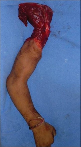 · 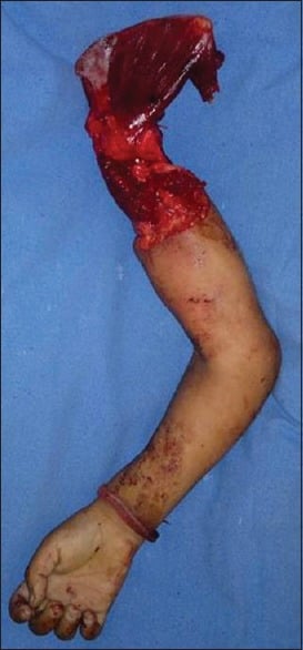 · 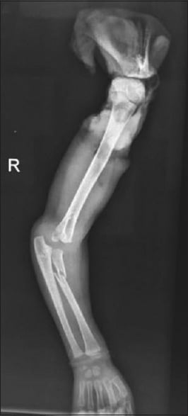 · 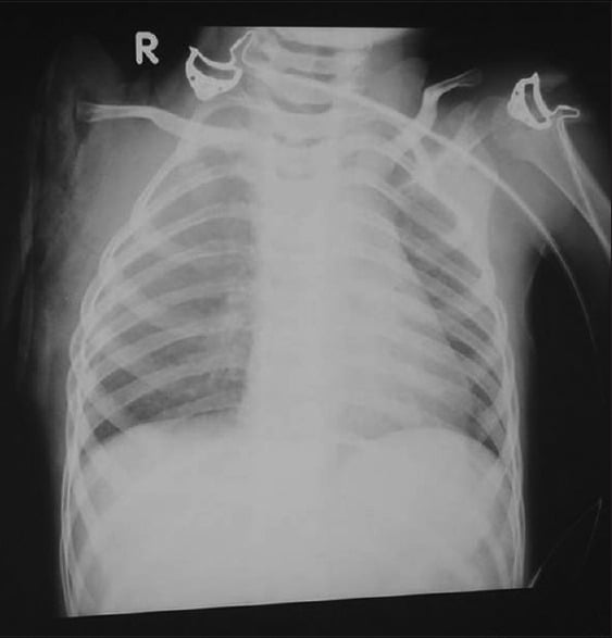 · 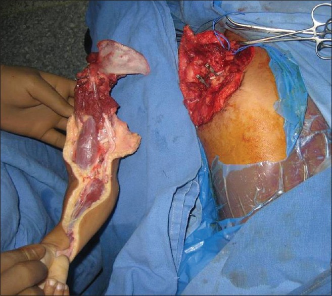 · 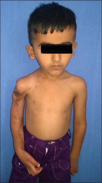 · 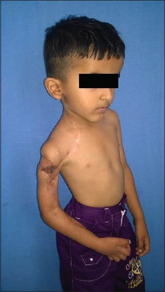 · |
|
#5
●
07-24-2024, 03:30 AM
| ||||||||
| ★ Legacy Member ★ Poster Rank:119 Secret Agent Join Date: Dec 2009 Posts: 13,215 Mentioned: 6 Post(s) Quoted: 2789 Post(s)
| ||||||||
|
Re: Thresher Accident
Such wonderful and amazing posts you bring us SG. Thank you so much for posting such amazing content here. You are truly an asset to the site. I love your posts! (Side note, when I read “Thresher” I first thought “Oh, shark attack” until I started reading.) |