|
#1
●
04-08-2023, 01:28 AM
| ||||||||
 These are the rooms Poster Rank:26 of ruin. Join Date: Sep 2014 Posts: 54,011 Mentioned: 145 Post(s) Quoted: 30403 Post(s)
| ||||||||
|
Reoccurring Tumor
Source: A 70-year-old woman with an enlarging tumor on the dorsum of her right second finger presented to our clinic. She had been diagnosed with hairy cell leukemia 5 years ago. The patient had received rituximab and cladribine and was in complete remission. At the present time, she was not receiving any treatment for hairy cell leukemia. She had a history of cutting her finger while cleaning and preparing a sheep's head and one week later, a non-healing lesion developed on her right second finger. Four months after the lesion's appearance, she visited a surgeon. The lesion was removed with a shave excision, and the pathology reported it as granulation tissue. One month later, a rapidly growing, lobulated mass recurred at the site of the shaved biopsy, which grew to approximately 6x10 cm after several weeks (Figure 1). She visited a family medicine physician and was referred to a surgeon for an incisional biopsy to rule out possible malignancies. However, during the operation, the surgeon decided to amputate the digit due to the broad base of the tumor and tissue fragility and sent it to a pathology laboratory. The histopathologic examination revealed vacuolated epidermal cells with eosinophilic inclusion bodies in some keratinocytes (Figure 4). The pathology result in addition to the patient's history of the previous contact with sheep was consistent with the diagnosis of orf disease. Two weeks after the surgery, signs of recurrence were noticed, and the patient was referred to a dermatologist (Figure 2). Recurrent lesions were treated with cryotherapy (every two weeks) and topical imiquimod cream (three times a week). Despite treatment with topical imiquimod cream and two sessions of cryotherapy, the lesions enlarged after one month, and new lesions began to appear at the site of the amputation. She was prescribed systemic subcutaneous injections of interferon alfa-2a (3 million IU twice weekly) and topical imiquimod cream 5% daily. Treatment was well tolerated by the patient with no complications except for a mild fever the first night after each injection. Four weeks after starting the interferon, remarkable shrinkage of lesions was observed. As a result, the interferon injections were reduced to 3 million IU weekly and continued for 6 more weeks when all lesions were resolved (Figure 3). No new lesions were noted during the six months of follow-up. 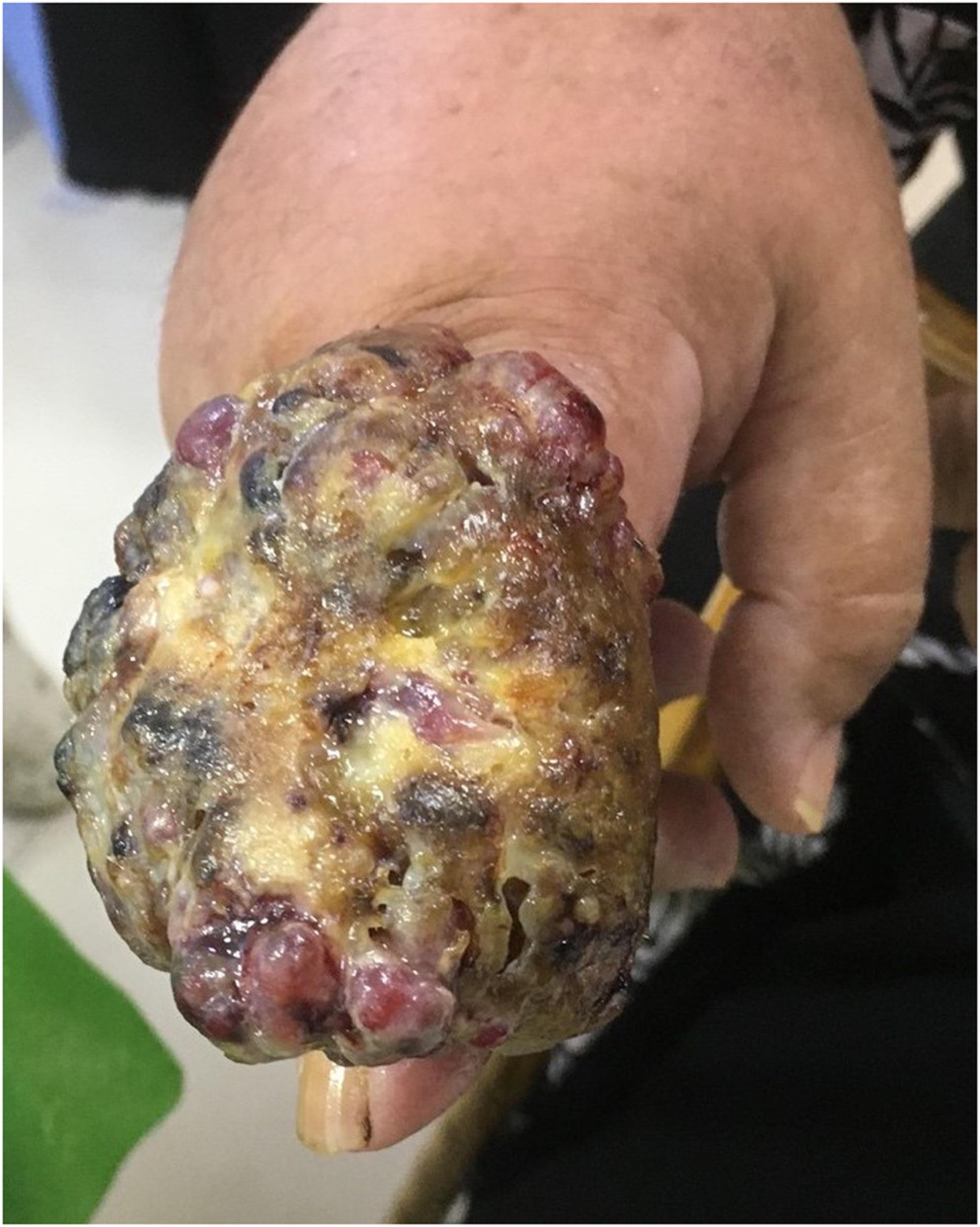 · 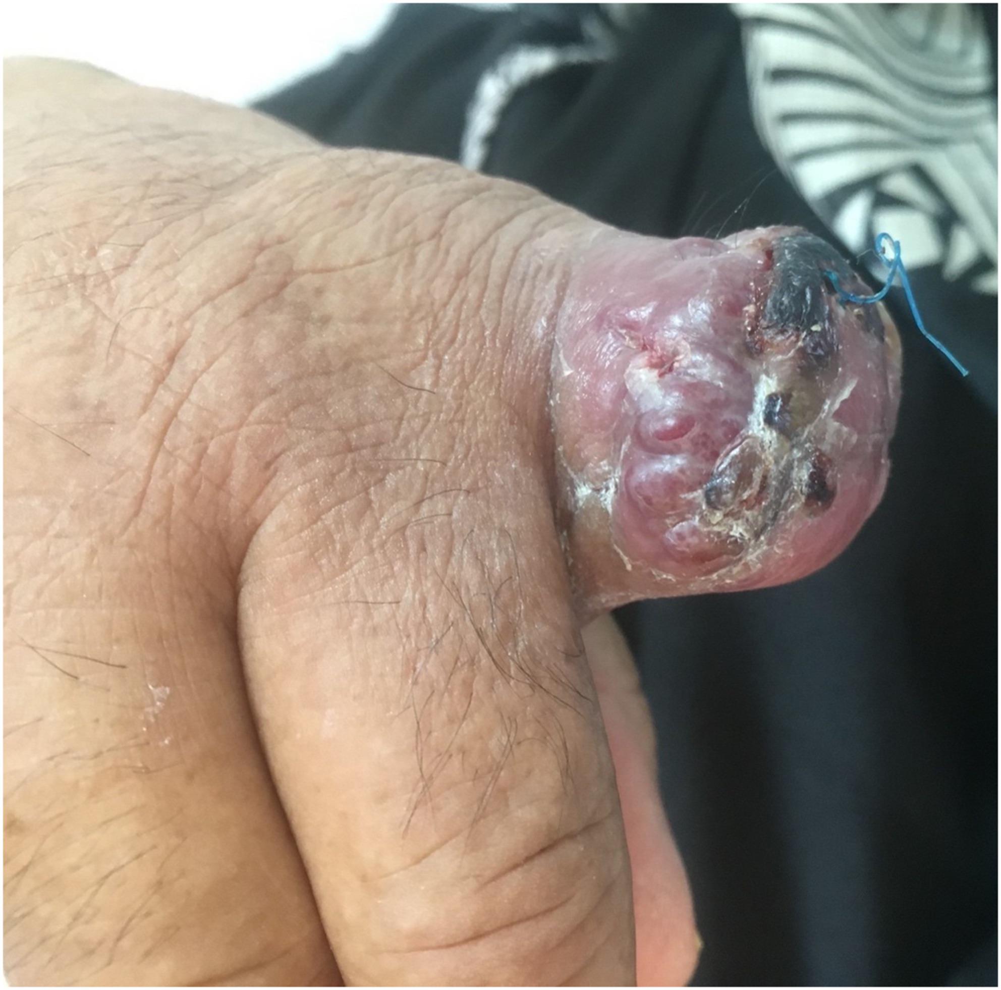 · 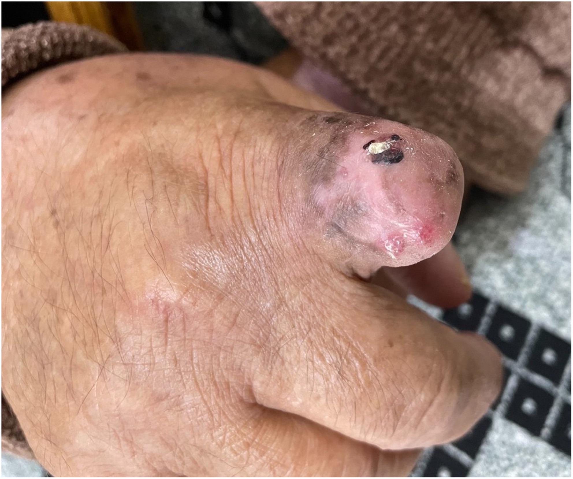 · |
|
#5
●
04-09-2023, 05:04 PM
| ||||||||
 ☆☆☆Jumbo Lesbian Cunt☆☆☆ Poster Rank:57 My pronouns are thin/skinny Join Date: Nov 2012 Posts: 25,933
Contributions: 1
Mentioned: 160 Post(s) Quoted: 13921 Post(s)
| ||||||||
|
Re: Reoccurring Tumor
The Kyrgyzstanis (central Asians) like their roasted sheep's head as a main course. She should've worn gloves; orf is a nasty disease.  |