|
#1
●
01-30-2023, 01:50 AM
| ||||||||
| These are the rooms Poster Rank:25 of ruin. Join Date: Sep 2014 Posts: 54,011 Mentioned: 145 Post(s) Quoted: 30403 Post(s)
| ||||||||
|
Pyoderma Gangrenosum (ulcers) Being Treated
Source. This case is out of Saudi Arabia: A 27-year-old male patient, medically free, with a positive surgical history for gastric bypass surgery as well as laparoscopic gastrectomy three years back, presented to our institute with an eight-day history of generalized edema, fatigue, dizziness, decreased urine output in addition to left leg nonhealing ulcers. The patient had been complaining of a leg ulcer for around six months, which had failed to be treated by various topical medications, including topical antibiotics and antimycotic creams that have been prescribed by general practitioners. The patient is a non-smoker and a non-alcoholic. There was no family history of such a complaint, as well as no family history of ulcerative colitis or Crohn's disease. Upon physical examination, the patient was afebrile, tachycardic, and tachypneic, with bilateral pitting lower limb edema, which extended up to the lower abdomen. While evaluating blood supply, a distal pulse could not be appreciated, with multiple large ulcers in the lower part of the left leg and foot (Figure 1). Laboratory investigations showed random blood glucose: 5 mmol/L (normal range 5.5-7.7), serum potassium: 3.66 mmol/L (normal range is 3.4-5.1), lactic dehydrogenase: 319 U/L (normal range is 100-200), white blood cells: 13.91 (normal range is 4-10), hemoglobin: 7.8 g/dL (normal range is 13-17), and P folate serum: 2.98 ng/mL (normal range is 3-18). The patient was admitted to the surgical ward and found to be malnourished with multiple organ failure. A biopsy of the non-healing ulcer was taken, with a histopathological result showing diffuse necroinflammatory and suppurative “neutrophilic” inflammation confirming pyoderma gangrenosum. A CT scan of the abdomen and pelvis with oral contrast demonstrated no extraluminal contrast leak, mild diffuse pancreatic atrophy, and diffuse gallbladder wall thickening with no pericholecystic fat stranding or fluid. The patient was managed with surgical debridement (Figure 2), vacuum-assisted closure, and dressing with silver-cell and intravenous (IV) antibiotics. As the patient was malnourished, he was managed with IV supplements initially and then shifted to oral. The patient has improved dramatically and was instructed to follow up in the outpatient clinic. Upon discharge, the patient was given vitamin B complex, vitamin D 5000 IU, zinc sulfate 30 mg orally (PO) once daily, folic acid 5 mg PO, infliximab 200 mg IV injections every two weeks for six weeks, and vitamin B12 IM weekly for three consecutive doses. The leg ulcer was improved with a satisfactory result (Figures 3, 4). 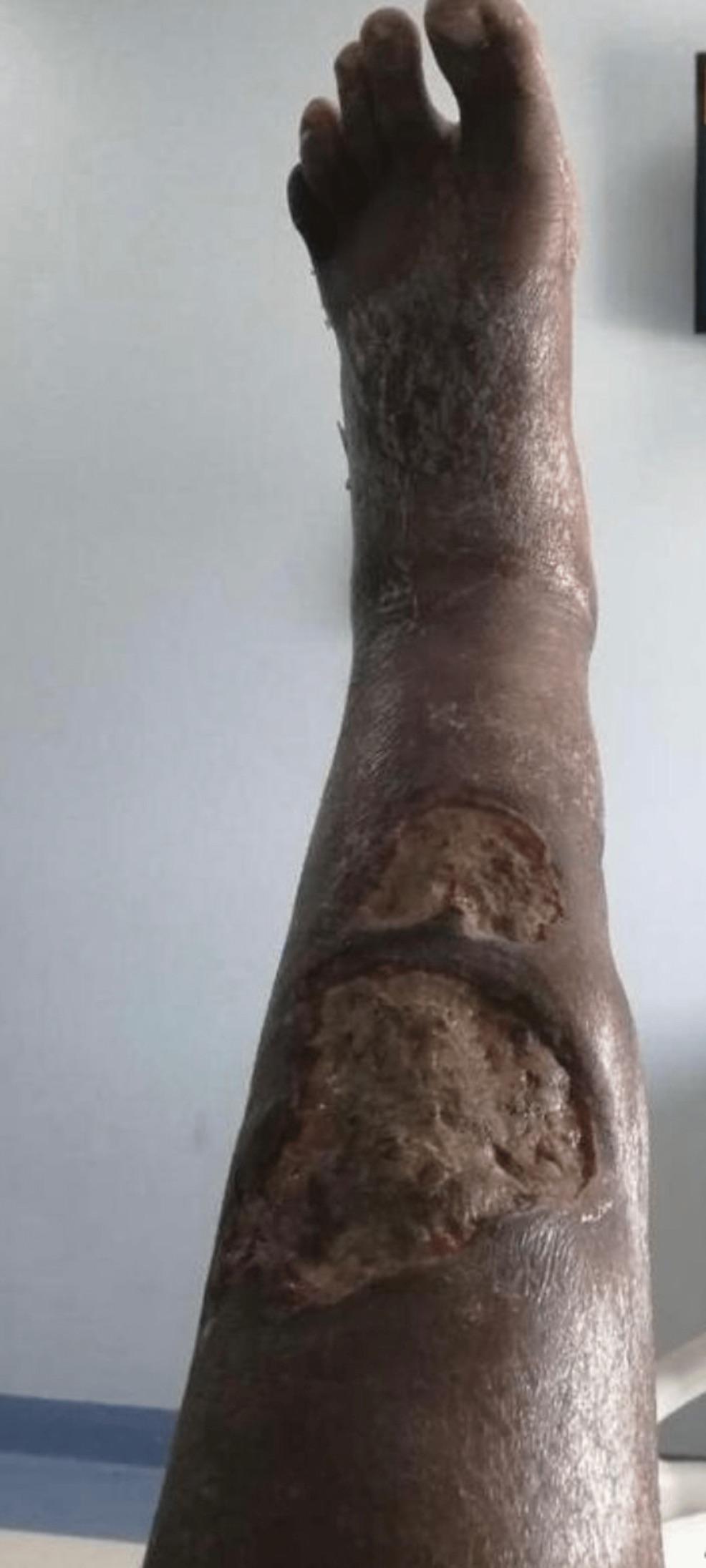 · 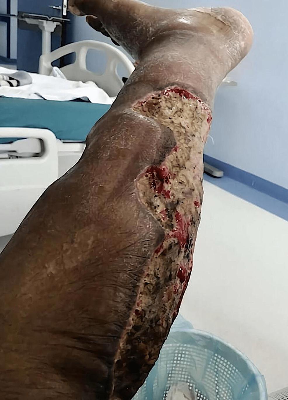 · 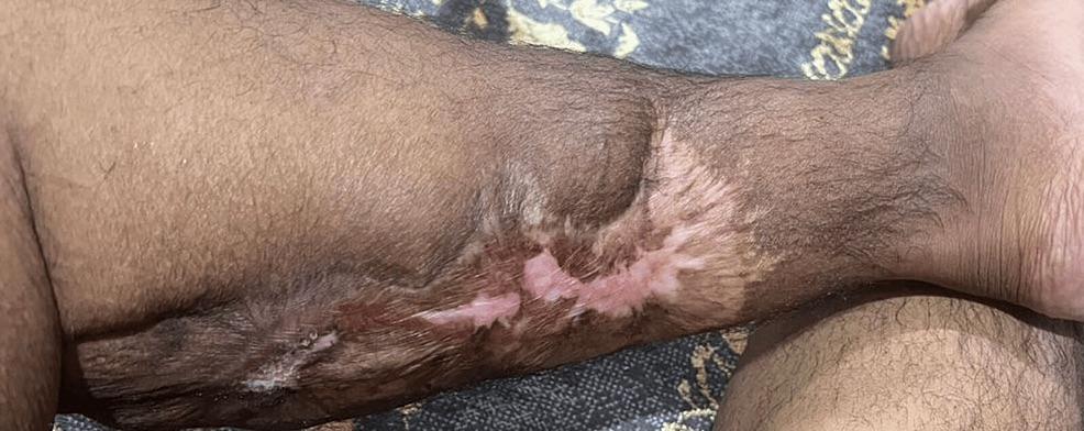 · 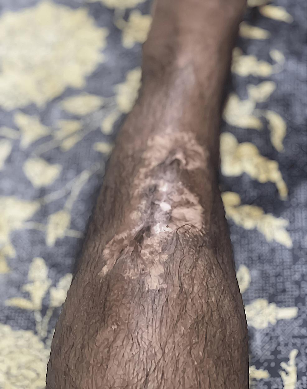 · |
|
#2
●
01-31-2023, 09:28 PM
| ||||||||
| ★ Legacy Member ★ Poster Rank:119 Secret Agent Join Date: Dec 2009 Posts: 13,214 Mentioned: 6 Post(s) Quoted: 2789 Post(s)
| ||||||||
|
Re: Pyoderma Gangrenosum (ulcers) Being Treated
Seeing the first photo I would have thought the leg would ge amputated. Was pretty surprised to see how well the leg looked after healing. |