|
#1
●
06-17-2024, 05:06 PM
| ||||||||
| These are the rooms Poster Rank:25 of ruin. Join Date: Sep 2014 Posts: 54,011 Mentioned: 145 Post(s) Quoted: 30403 Post(s)
| ||||||||
|
Prune Belly Syndrome
A 2-day-old boy was admitted for right bulging flank in November 2011. Gestational age was 39 weeks, with no fetal distress or asphyxia. The birth was by Cesarean section. Apgar scores were 8 at 1 minute and 9 at 5 minutes. Birth weight was 2900 g, birth length 50 cm, head circumference 34 cm, and chest circumference 33 cm. The infant was born to a 20-year-old uncomplicated mother, Gravida 1 Para 1. Prenatal sonography showed an intra-abdominal cystic collection in the fetus at 28 weeks' and 34 weeks' gestation. There was no prenatal intervention. After admission, physical examination showed bulging flanks, especially the right side, thin flabby, wrinkled skin (Figure 1), and bilateral testes impalpable within the scrota. There were no other issues, and urine output was normal. The chest and abdominal X-ray showed normal lung volume, hypoplasia of the upper mediastinum and lung, and no lung marking in the upper right lung, upper or middle left lung. Diffusely distended flanks were seen, with mass-like areas in the lower abdomen. Voiding cystourethrogram showed bladder diverticulum and bilateral vesicoureteral reflux, and the bilateral ureters were tortuous and dilated (Figure 2). An abdominal computed tomography (CT) scan showed small and irregular bilateral renal shape, renal parenchymal density, mild discordance of part of the renal pelvis, and dilatation of the renal calices and superior segments of both ureters. Cystic outward protrusion on the right side of the dilated bladder was also noted (Figure 3). Echocardiography showed mesocardia, branching of the coronary artery–right ventricular fistula, and mild tricuspid reflux. The urinalysis showed urine occult blood (+), protein (+), and white blood cell count (WBC) 32.8/μL (0–25); renal function showed blood urea nitrogen 12.94 mmol/L and creatinine 284.4 μmol/L on day 5. The baby had fever from day 7. Urine analysis showed urine occult blood (+), protein (+), and WBC 1997/μL (0–25); renal function showed blood urea nitrogen 21.83 mmol/L and creatinine 349.1 μmol/L on day 7. Urine culture was negative. Chromosome analysis was normal. The clinical diagnoses were PBS, coronary artery branch fistula, urinary tract infection, and renal inadequacy. The parents chose to cease treatment, and the patient died subsequently at 28 days due to septicemia from a severe urinary tract infection. 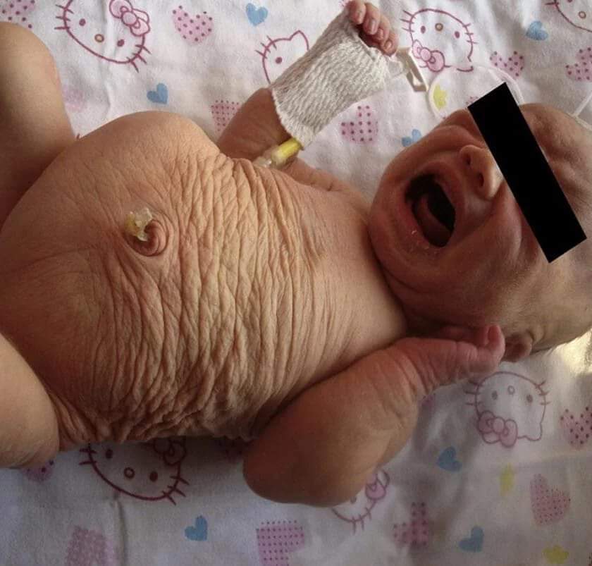 · 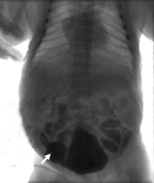 · |
|
#4
●
06-17-2024, 10:39 PM
| ||||||||
| ♚ Legacy Gold Member ♚ Poster Rank:130 Happy Happy Happy Join Date: Dec 2009 Posts: 12,404 Mentioned: 20 Post(s) Quoted: 4087 Post(s)
| ||||||||
|
Re: Prune Belly Syndrome
Something about dead children always put a shitty taste of cringe in my mouth. and I've seen some shit
|