|
#1
●
05-16-2024, 08:09 PM
| ||||||||
| These are the rooms Poster Rank:25 of ruin. Join Date: Sep 2014 Posts: 54,011 Mentioned: 145 Post(s) Quoted: 30403 Post(s)
| ||||||||
|
Necrotizing Fasciitis
>A 57-year-old obese female presented to the emergency department with worsening pain and infection of the right inguinal region, right thigh, and lower abdominal wall. A CT scan confirmed the likely necrotizing fasciitis, and the patient was also found to have poorly controlled diabetes mellitus and sepsis on admission. She was admitted by the Critical Care team and emergently taken to the operating room for incision and drainage, with extensive debridement of her necrotizing fasciitis. The original wound size following this debridement was 60 cm x 30 cm x 15 cm down to the muscle. Her sepsis failed to resolve, and she was, therefore, taken back to the operating room 36 hours later for further debridement down to the muscle with a long segment of visible femoral artery and sciatic nerve exposed. She was taken back to the operating room again four days later for further debridement as she was too unstable during the previous procedures for prolonged anesthesia and blood loss. >>Throughout the above-mentioned procedures, her wound was also thoroughly irrigated via pulsavac lavage. After the second debridement due to the severity of her wound, an orthopedic consult was obtained for an opinion on a hip disarticulation; they recommended proceeding with the current wound care and would proceed with disarticulation if she failed with the current therapy. Forty-eight hours following the 3rd OR debridement (Figure 1), an NPWTi-d Veraflo device was placed on the wound with normal saline instillation solution with the following settings: 325 ml normal saline was instilled for a 10 minute soak every three and a half hours under -125 mmHg continuous pressure per the recommendations of the clinical panel [1-4] (Figure 2). Use of normal saline for instillation was chosen as studies have shown that it is as effective as other topical wound cleaning solutions [5-6]. Similar to the use in this patient, multiple clinical trials have shown that NPWTi-D versus NPWT achieves faster wound closure and better skin perfusion when used with -125 mmHg [7-9]. >>The dressings were changed on a 3-4 day schedule. After four days of treatment with the NPWTi-d Veraflo, the wound showed improvement with increased granulation tissue and viable skin edges, with granulation over the exposed artery and nerve (Figure 3). >>The patient had a prolonged hospital course. Once she was off sedation and medically stable, due to the size and extent of her wound, she was taken back to the OR three days later for a dressing change, and the overall wound showed great improvement with increased granulation throughout (Figure 4). >>NPWTi-d Veraflo therapy was continued with one more subsequent dressing change seven days later and then returned to NPWT -125 mmHg until discharge from the hospital to the LTAC facility. NPWT was continued with dressing changes three times weekly at the outside LTAC facility as discussed for continued therapy for expedited granulation. The patient was then readmitted 20 days later for other medical complications; the wound was evaluated and exhibited almost complete granulation over all exposed muscle, vasculature, and nerve structures—almost superficial to surrounding peri-wound skin (Figure 5). >>She was once again discharged to the LTAC facility with NPWT. The patient was then readmitted about 30 days later for VRE pneumonia and expired from this, therefore complete closure of her wound was not able to be achieved. Informed consent was waived, and no reference to the patient's identity was made at any stage during data analysis or in the report. 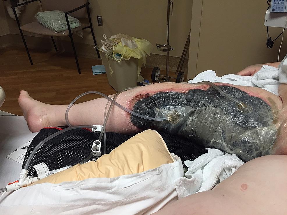 · 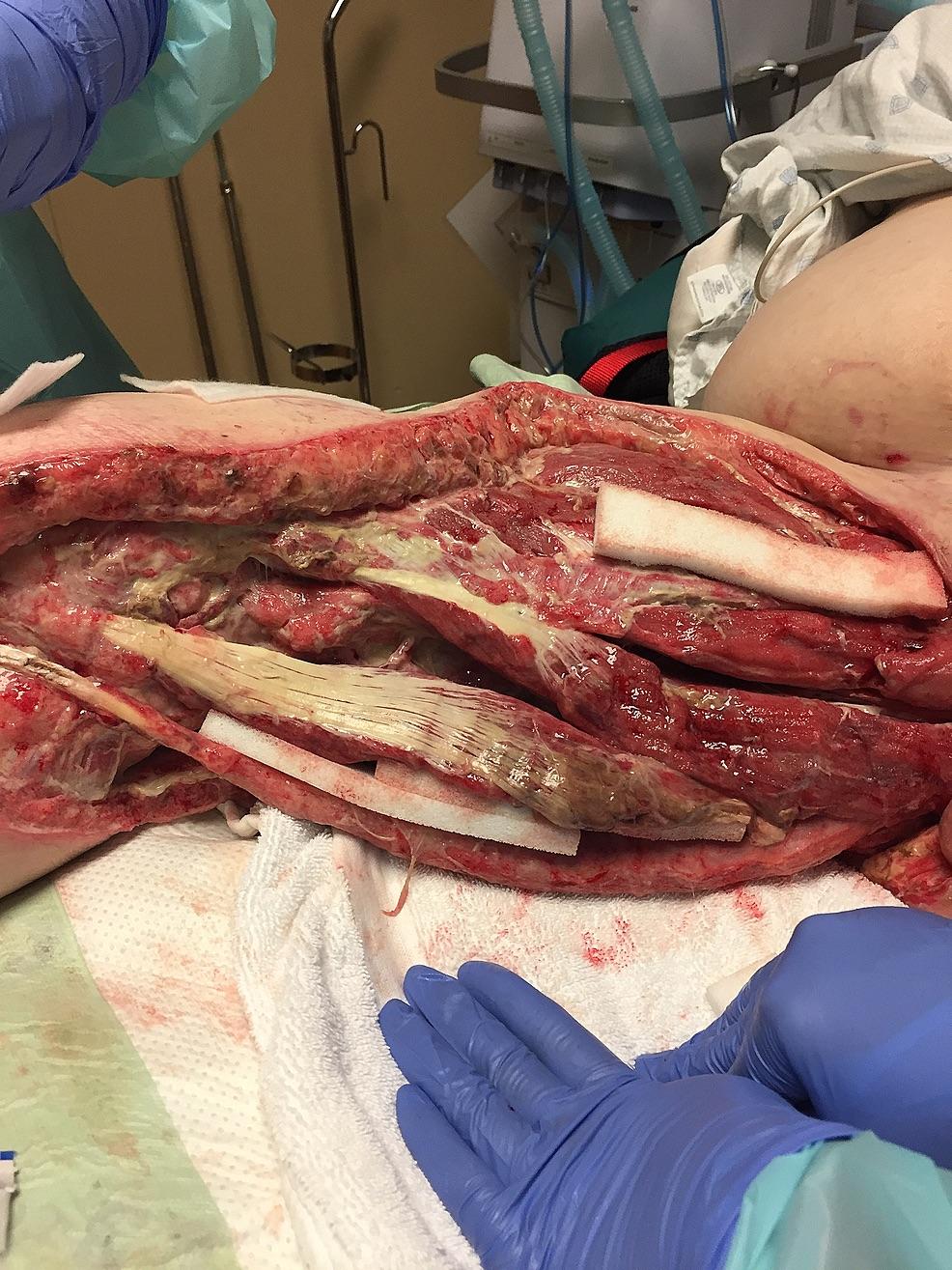 · 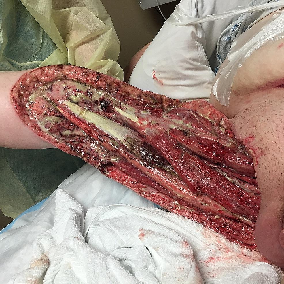 · 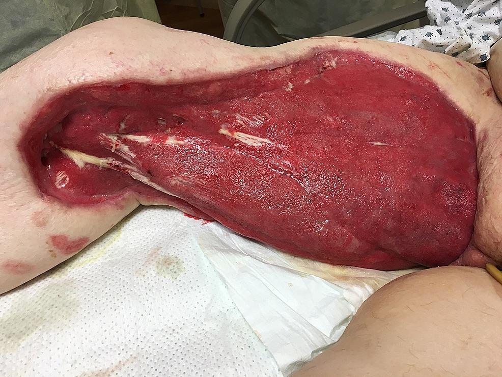 · 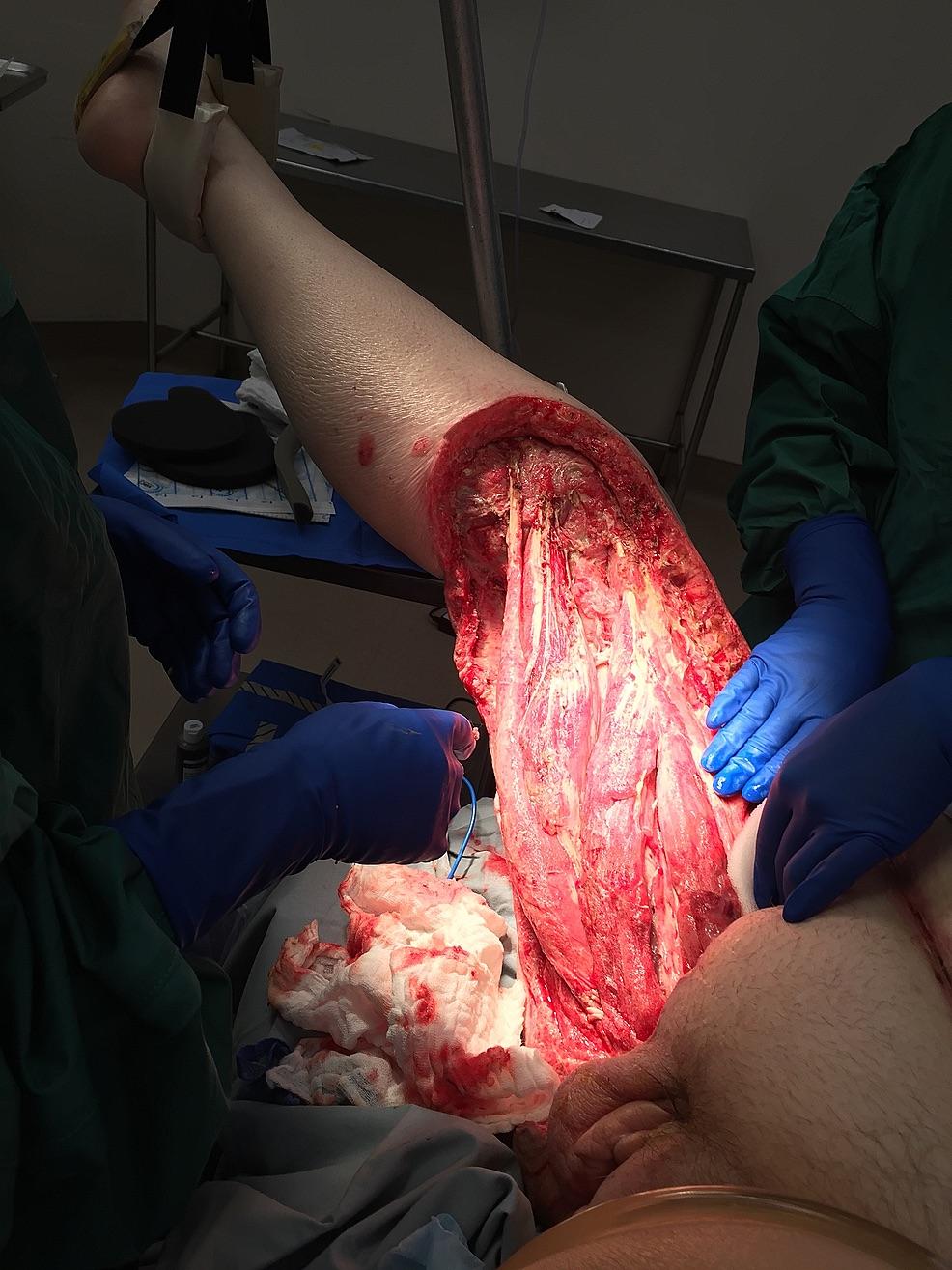 · |
|
#5
●
05-17-2024, 08:16 PM
| ||||||||
| ★ Legacy Member ★ Poster Rank:248 So many choices now Join Date: Jul 2015 Posts: 5,548 Mentioned: 15 Post(s) Quoted: 2120 Post(s)
| ||||||||
|
Re: Necrotizing Fasciitis
Well, at least now she won't have to worry about her thighs rubbing together...
|