|
#11
●
04-29-2023, 07:48 PM
| ||||||||
| ✖ The Antique Christ ✖ Poster Rank:105 Gonzo Punk Join Date: Jan 2009 Posts: 15,488
Contributions: 58
Mentioned: 23 Post(s) Quoted: 2046 Post(s)
| ||||||||
|
Re: Lithopedion - Calcified Dead Fetus or Stone Baby
Zahra Aboutalib, a 26 year old lady from a small village outside Casablanca, was rushed to her local hospital in 1955 after 48 hours of excruciating labour. The doctors told her she would need a caesarian section. Whilst she was waiting on the ward for her operation, she witnessed the painful death of a woman in childbirth. After seeing this, Zahra ran away from the hospital and back to her village. She suffered further terrible pains, but after a few days, they stopped. Her baby had died. But there was no miscarriage. There was a myth in her village that a baby can go to sleep inside its mother, and Zahra chose to believe this. Nearly 50 years later, when Zahra was 75, she went to hospital again with similar pains. She was referred to a specialist, Professor Taibi Ouazzani, in Rabat, who at first thought she was suffering from an ovarian tumour. What Prof. Ouazzani discovered was a large, calciferous lump. He sent her for an MRI scan, which revealed the lump to be Zahra's dead baby from nearly 50 years before. It was removed after a five hour operation, which revealed the stone baby. Below are picture of it before and after dissection. 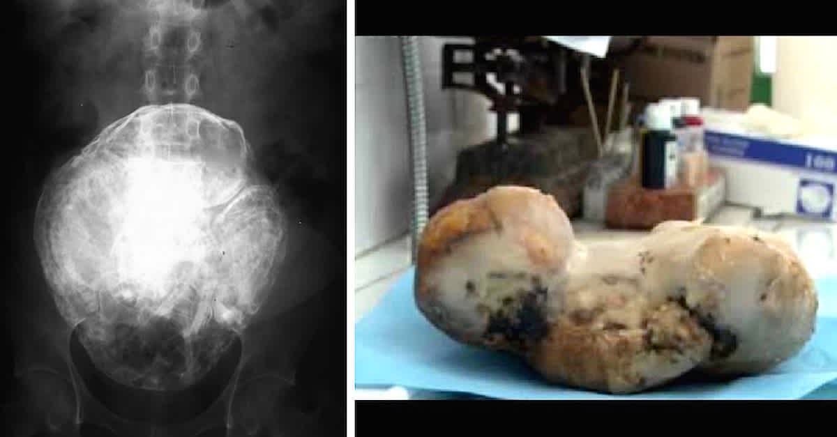 · 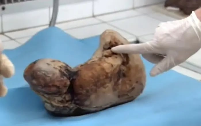 · 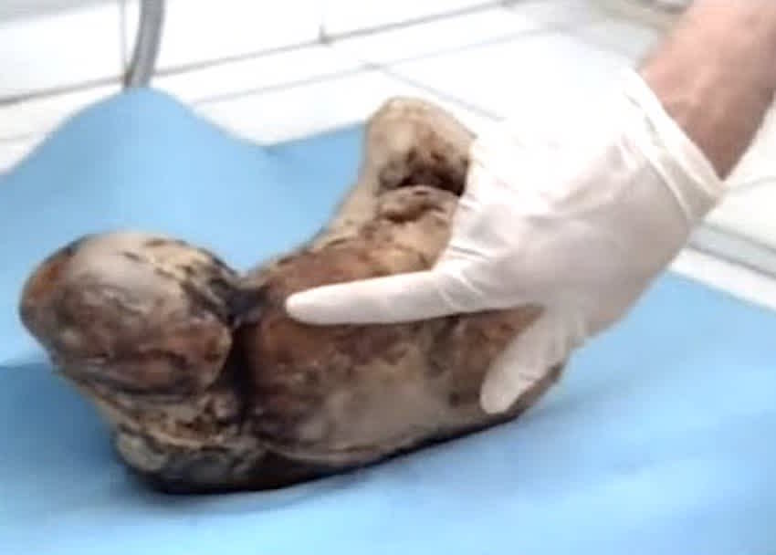 · 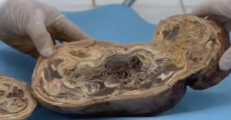 · 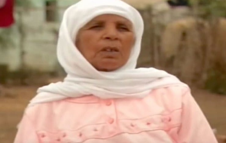 · |
|
#12
●
04-29-2023, 07:51 PM
| ||||||||
| ✖ The Antique Christ ✖ Poster Rank:105 Gonzo Punk Join Date: Jan 2009 Posts: 15,488
Contributions: 58
Mentioned: 23 Post(s) Quoted: 2046 Post(s)
| ||||||||
|
Re: Lithopedion - Calcified Dead Fetus or Stone Baby
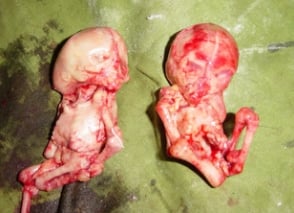
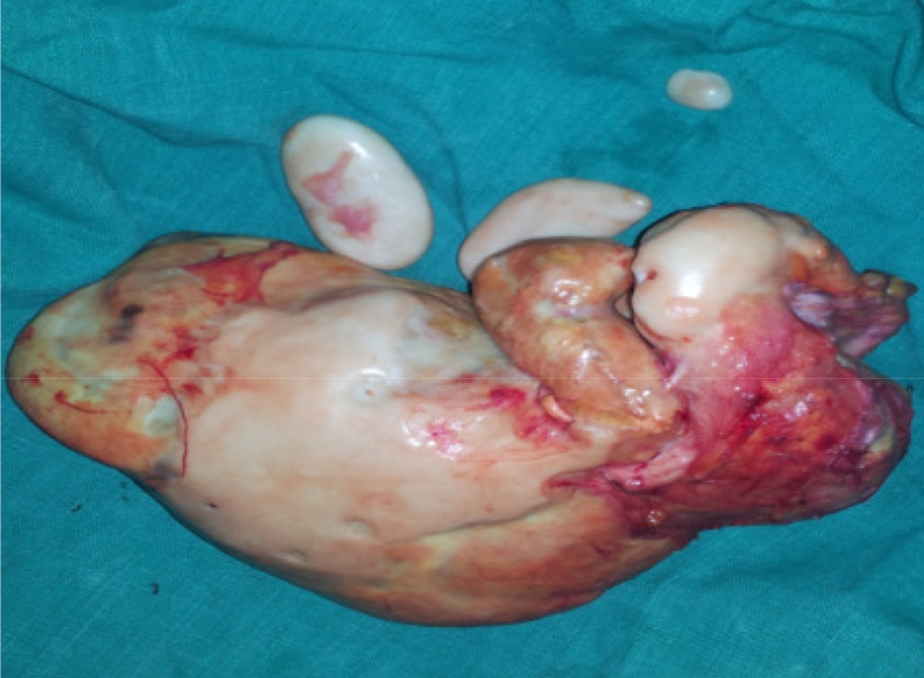
In India, a case of Twin Lithopedia was reported in a 40 year old woman who had arrived at a hospital presenting symptoms of “acute intestinal obstruction”[xxvi]. “She had abdominal distension, vomiting and absolute constipation.”[xxvii] An internal exam showed signs of a pregnancy which had somehow terminated around the fifth month of pregnancy, but nothing unusual. However, no product of the pregnancy was ever expelled from the uterus.[xxviii] Radiography showed “[t]wo radiopaque, calcified, globular shadows…on both sides of the lower abdomen”[xxix] while ultrasonography “showed two oval calcified areas on both sides of the lower abdomen.”[xxx] During the laparotomy, one Lithopedion was “morbidly adhered” to “a devitalised portion of the ileum” while the other was attached to the greater omentum.[xxxi] When the two oval masses were dissected, “two mummified and calcified foetal skeletons were recovered. Both skeletonised foetuses were of the same age (around 5 months old).”[xxxii] This is the only recorded example of Twin Lithopedia.
 · |
|
#13
●
04-29-2023, 07:57 PM
| ||||||||
| ✖ The Antique Christ ✖ Poster Rank:105 Gonzo Punk Join Date: Jan 2009 Posts: 15,488
Contributions: 58
Mentioned: 23 Post(s) Quoted: 2046 Post(s)
| ||||||||
|
Re: Lithopedion - Calcified Dead Fetus or Stone Baby
The 52-year-old, who does not want to be named, terminated her pregnancy back then as her family allegedly did not approve of her having another child. At the time, her obstetrician and gynaecologist treating her confirmed the baby was successfully aborted. However, she went on to suffer abdominal pain over the years and visited several doctors, who only gave her painkillers and pills for acidity. And for the last three years, she had been continually vomiting, raising alarm. She then went to see a specialist who discovered the rare find – and has had a two-hour operation to remove the 'fully grown stone baby' as a medic described it. Eventually, the woman, from a small village near Nagpur, in Maharashtra in the northern part of the country, met with a specialist. Medics at Junankar Surgical Nursing Home, in Nagpur city, did a CAT scan and found an obstruction in her intestines, blocking the digestive system. Dr Nilesh Junankar, a laparoscopic surgeon, said: ‘Her scan showed intestinal obstruction, a blockage in her food pipe along with a stone like structure. 'Due to this unusual report, a laparoscopy was done and to our utter surprise, there was a four-month old baby in the abdomen. We were very shocked. It’s extremely rare.’ A team of two doctors decided to immediately operate and Dr Nilesh removed the dead baby in a two-hour operation on November 23. Dr Nilesh explained: ‘It was a shock for everyone present in the operating theatre. 'After opening the abdomen, a fully grown ‘stone baby’. However, her uterus, ovaries and fallopian tubes were totally normal. 'The patient had stopped menstruating five years back and was not in a child bearing age.’ ‘Since no sonography was done, neither she nor the doctors thought the baby was still there,’ Dr Nilesh added. ‘The chance of abdominal pregnancy is one in 11,000 pregnancies and only between 1.5 to 1.8% of abdominal pregnancies develop into Lithopedia. 'We have removed the stone baby from woman’s body and her four feet intestines have also been removed.’ The woman thanked the hospital for freeing her from a decade of pain and has since been discharged from the hospital. 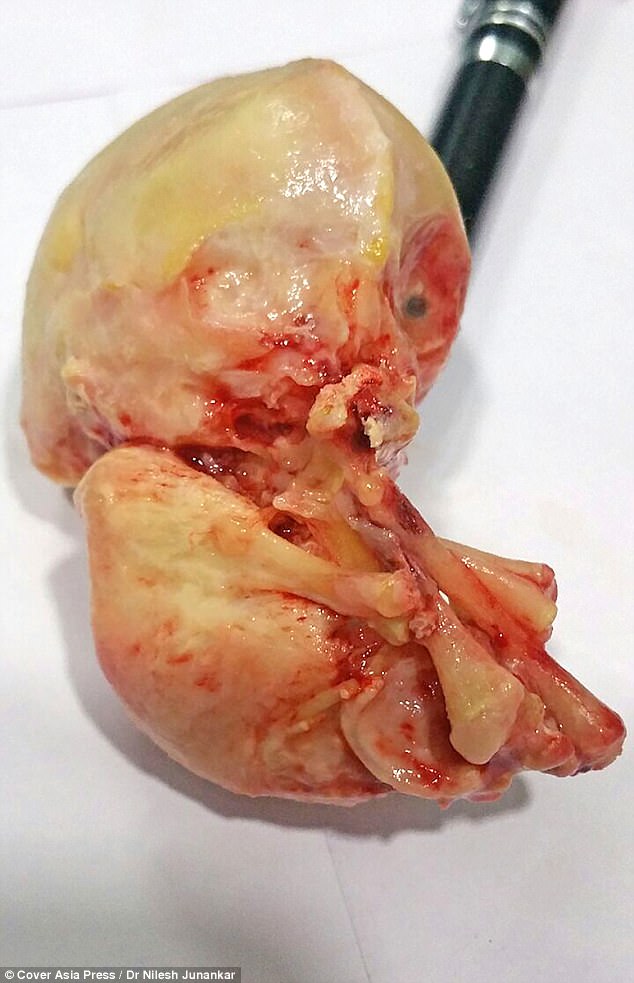 · 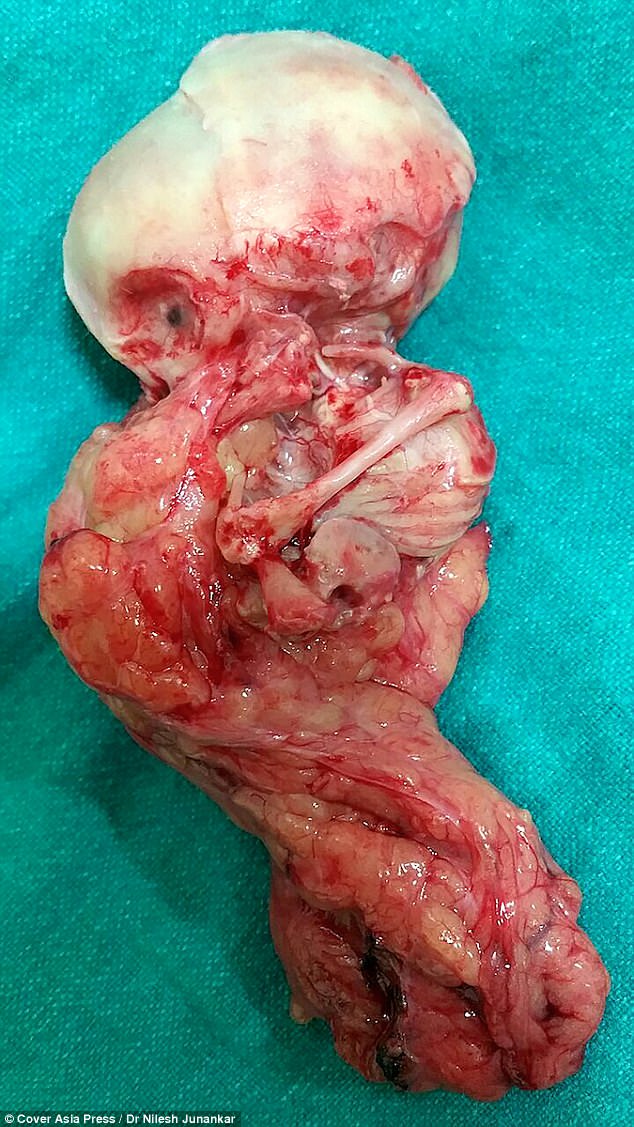 · 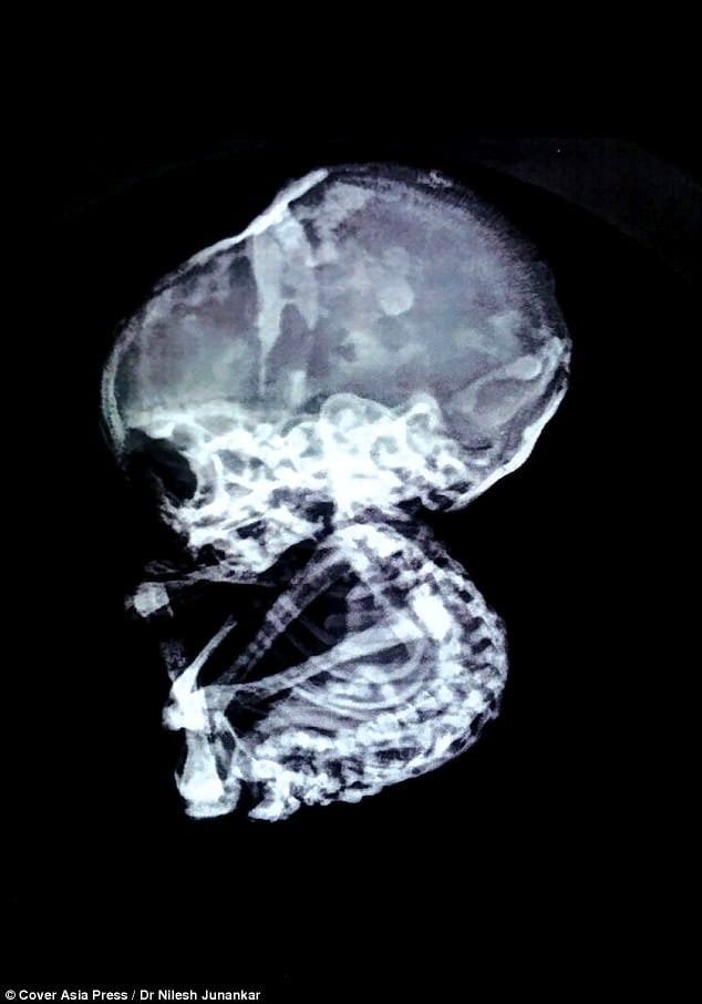 ·  · |
|
#14
●
04-29-2023, 08:12 PM
| ||||||||
| ✖ The Antique Christ ✖ Poster Rank:105 Gonzo Punk Join Date: Jan 2009 Posts: 15,488
Contributions: 58
Mentioned: 23 Post(s) Quoted: 2046 Post(s)
| ||||||||
|
Re: Lithopedion - Calcified Dead Fetus or Stone Baby
The case is a 39-year-old parity-5 woman who was admitted with complaints of an excruciating right iliac fossa pain which was associated with nausea and vomiting. At the time of admission, she was feverish and moderately dehydrated. Medical history revealed that her previous deliveries were all village births and her last child had been born 5 years before. Following this, she recalled being amenorrhoeic for a period of time but could not recall for how long or the exact dates. Then about 2 years before, she re-established her periods, which had since been regular. She had never used any family planning. She often had pains in her abdomen but the pains were described as mild. However, in recent months the pains had become severe and prolonged. Over the past 2 weeks before she presented, the pains became excruciating. She did not have compressive symptoms of pelvic pains, urinary bladder or rectum. Physical examination performed by the admitting surgical team revealed a vague mass in her right iliac fossa, which was firm and tender. A provisional diagnosis of appendicular mass was made. Her haematological profile included haemoglobin, haematocrit and platelets, which were all within the normal range, and a slight degree of leukocytosis. She was taken to the operating theatre and surgery revealed a hard mass that was covered with omentum and fascia. It felt solid and gritty. Further dissection finally revealed a calcified fetal skeleton (Figure 1). At that stage intraoperatively, the gynaecological team took over the management of the case. The lithopedion was further dissected off the right tube which was firmly adhered and had formed fibrous tissue around it together with omentum and had some attachment to large bowel. Further careful dissection resulted in complete removal of the lithopedion (Figure 2). The right ovary was also removed together with the fallopian tube as they were tightly adhered and the lithopedion had grown into them. The uterus was grossly normal as well as the contralateral tube and ovary. Left tubal ligation was also performed. The patient recovered uneventfully and was discharged in a stable condition after a few days in hospital. 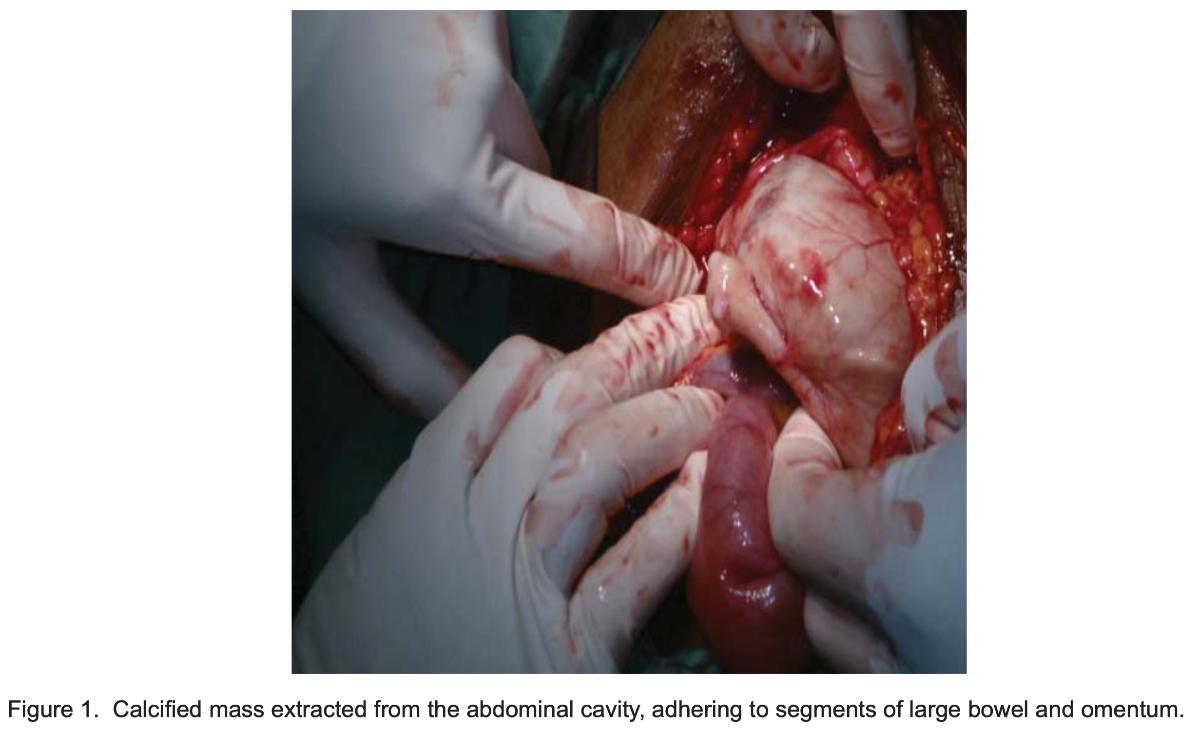 · 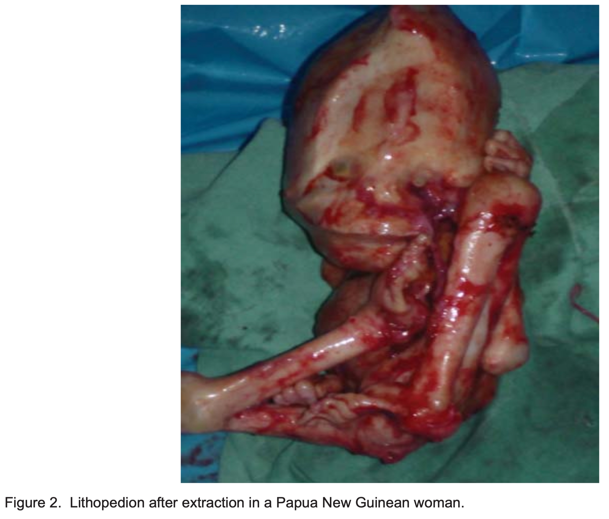 · |
|
#15
●
04-29-2023, 08:25 PM
| ||||||||
| ✖ The Antique Christ ✖ Poster Rank:105 Gonzo Punk Join Date: Jan 2009 Posts: 15,488
Contributions: 58
Mentioned: 23 Post(s) Quoted: 2046 Post(s)
| ||||||||
|
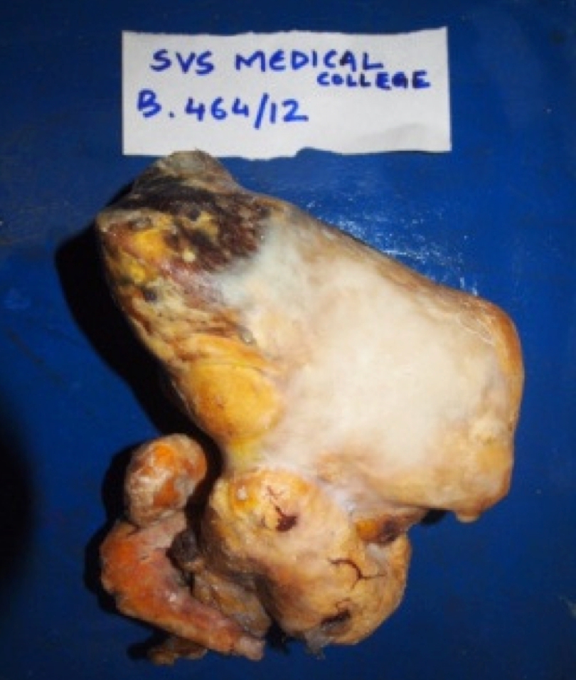
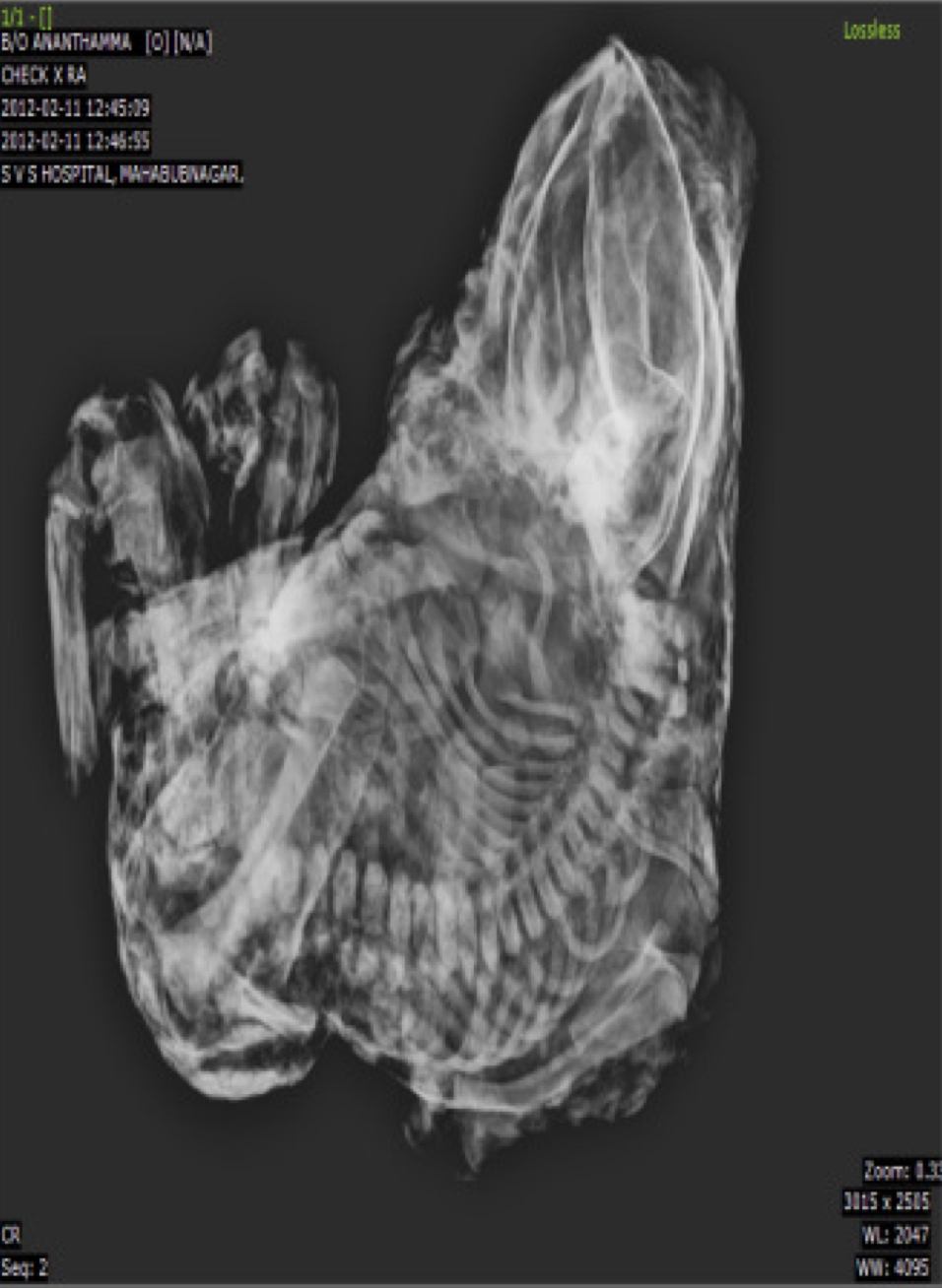
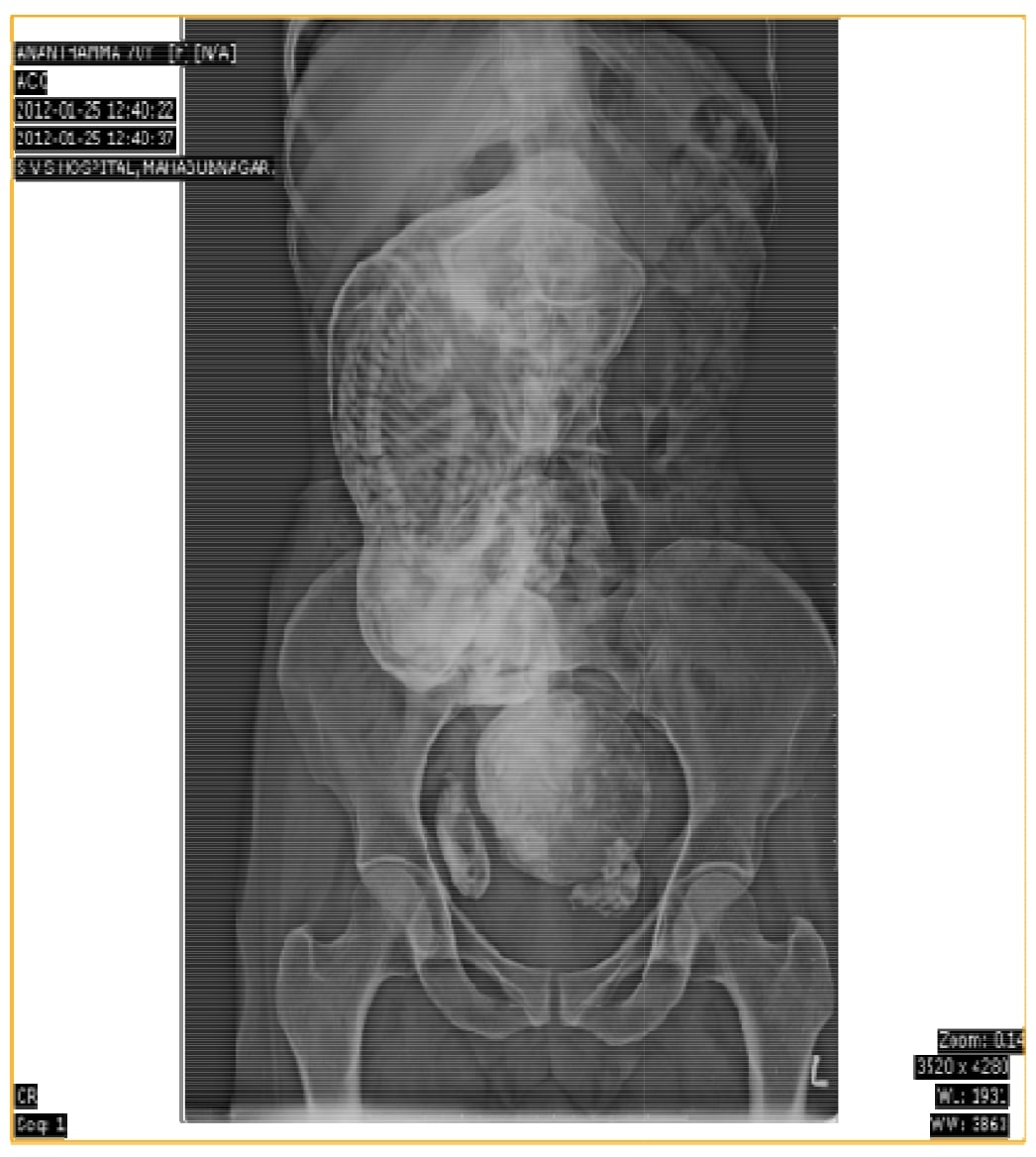
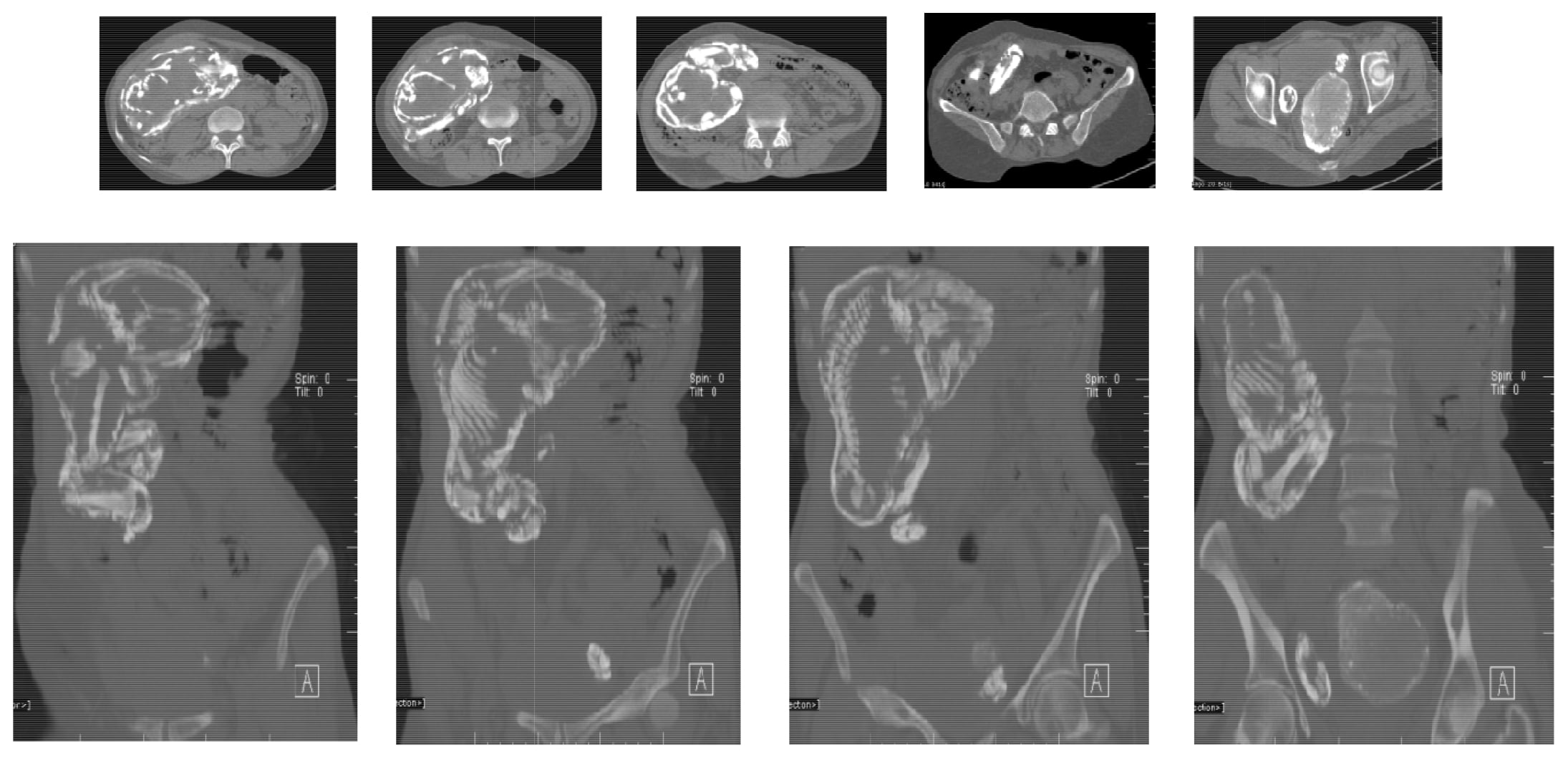
Re: Lithopedion - Calcified Dead Fetus or Stone Baby
We report the rare case of a lithopedion in 70 year old female with chief complaints of mass per vaginum. Diagnosis was confirmed by plain abdominal X-ray and computed tomography and patient consequently undergone laportomy. Women have retained the calcified fetus for 37 years.
 ·  ·  ·  ·  · |
|
#16
●
04-30-2023, 02:36 AM
| ||||||||
| My Rank: MASTER GUNNERY SERGEANT Poster Rank:347 Manly Man Join Date: Sep 2017 Posts: 3,601 Mentioned: 0 Post(s) Quoted: 700 Post(s)
| ||||||||
|
Re: Lithopedion - Calcified Dead Fetus or Stone Baby
That's crazy. Where can I buy one of these?
|
|
#18
●
04-30-2023, 06:40 PM
| ||||||||
| My Rank: SERGEANT Poster Rank:1228 Join Date: Dec 2015 Posts: 511 Mentioned: 0 Post(s) Quoted: 182 Post(s)
| ||||||||
|
Re: Lithopedion - Calcified Dead Fetus or Stone Baby
Incredible! Thanks for sharing! Also, it's more proof that the Abrahamic God doesn't exist. |
|
#19
●
04-30-2023, 09:57 PM
| ||||||||
 My Rank: MASTER SERGEANT Poster Rank:522 Join Date: Apr 2010 Posts: 1,957 Mentioned: 3 Post(s) Quoted: 220 Post(s)
| ||||||||
|
Re: Lithopedion - Calcified Dead Fetus or Stone Baby
Um ,,,,,,,, This is so extraordinary that I have to comment .................... What if ,,,,, these cases are because of Extraterrestrial impregnation . -----
|