|
#1
●
06-07-2024, 04:36 PM
| ||||||||
 These are the rooms Poster Rank:26 of ruin. Join Date: Sep 2014 Posts: 54,011 Mentioned: 145 Post(s) Quoted: 30403 Post(s)
| ||||||||
|
Eye Avulsion
A 40 years old male presented with traumatic expulsion of right eyeball with optic nerve avulsion following motorbike accident. Patient was conscious, well oriented to time, place person with no other vital injury of other parts. On ocular examination right globe was protruding out of orbit with minimal tissue attachment. (Figure 1) Wooden foreign body was present between right lateral orbital wall and right globe. (Figure 2) The cornea of injured eye was clear but pupil was dilated and non reacting without any perception of light. Lid laceration of 4 x 2cm was present on right upper eyelid. Ecchymosis was present in both upper and lower eyelids. Visual acuity in left eye was 6/6 with normal pupil reactions. Ocular movements and fundus were normal. The CT scan showed protrusion of right globe with optic nerve avulsion. Patient was started on intravenous antibiotics and enucleation of right eye was done along with removal of 5×2 cm size foreign body. The right eyelid laceration was repaired. Cornea of patient was preserved after written consent of the patient and used for keratoplasty on other patient having pseudophakic bullous keratopathy right eye. Case 2, the recipient: A 75 years old male presented with painless progressive loss of vision in right eye for 12 years. The patient had history of trauma in left eye following which he lost vision in this eye. He underwent penetrating keratoplasty in left eye but 6 months later patient developed pain, redness,watering and diminution of vision left eye. The patient was diagnosed of as graft rejection and put on oral prednisoloneand topical prednisolone, G homatropine, G Timolol. The patient was lost to follow up and came after 4 years. Onocular examination there was no perception of light in righteye with afferent pupillary defect. The intraocular pressure was high 32mm of Hg and anterior segment was normal. Fundus examination showed complete glaucomatous opticatrophy. Visual acuity in left eye was HMCF. Cornea was almost opaque due to failed penetrating keratoplasty. Theintraocular pressure was normal and B scan showed no gross abnormality of posterior segment. After informed written consent the patient was planned for autograft from opposite absolute eye having clear cornea. But following graft excision from right eye the expulsive haemorrhage developed despite preoperative I/V Mannitol and Acetazolamide and eye has to be eviscerated.The full thickness corneal graft taken from this eye was puton the other eye. Intraoperative procedure was uneventful in this eye. Post-operatively patient had normal graft host junction, some Descemet’s folds on the graft, well formed anterior chamber in left eye and visual acuity of 6/60.(Figure 4) The patient could move independently, which was not possible before surgery. He was started on standard post operative treatment in left eye for keratoplasty and antibiotics in right eye. At 6 weeks follow up visual acuity in the left eye was 6/24. The cornea was clear and the graft host junction was well healed up and intraocular pressure was normal. 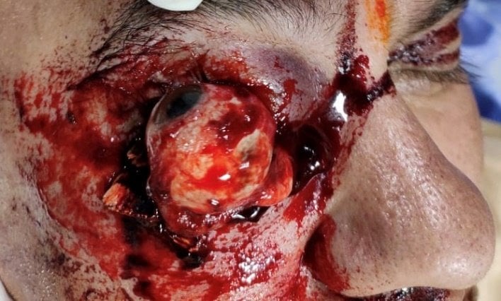 ·  · 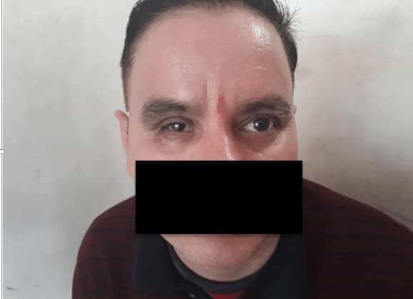 · 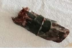 · |
|
#3
●
06-07-2024, 11:20 PM
| ||||||||
 My Rank: MASTER GUNNERY SERGEANT Poster Rank:352 Manly Man Join Date: Sep 2017 Posts: 3,601 Mentioned: 0 Post(s) Quoted: 700 Post(s)
| ||||||||
|
Re: Eye Avulsion
They have his mouth blacked out in the photos...they afraid he's going to talk?
|