|
#1
●
05-17-2024, 05:24 PM
| ||||||||
 These are the rooms Poster Rank:24 of ruin. Join Date: Sep 2014 Posts: 54,011 Mentioned: 145 Post(s) Quoted: 30403 Post(s)
| ||||||||
|
Crushed Foot
22-year-old male patient was brought to R L Jalappa Hospital & Research Centre, affiliated with Sri Devaraj Urs Medical College, Kolar, India. The patient presented with an alleged history of a road traffic accident. He sustained a crush injury to his right heel pad, resulting in an open comminuted fracture of the right calcaneum with bone loss (Figure 1). The range of motion at the right ankle and the subtalar joint was painful and restricted. Dorsalis pedis artery pulsation was palpable, and distal sensation was intact. Active toe movements were present. The capillary refill time was normal. All other long bones and joints were clinically normal. There were no other injuries anywhere in the body. >>At the emergency department, triple antibiotic prophylaxis was administered. It consisted of intravenous (IV) administration of augmentin 1.2 g, amikacin 500 mg, and metronidazole 100 ml. Wound wash was given with six liters of sterile saline at the bedside and the wound site dressed with moist gauze. The patient was stabilized with a below-knee splint and sent for radiographs. Preoperative radiographs are provided in Figure 2. After imaging was completed and informed consent obtained, the patient underwent thorough wound debridement and NPWT was given. The initial operative encounter consisted of the patient being operated under spinal anesthesia. Gross wound contaminants were removed, including all devitalized bone. Three liters of sterile saline were then used to irrigate the wound. Once complete, all wounds were left open and a wound VAC was applied. Figure 3 shows the clinical picture of the right heel pad after three days following initial debridement. >>The wounds were then lightly packed with sponge and sealed with adhesive drapes. The sterile sponge was applied over the exposed part of the bone and hardware. When sealing the sponge, care was taken to lay drapes over the skin with minimal skin tension. Suction was set at -125 mm of Hg. A total of seven days of NPWT was allowed [2]. The vacuum-assisted closure device is shown in Figure 4. >>Postoperatively, the patient received intravenous augmentin 1.2 g twice daily for seven days, amikacin 500 mg twice daily for seven days, and metronidazole 100 ml thrice daily for three days. It was followed by oral Amoxiclav 625 mg twice daily for seven days. The patient was taken to the operating room one more time for wound debridement (Figure 5), followed by reverse sural artery flap placement (Figure 6). Assessment of the wound during the time of secondary debridement demonstrated a progressive wound bed with granulation tissue and a decrease in overall wound size. Postoperatively, the patient stayed in the hospital for seven days for flap monitoring. The surgical site healed well (Figures 7-10). Active dorsiflexion of the right ankle was present. The patient is being followed at regular intervals. Outpatient follow-up ensued at two weeks, one month, and three months. 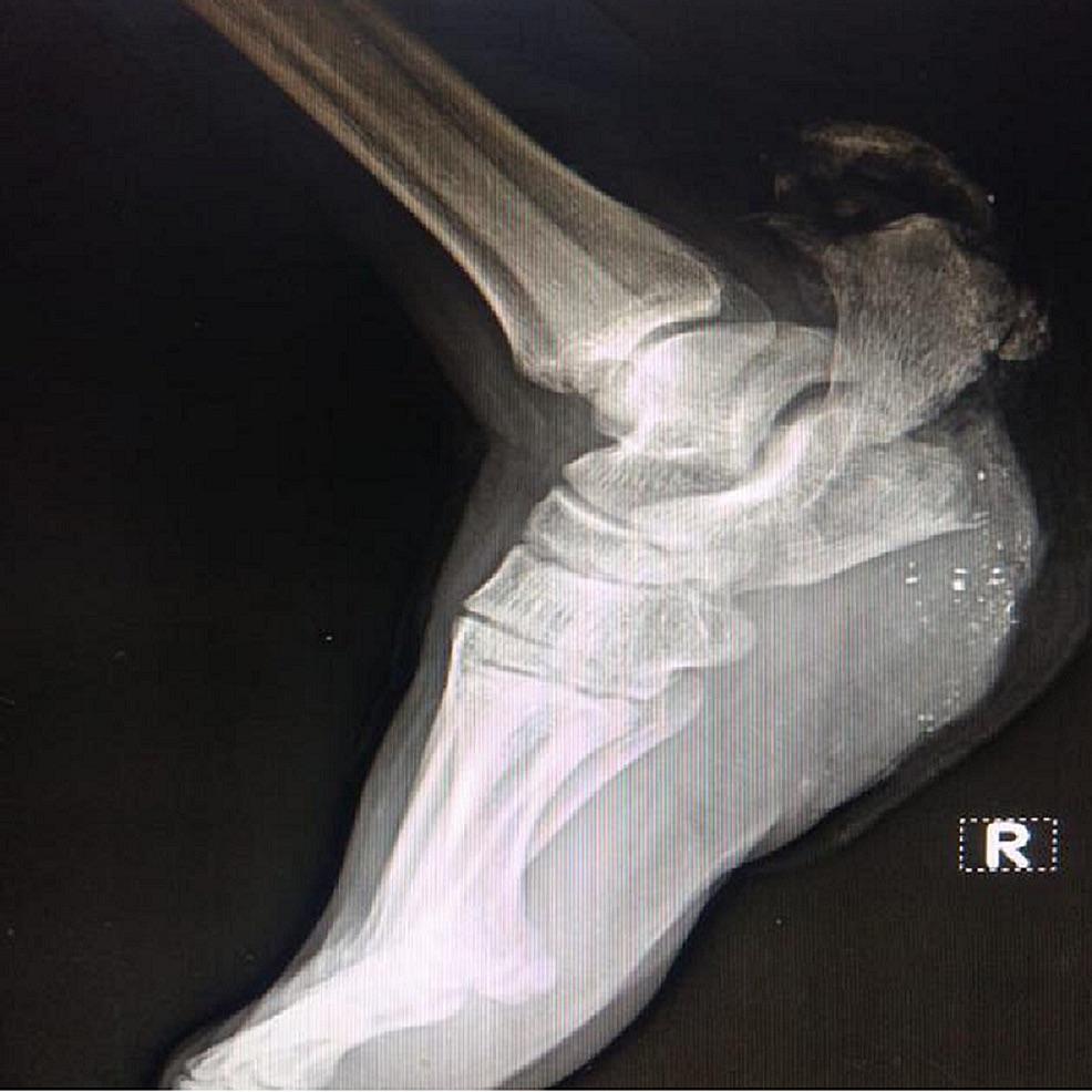 · 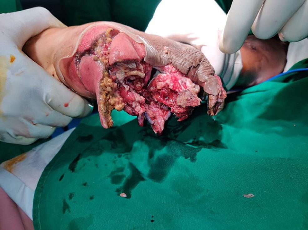 · 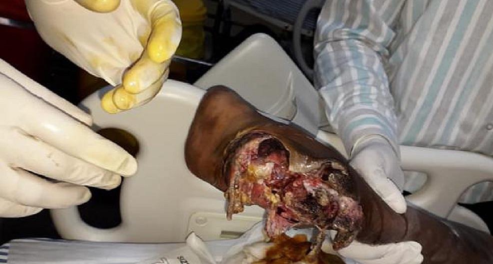 · 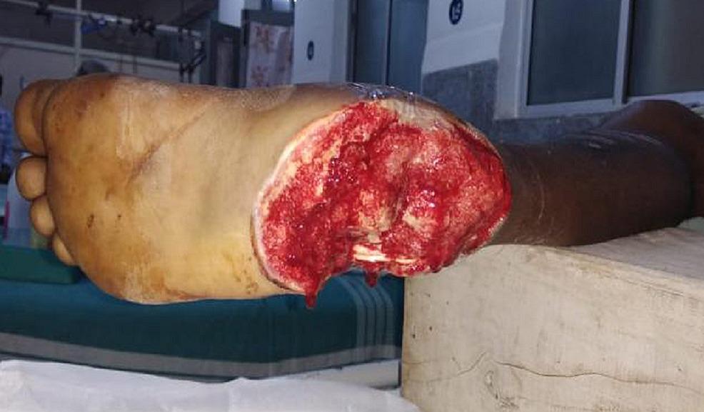 · 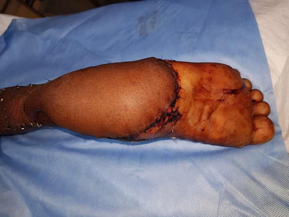 · 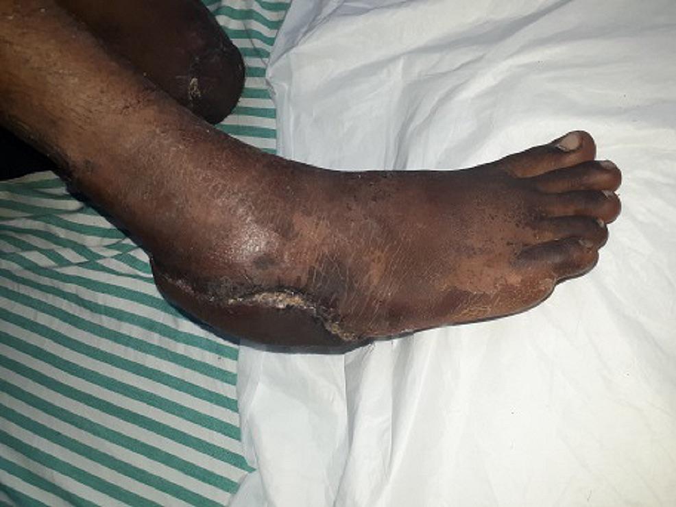 · 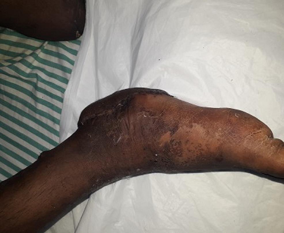 · 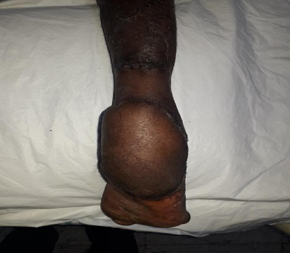 · 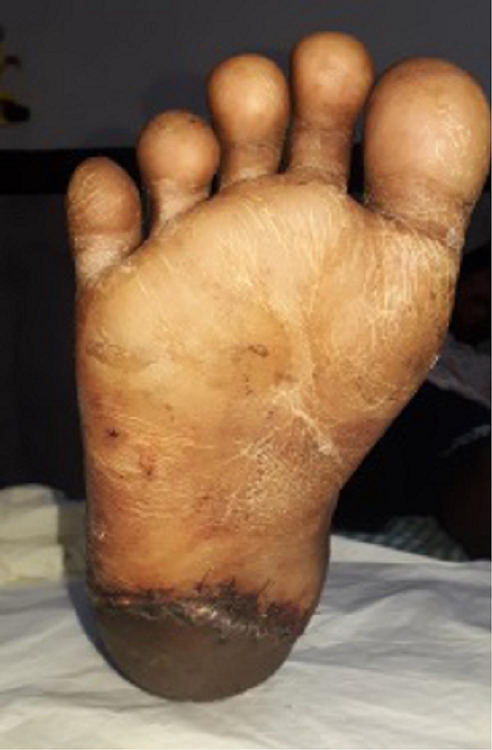 · |
|
#3
●
05-18-2024, 12:51 AM
| ||||||||
 ♚ Legacy Gold Member ♚ Poster Rank:265 ꓕSIꓤIꓕ∀S ϽINOᗡꓤ∀S Join Date: Feb 2010 Posts: 5,156
Contributions: 6
Mentioned: 42 Post(s) Quoted: 1435 Post(s)
| ||||||||
|
Re: Crushed Foot
He'll be back to riding his motorbike with flip-flops in no time...
|