|
#1
●
08-31-2024, 06:44 PM
|
 These are the rooms Poster Rank:26 of ruin. Join Date: Sep 2014 Posts: 54,008 54,008Posts 87,658Thanked 30,403Quoted 145Mentioned 0Contrib. |
|
Atv Accident
A 28-year-old male sustained an open injury to his right ankle while riding an all-terrain vehicle (ATV) on the evening of October 5, 2019. The patient recalls that the injury was a result of the ATV rolling over while accelerating between 15 and 20 miles per hour (mph). His right foot was caught under the ATV as the vehicle flipped. He had an immediate onset of pain, deformity, and inability to bear weight. He was brought to the emergency department for an initial assessment. A full examination of the ankle was not possible due to significant hemarthrosis. A large transverse laceration was noted over the medial ankle with gross contamination (Figure 1). Plain radiographs of the right ankle revealed a comminuted displaced medial malleolus fracture (Figure 2). He proceeded to the operating room the same night for incision and debridement of the open fracture and wound. The patient was placed supine and draped in a sterile manner. Examination showed a 10-cm transverse incision extending from the anteromedial to the posteromedial aspect of his right ankle. Also, there was a segmental portion missing from the anterior shoulder to the posterior colliculus which extended for approximately 3 x 1.5 cm (Figure 3). The deltoid origin was still intact and there was a very small fragment of bone present distally. Examination showed that the deep peroneal nerve was intact. There was a complete transection of the posterior tibial tendon, avulsion of the flexor digitorum longus muscle belly, and the posterior tibial artery was avulsed and occluded. There was an intact posterior tibial vein, posterior tibial nerve, flexor hallucis longus, and Achilles tendon. We found grass, dirt, and gravel in the wound. Sharp excision was used to remove devitalized skin, subcutaneous tissue, muscle, and bone. Following the debridement, the wound was thoroughly irrigated with 9 L of normal saline. Next, the posterior tibial tendon ends were tagged with a PDS II (polydioxanone) suture. In addition, the flexor digitorum tendon was preserved for a tenodesis during the reconstructive phase. The patient was taken back to the operating room for a total of three debridement procedures with splinting performed following each debridement. Following initial debridement, a peroneal nerve palsy was identified. A working diagnosis of type IIIA open medial malleolus fracture was made, and the patient was scheduled for open reduction internal fixation surgery with allograft reconstruction on November 13, 2019. The patient was taken back to the operating room, placed supine with a hip bump, and draped in a sterile fashion. A standard medial malleolus approach was used to access the defect. An anatomically aligned fresh osteochondral tibia allograft was sized for placement over the defect (Figure 4). It was then placed in the defect and temporarily fixated with 2.0 Kirschner wires. The graft was secured with two 2.7-mm screws that were placed using a lag screw technique. A distal radius plate was placed and bent to the shape of the allograft to provide stabilization and buttress to the medial joint line. Reconstruction of the medial complex was then performed with an anterior tibialis allograft. This was placed through the deltoid ligament around the allograft and remained extra-articular. Using a suture anchor, this was then stabilized to the tibia. Tensioning was performed of the deltoid and then an additional suture anchor was placed tensioning the deltoid/anterior tibialis allograft (Figure 5). An additional suture anchor was placed anteriorly. At the same time, the anterior capsule was applied to the native tissue and to the allograft bone. In addition, a posterior suture anchor was placed stabilizing the posterior structures to the native bone. The ankle was found to be stable and bone marrow aspirate from the iliac crest was added to the allograft. Upon closing, there was a notable 1.5 cm skin defect for which we used Integra to help close the gap. A wound vac was then applied. Intraoperative radiographs demonstrated good placement of the osteochondral allograft. The patient was placed into a short leg splint with stirrups and made non-weight bearing on the right lower extremity. At two weeks postoperatively, he was doing excellent. His pain was well controlled, and he had no complaints. His gross motor and sensory examination remained intact except for peroneal nerve palsy and mild foot drop on the right. Three views of radiographs of the right ankle demonstrated intact allograft and hardware with no interval displacement (Figure 6). Sutures were removed at this time, and he was instructed to begin range of motion exercises of the ankle and digits. He was placed into a controlled ankle motion (CAM) walker boot and told to remain non-weight bearing on the right lower extremity for another six weeks. When the patient presented at six-week postoperatively, he had no changes in his examination or new complaints. At one-year status post-allograft reconstruction, he was able to ambulate independently. He denies instability in the ankle. There was no fever or any sign of infection. Symptoms from the right peroneal palsy were improved. At two-year post-allograft reconstruction, he presented once more with no pain with ambulation or any other complaints. His exam showed an equal dorsiflexion range of motion to the contralateral side. Repeat radiographs of the right ankle demonstrated intact implants with significant callus formation, incorporation of the allograft, and no signs of osteoarthrosis (Figure 7). 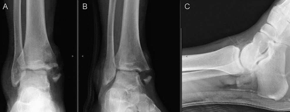 · 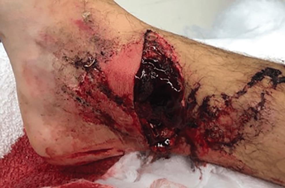 · 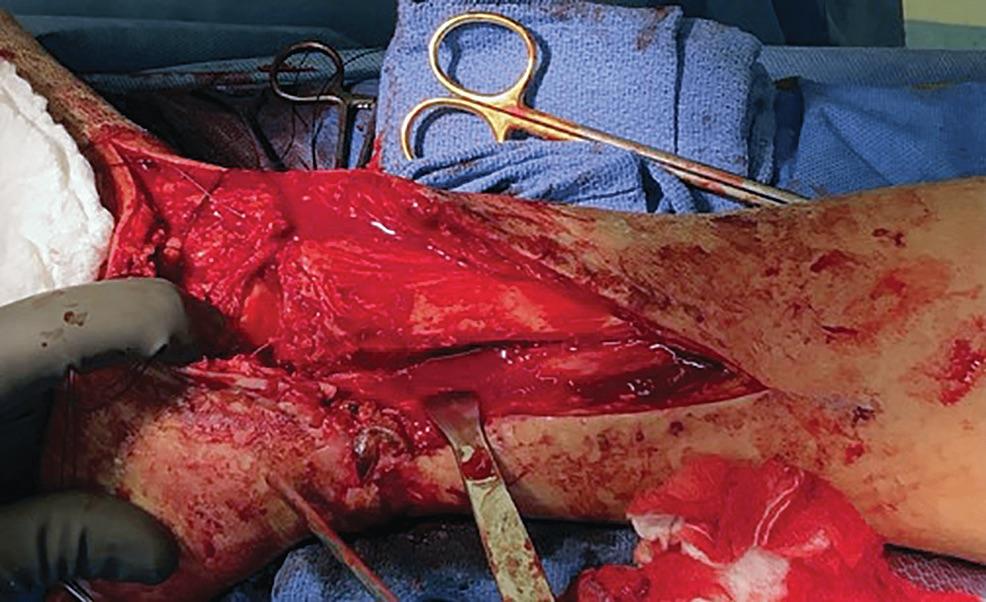 · 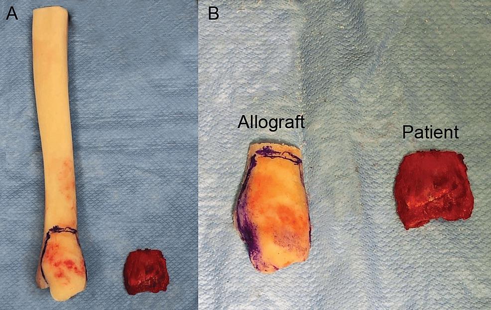 · 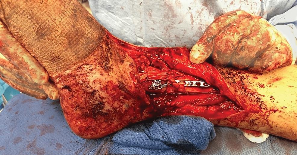 · 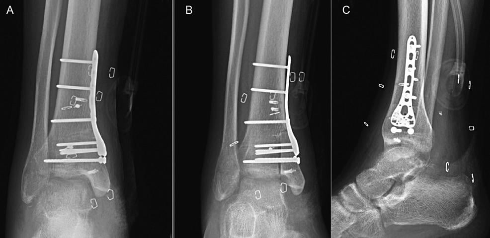 · 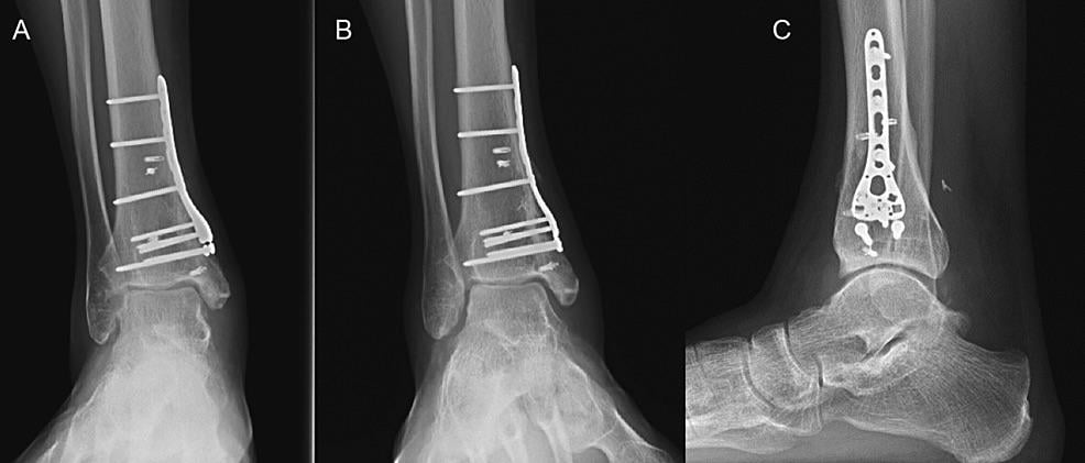 · |
|
#2
●
09-01-2024, 03:50 AM
|
 ★ Legacy Member ★ Poster Rank:110 Secret Agent Join Date: Dec 2009 Posts: 14,804 14,804Posts 49,175Thanked 3,055Quoted 6Mentioned 0Contrib. |
|
Re: Atv Accident
That little piece of surgical titanium screwed to his ankle bone probably cost in upwards of $25 to $30K just for the cool looking metal piece not including the screws to hold it in place.  |
|
#3
●
09-01-2024, 05:14 PM
|
 ♚ Legacy Gold Member ♚ Poster Rank:268 ꓕSIꓤIꓕ∀S ϽINOᗡꓤ∀S Join Date: Feb 2010 Posts: 5,179
Contributions: 8
5,179Posts 31,161Thanked 1,441Quoted 45Mentioned 8Contrib. |
|
Re: Atv Accident
Kinda reminds me of this guy... although his intoxication seems to make self-reassembly on the roadside virtually pain free.
__________________  200+ Additional Smilies Custom DR Search Engine 200+ Additional Smilies Custom DR Search Engine ✦ Live life to it's fullest and leave a sexy corpse ✦ |
|
#4
●
09-02-2024, 12:13 AM
|
| ★ Legacy Member ★ Poster Rank:110 Secret Agent Join Date: Dec 2009 Posts: 14,804 14,804Posts 49,175Thanked 3,055Quoted 6Mentioned 0Contrib. |
|
Re: Atv Accident
OhFuckOhFuckOhFuck NO-NO-NO!!!! I could only watch about 10 cringing seconds of that one!!! |