|
#1
●
11-10-2012, 04:33 PM
| ||||||||
| ♚ Legacy Gold Member ♚ Poster Rank:34 Female Join Date: Nov 2008 Posts: 43,403 Mentioned: 95 Post(s) Quoted: 2281 Post(s)
| ||||||||
|
Tortoise Autopsy
From a captive reptile forum: This post mortem concentrates on the coelomic viscera inside the shell since the rest of the body was preserved for burial afterwards. The plastron has been removed with a diamond edged circular saw (which we use to open flaps of the shell for surgery) and was replaced and glued with epoxy resin after the procedure had finished. The animal shortly after presentation at the surgery and permission was given for a respectful post mortem examination. In the first picture the paired ventral coelomic veins are visible(V) care must be taken in the surgery of these animals not to damage these vessels. If they are damaged they can safely be tied off. The coelomic membrane (M) is also visible. The green staining to the left has been caused by rupture of the gall bladder (a post mortem change). Note the large pectoral muscles, used for expiration in tortoises. 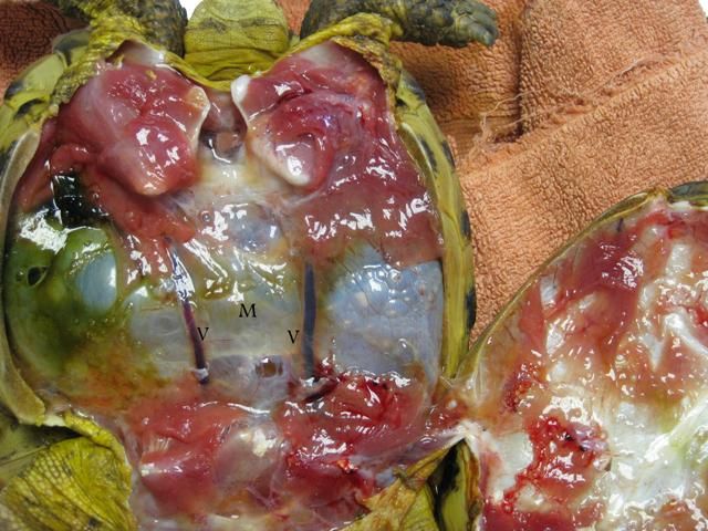 The Coelomic membrane has been incised and removed. The Heart (H), Liver(L) and Bladder (B) are visible. The heart is 3 chambered with the paired auricles lying above the single ventricle. The liver is large and separated into 2 lobes. The Gall bladder can be found in the right liver lobe. The bladder is very large and plays an active role in water storage and electrolyte balance in tortoises. In this picture it is clear what a large portion of the back half of the coeloem the bladder occupies. 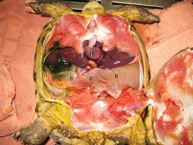 The Bladder has been reflected to show the Large Intestine (L) and small intestine (si). Urates (U) are visible within the bladder. The large intestine is the site for microbial (bacterial) digestion of plant material, the nutrients derrived from which are absorbed by the tortoise. It is important to note that the bladder contents are considered non sterile so surgery of the bladder (eg removing bladder stones) must be carried out with caution. 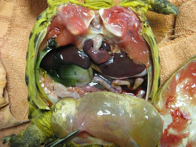 This image shows the stomach (St)and the spleen (Sp) tortoises have a simple stomach (cows have 4 chambers for herbage digestion). A lolly stick was found inside the stomach. 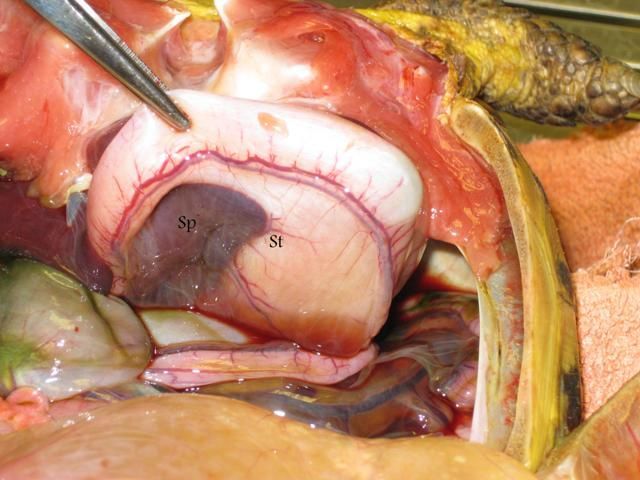 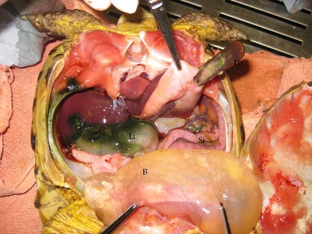 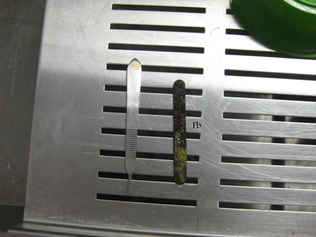 The kidneys (K) are retrocoelomic (ie they lie outside the coelomic membrane) in this tortoise. You can see that the coelomic membrane has been incised to access them. they are enlarged and grossly abnormal. Samples of kidney tissue had been submitted for histopathology but kidney failure/disease was the likley cause of death for this animal. 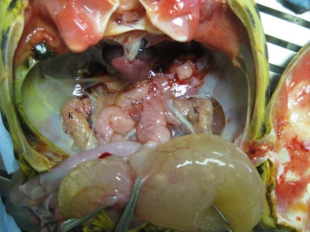 The lungs (L) have been opened to show the structure of the gaseous exchange surface. The short trachea and bronchi, combined with this open lattice structure, allows penetration of nebulised droplets as an aid to therapy in tortoise pneumonia. Chelonia are able to breath hold for long periods of time. in fact they have the highest bicarbonate levels of all vertebrates allowing them to buffer acids produced by respiration when they breath hold therefore enabling them to hold their breath for longer. Their lungs are reduced to 1/5 their size when the head is retracted. 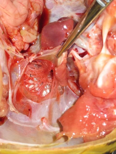 |
|
#6
●
11-10-2012, 10:26 PM
| ||||||||
| My Rank: STAFF SERGEANT Poster Rank:825 Inny Join Date: Sep 2009 Posts: 931 Mentioned: 1 Post(s) Quoted: 42 Post(s)
| ||||||||
|
Re: Tortoise Autopsy
Ok, that was gross. LOL I can handle people innards all day long. Guess I just can't stomach tortoise. |
|
#8
●
11-10-2012, 11:39 PM
| ||||||||
| Born in the USA Poster Rank:123 100% female Join Date: Jul 2012 Posts: 12,876 Mentioned: 81 Post(s) Quoted: 1799 Post(s)
| ||||||||
|
Re: Tortoise Autopsy
And the pop stick did not contribute to the death? He must have been hungry to consume that. Thanks Sharon |


