|
#1
●
05-03-2021, 08:12 AM
| ||||||||
| ♚ Legacy Gold Member ♚ Poster Rank:908 Join Date: Jul 2020 Posts: 820 Mentioned: 13 Post(s) Quoted: 173 Post(s)
| ||||||||
|
Elephantiasis Nostra Verrucosa at Autopsy
Elephantiasis nostra verrucosa (ENV) is a rare form of chronic lymphedema that causes progressive cutaneous hypertrophy. It can lead to severe disfiguration of body parts with gravity-dependent blood flow, especially the lower extremities. It is associated with obesity and chronic heart failure. A 58-year-old woman found dead sitting in a chair. There was a past history of mobility issues. She spent her time in the chair or rested in her bed which was very close by. She never left the house and took no medication. On examination, the chair appeared urine and fecal stained. Concerns were raised about whether she had been properly looked after. 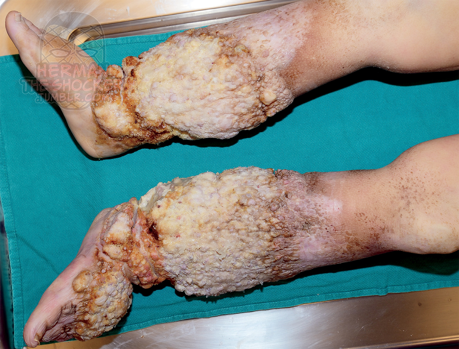 Figs.1-5 Gross appearance of the legs. 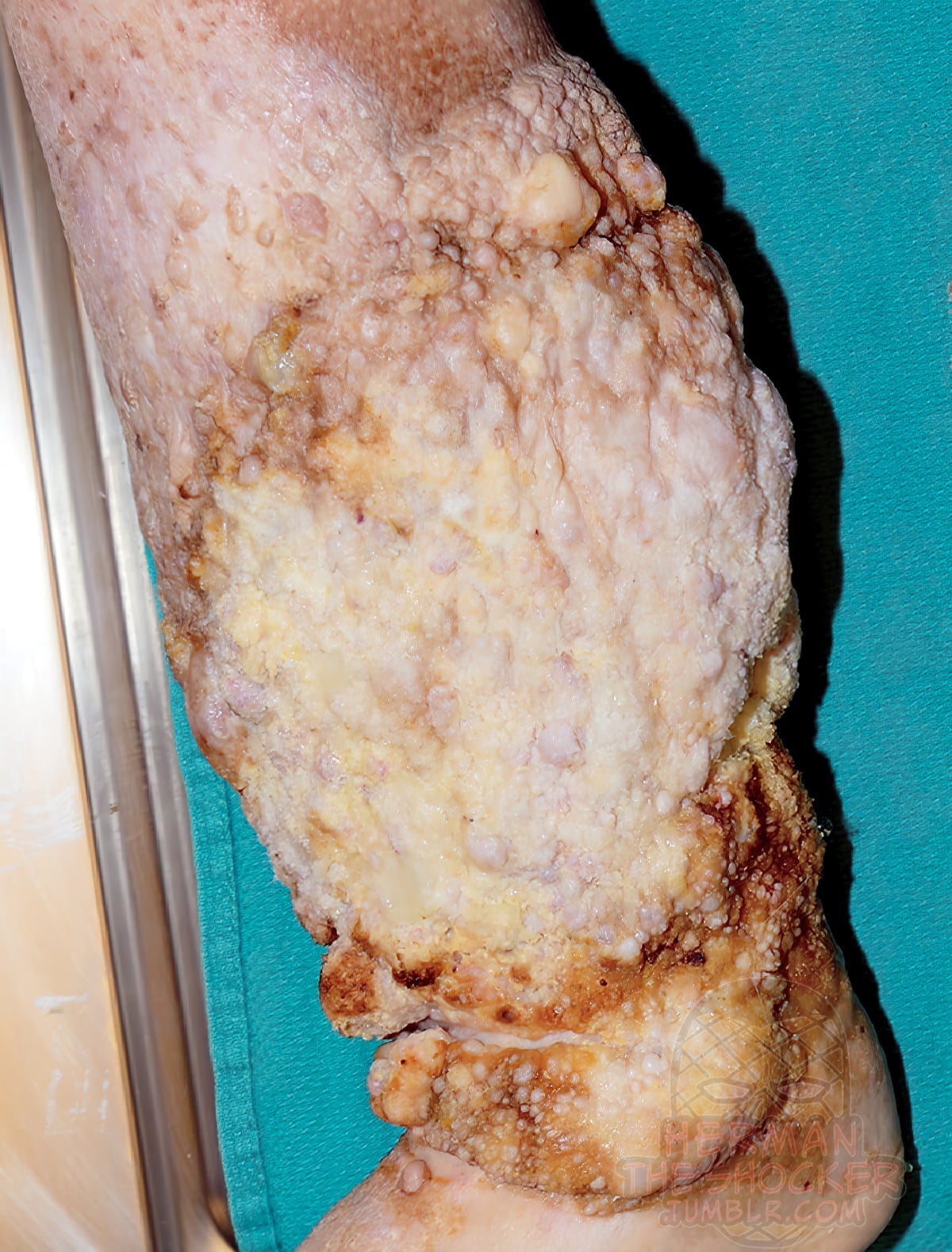 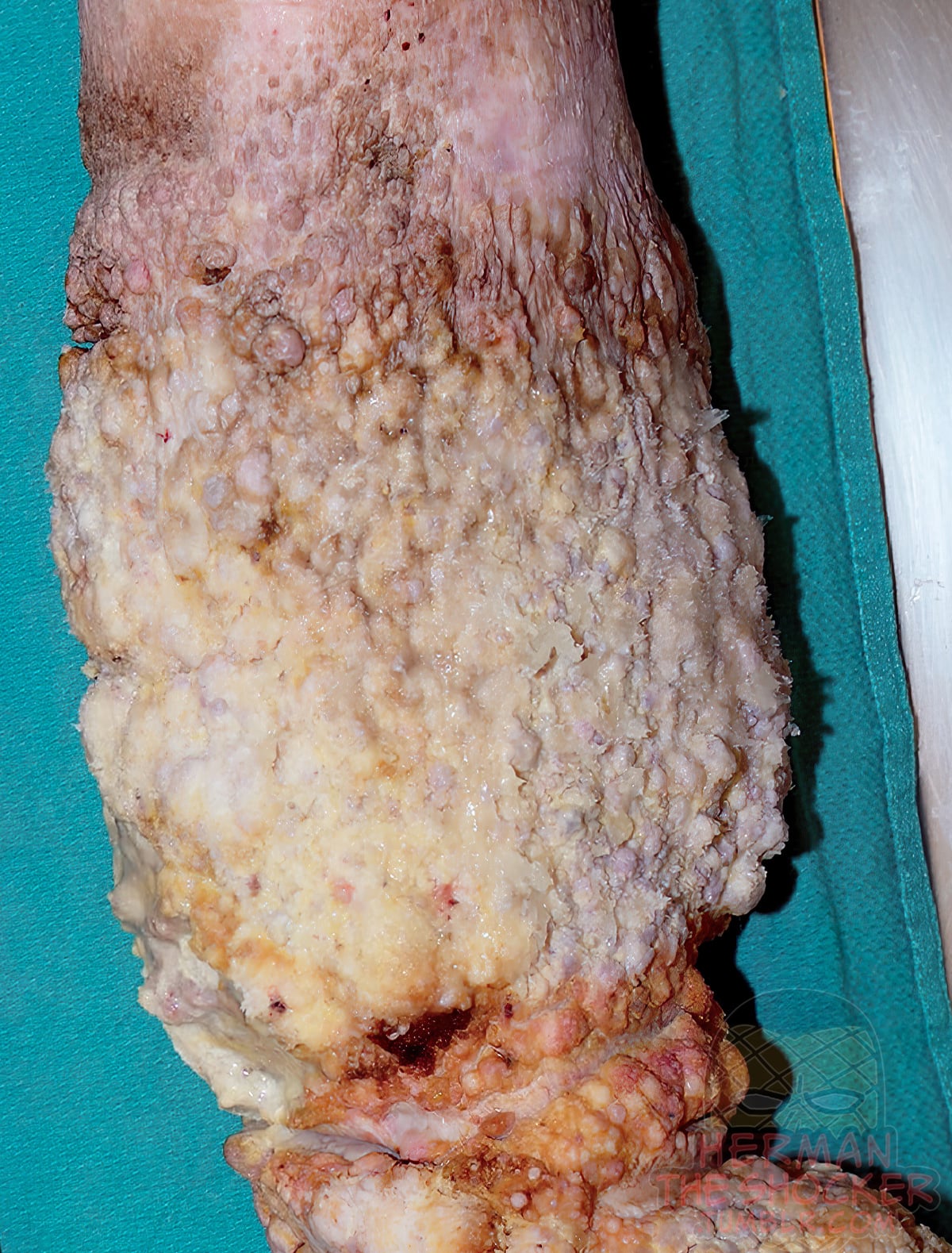 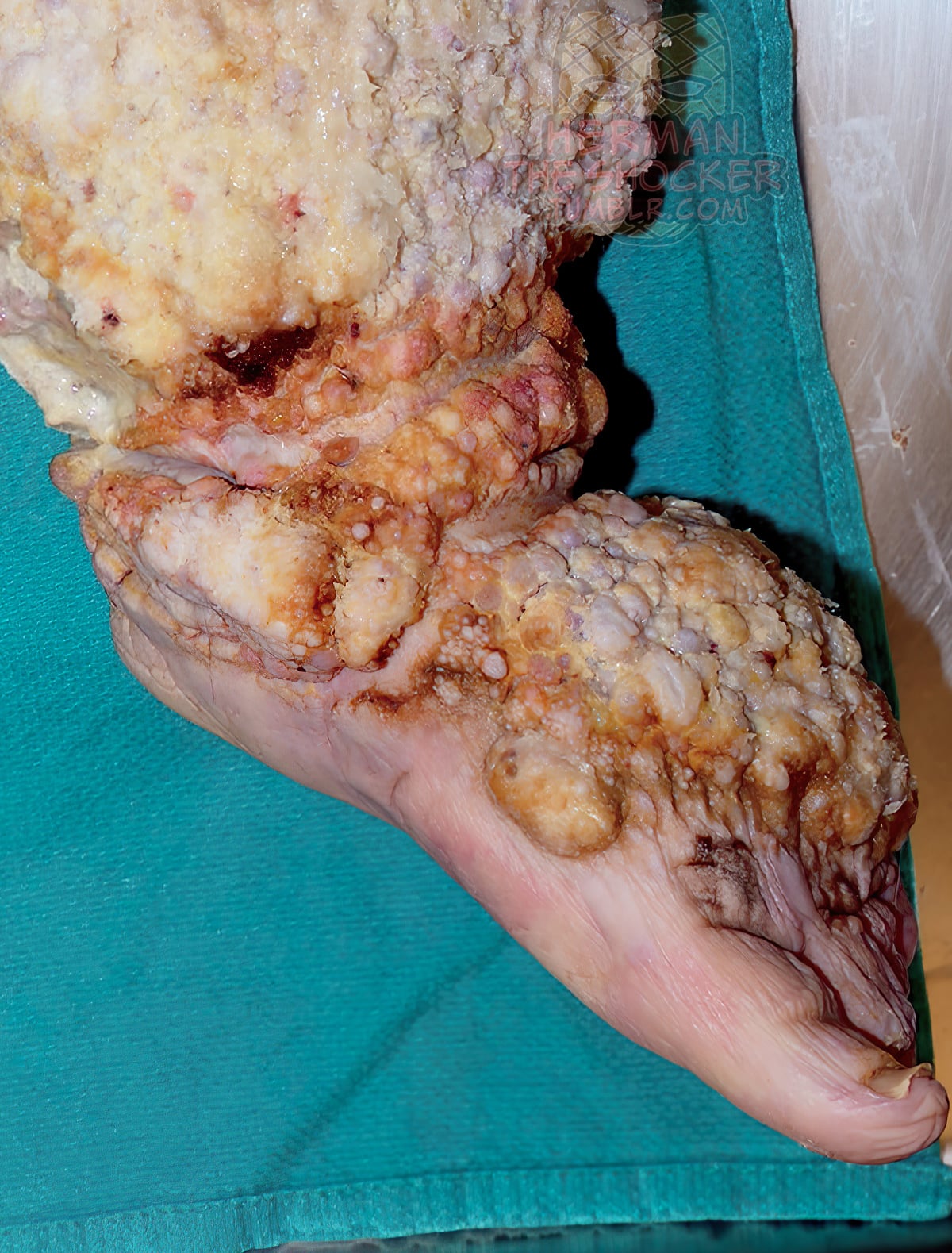 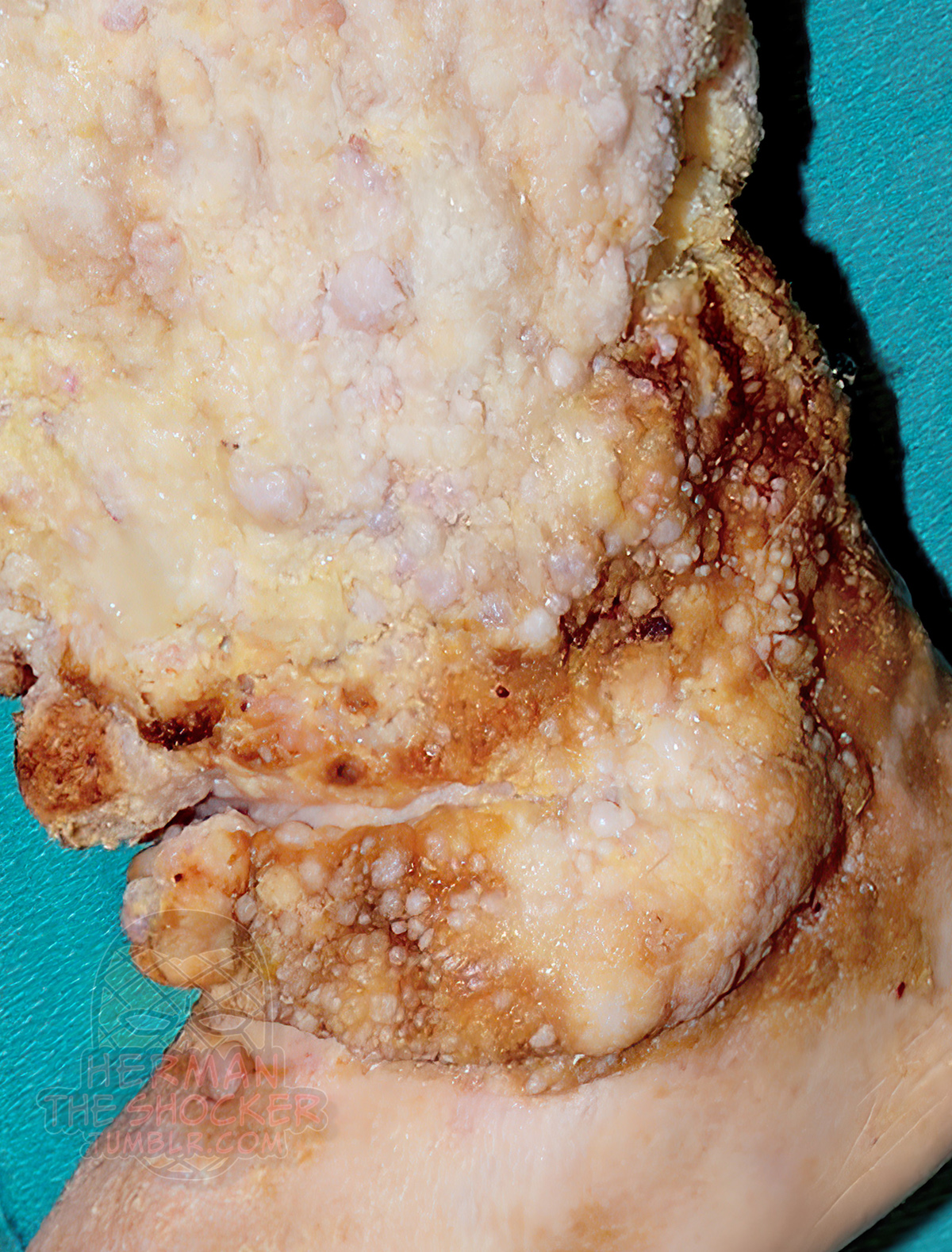 At autopsy, the woman was 149 cm (4.8 ft) tall and weighed 55 kg (121 lbs). She was dressed in heavily soiled clothing. No traumatic injuries were present. The legs and feet were covered in homemade bandages extending from just below the knees to the feet. On removing the bandages, the legs and feet were seen to be grossly distended from chronic edema and massive verrucous hyperplasia of the skin and had a cobblestoned appearance (Figs.1-5). There was ulceration of the inner aspect of the right ankle and further ulcers were present on the back of the right ankle. The ulcers were infected and contained pus. 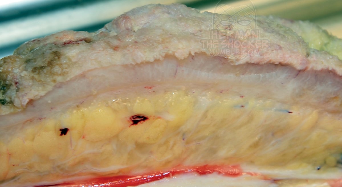 Fig.6 Appearance of thickened skin on dissection. On internal examination, the lungs showed no evidence of pneumonia. The liver, kidneys, and spleen were normal. The brain, spinal cord, and heart were submitted for detailed neuropathology and cardiac pathology consultation. There was no abnormality in the brain and spinal cord. The heart weighed 240 g. The myocardium and valves were normal. There was coronary artery atheroma with maximal stenosis of 50% in the left anterior descending coronary artery.  Fig.7 Microscopic appearance of the epidermis and dermis (H&E, x25). Toxicological examination was negative. Vitreous biochemistry did not show any significant findings. Microbiological cultures grew multiple bacteria from the ulcers. Group B streptococcus and viridins group streptococcus were found grown from blood cultures. 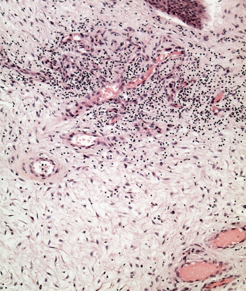 Fig.8 Appearance of the dermis (H&E, x100). Histology of the skin revealed verrucous hyperplasia of the epidermis with edema in the dermis and subcutaneous tissue (Fig.7). Fibroblastic proliferation was seen and there was chronic inflammation around some vessels (Fig.8). Lymphatic and vascular dilation was present, but no hemosiderin was seen (Fig.9). No filarial parasites were present. The features were those of ENV. 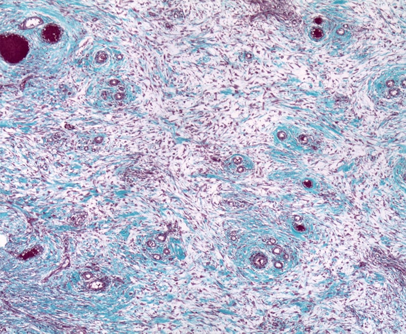 Fig.9 Appearance of the dermis (Masson trichrome, x100). The presence of ENV in cases like this woman raises the question of neglect. Elephantiasis nostra verrucosa is going to be more prevalent with the increasing incidence of morbid obesity and chronic heart failure. - This post is for educational purposes only and is nonprofit. Under Section 107 of the US Copyright Act of 1976; Allowance is made for "Fair Use" for purposes such as criticism, comment, news reporting, teaching, scholarship, and research. No copyright infringement intended. This post does not encourage or glorify violence or harassment. All/some of the images have been upscaled and sharpened/enhanced. The text might have been shortened and simplified, and/or reorganized for online view. Original case report by Milroy C.M. Ottawa Hospital - Acad Forensic Pathol 2019. |
|
#2
●
05-04-2021, 06:38 AM
| ||||||||
| My Rank: LANCE CORPORAL Poster Rank:1846 Male but Gay and if someone Hate´s me: Excuse me ich havent Choose it freely. Join Date: May 2017 Posts: 278 Mentioned: 1 Post(s) Quoted: 79 Post(s)
| ||||||||
|
Re: Elephantiasis Nostra Verrucosa at Autopsy
Very Interesting Case! Thanks for Upload.
|
|
#3
●
05-05-2021, 08:06 AM
| ||||||||
| ♚ Legacy Gold Member ♚ Poster Rank:195 Female Join Date: Nov 2009 Posts: 7,761 Mentioned: 5 Post(s) Quoted: 708 Post(s)
| ||||||||
|
Re: Elephantiasis Nostra Verrucosa at Autopsy
Wow. Looks like some of the legs you see on My 600lb life. One guy was 1003lbs at his heaviest. But this woman was small compared to them. |
|
#4
●
05-30-2021, 07:07 PM
| ||||||||
 ★ Legacy Member ★ Poster Rank:772 I'm not A bitch.... I'm THE bitch. Join Date: Jul 2012 Posts: 1,033 Mentioned: 4 Post(s) Quoted: 174 Post(s)
| ||||||||
|
Re: Elephantiasis Nostra Verrucosa at Autopsy
“Gross appearance of the legs“........no truer statement has ever been uttered.
|
|
#5
●
07-08-2021, 04:12 AM
| ||||||||
| Snow Tan Poster Rank:44 SPYCOSIS' ONLY FRIEND IS A BALD DEAD CUNT Join Date: Dec 2009 Posts: 32,693 Mentioned: 80 Post(s) Quoted: 13440 Post(s)
| ||||||||
|
Re: Elephantiasis Nostra Verrucosa at Autopsy
Ok, no more macaroni and cheese for me.
|
|
#8
●
02-03-2022, 07:30 PM
| ||||||||
| My Rank: CORPORAL Poster Rank:1347 Male Join Date: Jun 2021 Posts: 448 Mentioned: 0 Post(s) Quoted: 90 Post(s)
| ||||||||
|
Re: Elephantiasis Nostra Verrucosa at Autopsy
his doctor should've just got a cheese grater. Zip that crap right off. |


