|
#1
●
04-01-2023, 12:21 AM
| ||||||||
 These are the rooms Poster Rank:26 of ruin. Join Date: Sep 2014 Posts: 54,011 Mentioned: 145 Post(s) Quoted: 30403 Post(s)
| ||||||||
|
Veruccous Carcinoma on the Foot
Source: A 52-year-old man presented with a 10-year-old, progressive, painful, pruritic, exophytic growing, hyperkeratotic, haemorrhagic plaque of 5.2 x 3.5cm on the right calcaneus (Figures 1-2). The lesion emerged after extensive burns following a blast trauma during childhood. The patient had a significant medical history of intellectual disability, left-sided transtibial amputation (as a result of a non-healing burn ulcer) and cholesteatoma. Excisional biopsy with 2mm margin in combination with clinical presentation favoured a verrucous carcinoma. An ultrasound showed a superficial tumour. No further diagnostic tests were done to assess lymph node status, due to the explicit wishes of the patient. The wound was closed ad secundam. The pathology report showed that the tumour was not excised radically; therefore re-excision with 5mm margin was required and was performed two months later. The wound was closed with a medial plantar artery perforator (MPAP) flap. The donor site was closed with a full-thickness skin graft (FTSG) from the medial side of the right upper leg and was covered by a tie-over bandage. After the operation, the leg was kept in an elevated position. Two days post-operatively, the MPAP-flap showed venous congestion at the distal periphery. Locally, the epidermis was excised and covered with heparin gauze to improve venous drainage for five days, after which normal vascularization recurred. At the lateral side of the inner foot (donor site of the MPAP flap), the FTSG only had partial take and was treated with Betadine gauze (Figure 3). Six weeks after surgery, all wounds were healed (Figure 4). The patient was allowed to walk freely after 10 weeks. 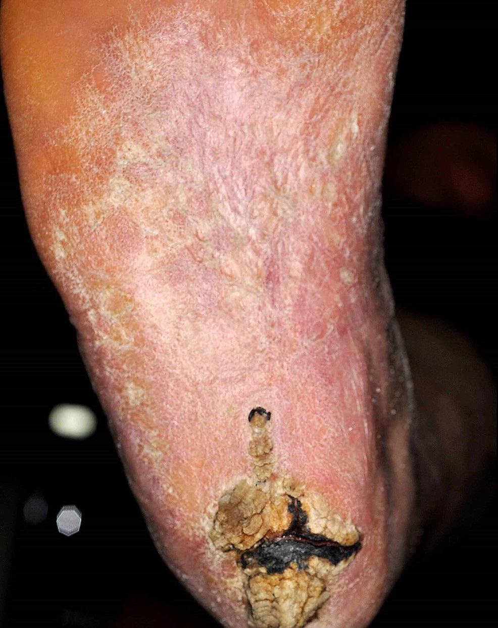 · 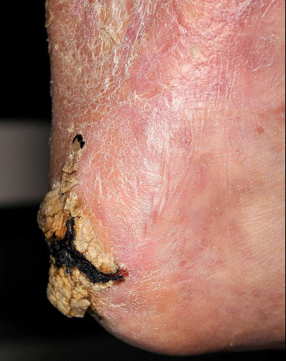 · 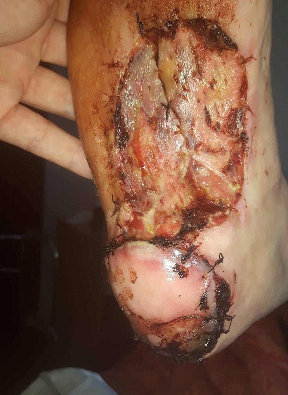 · 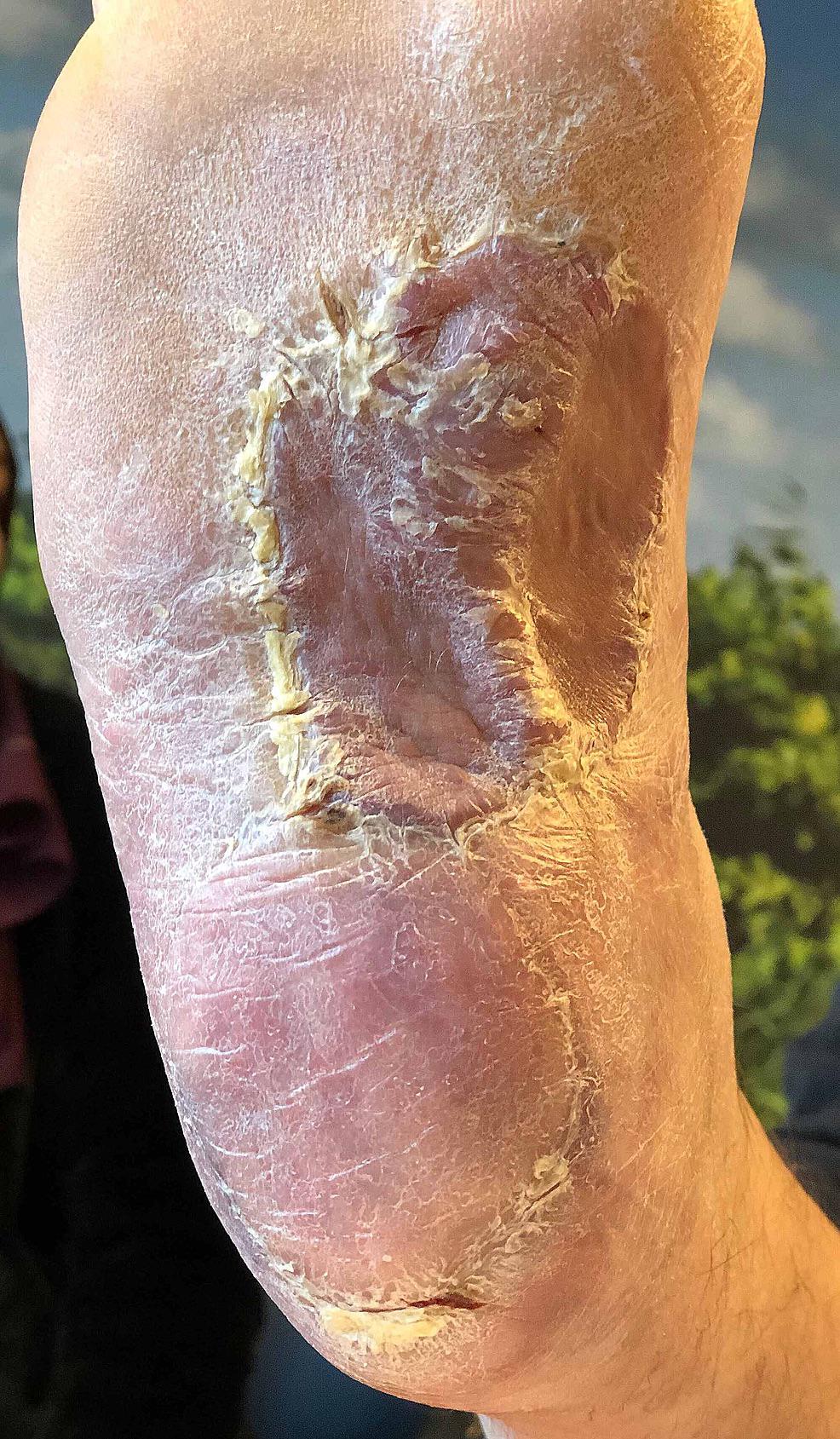 · |
|
#2
●
04-01-2023, 12:32 AM
| ||||||||
 My Rank: FIRST SERGEANT Poster Rank:399 Join Date: Apr 2011 Posts: 3,054 Mentioned: 2 Post(s) Quoted: 276 Post(s)
| ||||||||
|
Re: Veruccous Carcinoma on the Foot
Nasty  Fortunately he survived and was able to walk again. Fortunately he survived and was able to walk again. This was in fact the first time I heard about such a condition. Browsing and devouring all these informative threads here will turn me into a walking medical textbook in no time  |
|
#3
●
04-01-2023, 12:40 AM
| ||||||||
 ★ Legacy Member ★ Poster Rank:252 So many choices now Join Date: Jul 2015 Posts: 5,548 Mentioned: 15 Post(s) Quoted: 2120 Post(s)
| ||||||||
|
Re: Veruccous Carcinoma on the Foot
A scab picker's wet dream.
|
|
#4
●
04-01-2023, 02:26 AM
| ||||||||
| These are the rooms Poster Rank:26 of ruin. Join Date: Sep 2014 Posts: 54,011 Mentioned: 145 Post(s) Quoted: 30403 Post(s)
| ||||||||
|
Re: Veruccous Carcinoma on the Foot
I've learned a lot of surprising things about the human body. |
|
#5
●
04-01-2023, 06:33 AM
| ||||||||
 ✝Mudderator from Hell✝ Poster Rank:10 e-mail Join Date: Oct 2006 Posts: 96,644
Contributions: 817
Mentioned: 476 Post(s) Quoted: 10344 Post(s)
| ||||||||
|
Re: Veruccous Carcinoma on the Foot
that's a bat spreading its wings or the 2 horned devil spreading its wings  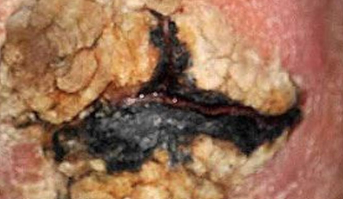 · |