|
#1
●
04-02-2023, 04:23 PM
| ||||||||
| These are the rooms Poster Rank:25 of ruin. Join Date: Sep 2014 Posts: 54,011 Mentioned: 145 Post(s) Quoted: 30403 Post(s)
| ||||||||
|
Verrucous Hemangioma Treated with Staged Skin Grafting
Source: A 20-year-old male from Kathmandu, Nepal presented to the plastic surgery clinic with extensive, warty, brownish lesions, or plaques involving the dorsum of foot, lateral aspect of the lower leg, lateral and posterior aspects of upper leg, and lateral thigh. The lesions were present since birth and enlarged gradually. The lesions were painless to begin with and sometimes bled with trivial trauma. The patient was mainly concerned about cosmesis and frequent bleeding episodes. Examination of the plaques revealed raised verrucous areas of red/violet discoloration with hemorrhagic crusts on the lower leg and foot lesions (Figure 1). Moreover, extension contracture could be seen at the base of middle three toes due to the disease process. Small satellite lesions were also seen in the periphery of larger, diffuse lesions. Lesions were non-tender, non-compressible, and had no pulsations. Bruits were not heard over the lesions and regional lymph nodes were not enlarged. The right lower extremity was normal and similar lesions were not found elsewhere. Magnetic Resonance Imaging (MRI) of the left lower limb revealed multiple T1 iso and T2 high signal intensity lesions of variable sizes in the subcutaneous plane without extension into muscular plane (Figure 2). The surgery of the verrucous hemangioma was performed in two stages: the first stage consisted of excision under tourniquet control and the second stage, split-thickness skin grafting. Intraoperatively deep infiltration of the tissue planes involving the skin, subcutaneous, and deeper tissues was seen. The excision was carried out in supra-fascial plane (Figures 3-5). After the first stage, alternate-day dressings were done until the wound was granulated thoroughly. After 10 days, a split-thickness split graft was harvested from the right thigh and applied on the post-excision raw areas. The first dressing of the skin graft was done on the fourth day, then every 2 days. The graft take was good with all the wounds healed in 2 weeks and with the good aesthetic outcome. Histopathology examination of the specimen revealed irregular papillomatosis, acanthosis, and hyperkeratosis of the epidermis with multiple, thin-walled, dilated blood-filled spaces in the dermis. 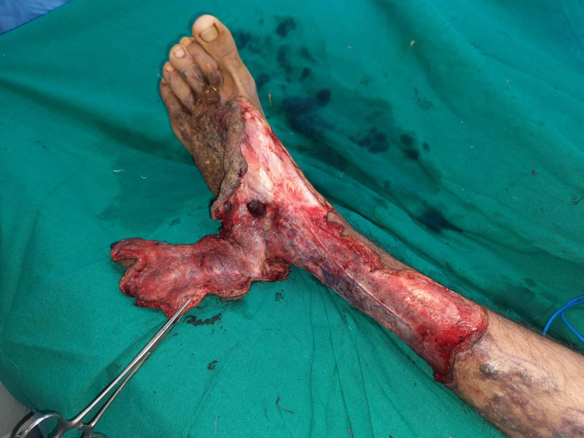 · 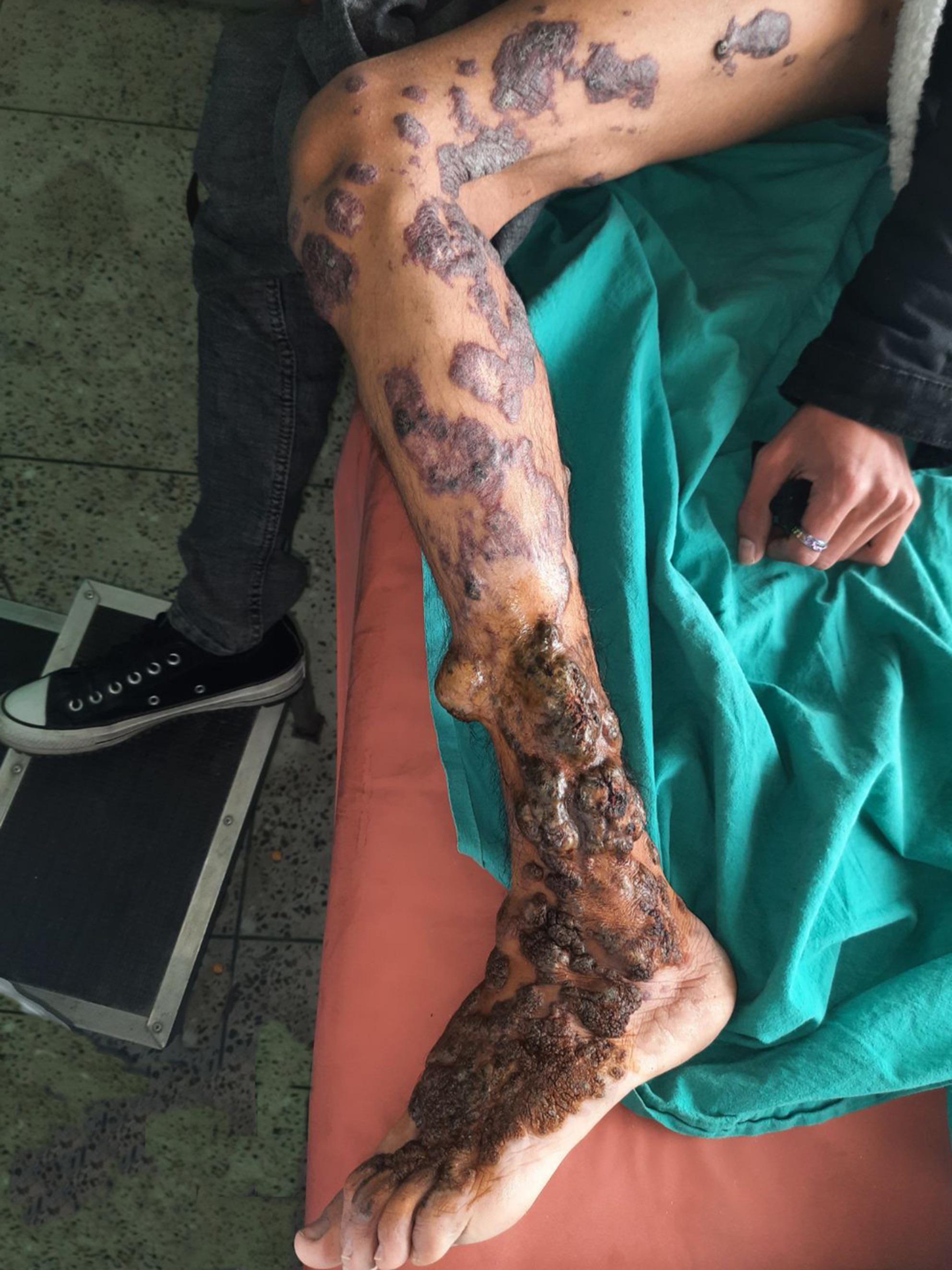 · 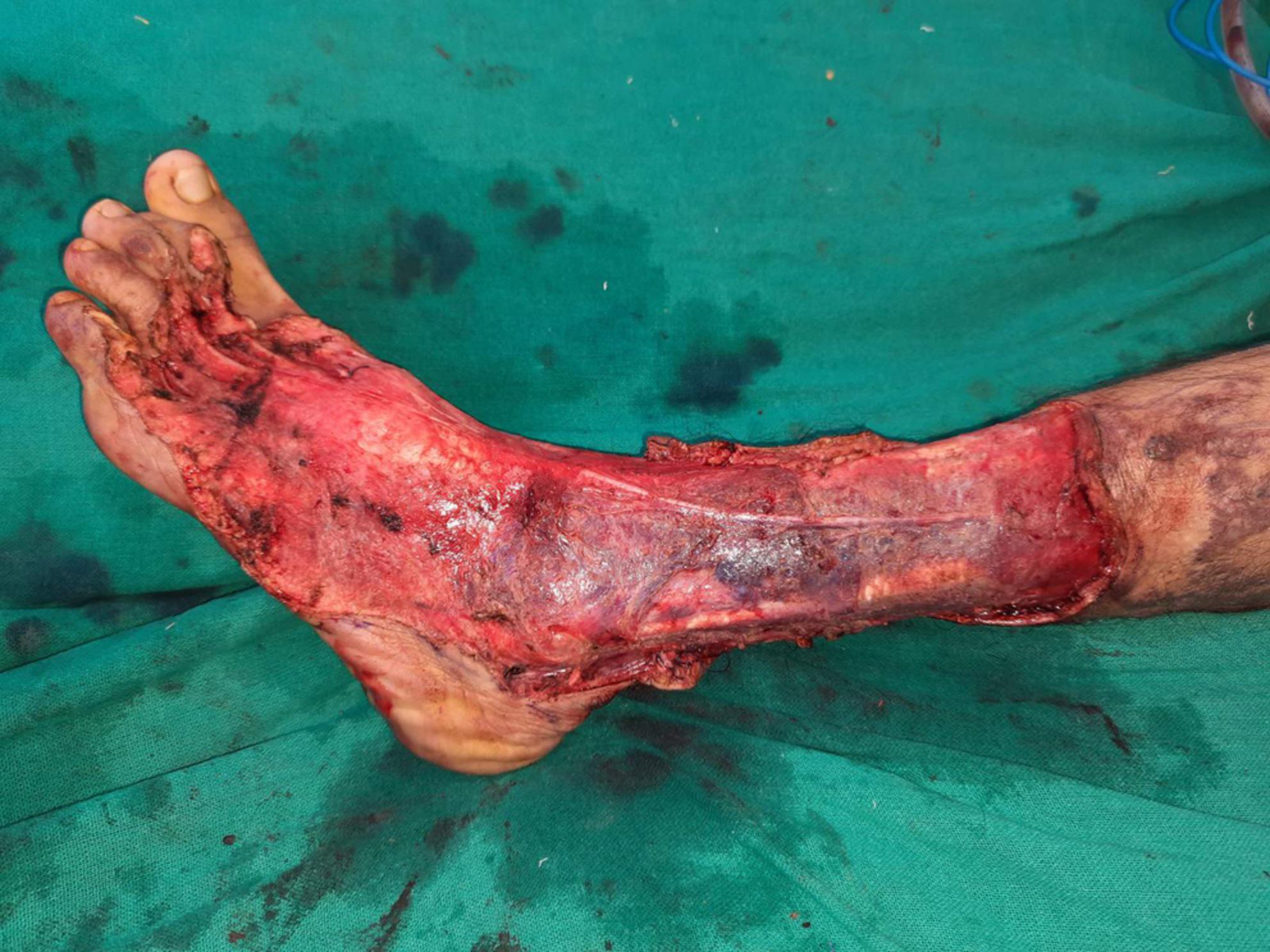 · 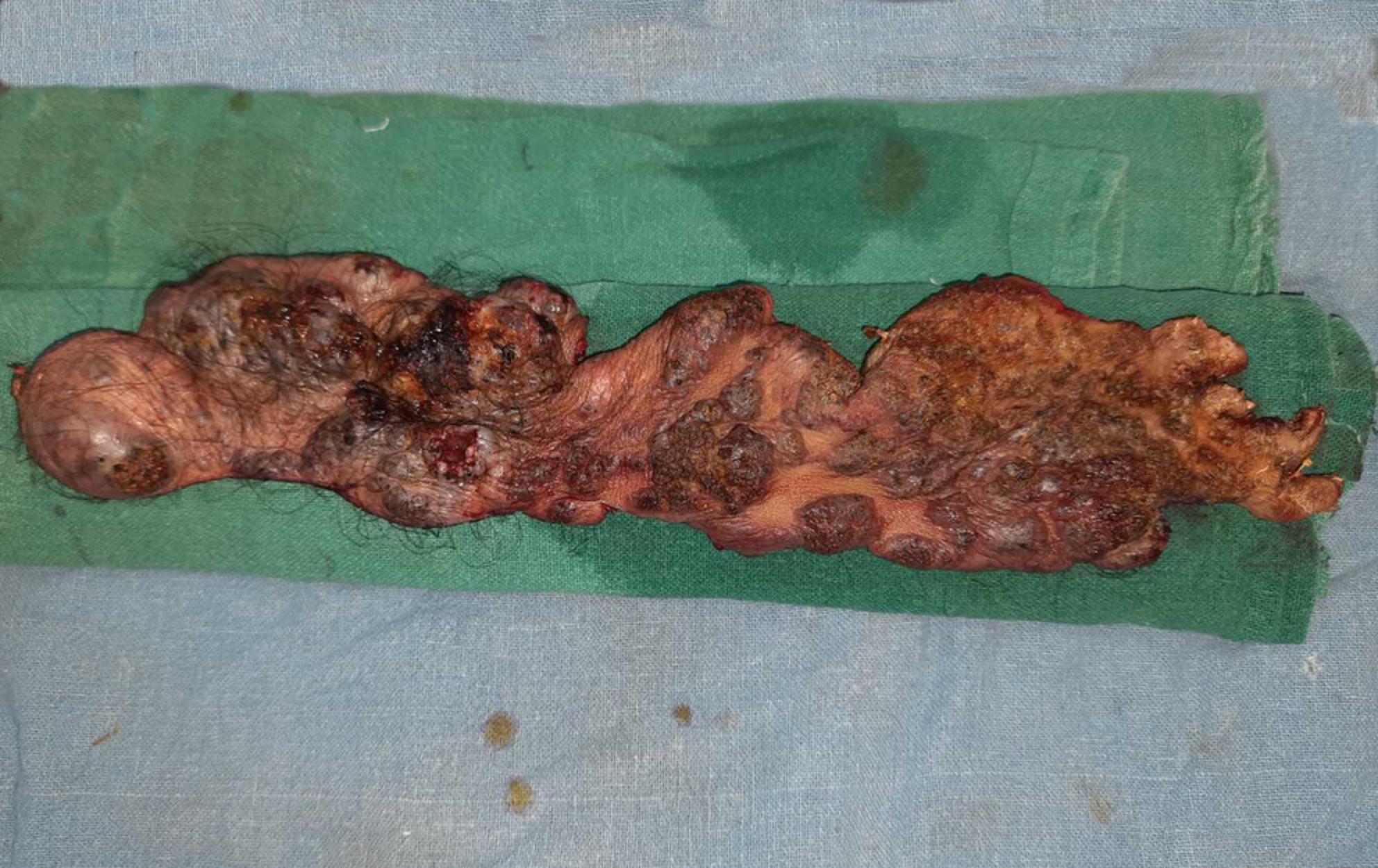 · 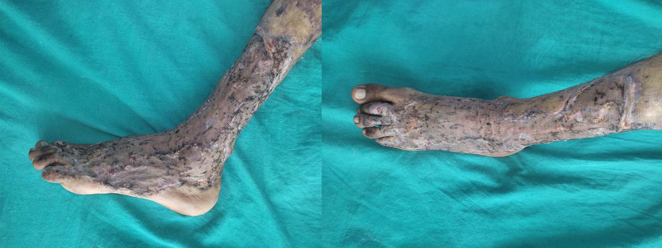 · |
|
#2
●
04-02-2023, 06:17 PM
| ||||||||
| ★ Legacy Member ★ Poster Rank:247 So many choices now Join Date: Jul 2015 Posts: 5,548 Mentioned: 15 Post(s) Quoted: 2120 Post(s)
| ||||||||
|
Re: Verrucous Hemangioma Treated with Staged Skin Grafting
looks like mud. Just wash it off.
|
|
#3
●
04-03-2023, 04:35 AM
| ||||||||
| ★ Legacy Member ★ Poster Rank:119 Secret Agent Join Date: Dec 2009 Posts: 13,201 Mentioned: 6 Post(s) Quoted: 2787 Post(s)
| ||||||||
|
Re: Verrucous Hemangioma Treated with Staged Skin Grafting
Strip the skin and toss some wet clay over the wounds. He’ll be fine. If the clay comes off we’ve got plenty more where that came from. |
|
#4
●
04-03-2023, 03:35 PM
| ||||||||
| ✝Mudderator from Hell✝ Poster Rank:10 e-mail Join Date: Oct 2006 Posts: 94,975
Contributions: 817
Mentioned: 472 Post(s) Quoted: 10078 Post(s)
| ||||||||
|
Re: Verrucous Hemangioma Treated with Staged Skin Grafting
hemorrhagic crusts  |
|
#5
●
04-04-2023, 01:57 AM
| ||||||||
| ★ Legacy Member ★ Poster Rank:192 All woman Join Date: Jun 2012 Posts: 7,782 Mentioned: 22 Post(s) Quoted: 1420 Post(s)
| ||||||||
|
Re: Verrucous Hemangioma Treated with Staged Skin Grafting
Why do I always look at this shit while I'm eating????
|