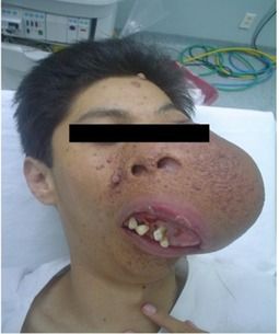
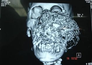
19 year old Mexican male with the diagnosis of tuberous sclerosis at the age of 8 years presented with multiple small cutaneous tumors, some pendunculated and others sessile, violet in color of bland consistency and vascular aspect, localization predominantly in left side of the face, as well as with a subcutaneous tumor in left maxillary region of progressive growth to complete deformation, occluding the left eye by over 70%, displacing the dental arch, the nasal pyramid, and extending to the ipsilateral preauricular region. The patient had been administered treatment with radiotherapy to this site in 2006 without tumor shrinkage of the tumor. It was decided to program the performance of left lateral rhinotomy and left maxillectomy in May 2007, prior to Magnetic resonance imaging (MRI) with reconstruction.
Pre-anesthetic evaluation scores for difficult airway were as follows: Mallampati, IV; Patil, 3; Interincisive distance (ICD), III, and Bellhouse Dore, I. From the patient’s arrival at the operating room, he was monitored with continuous Electrocardiography (EKG), T/A, and pulse oximetry, and equipment was prepared to include endotracheal catheters, guides, fibrolaryngoscopy, aspiration catheter, and adjunct anesthesiology.
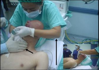
Previous oxygenation with mask and with the patient conscious under sedation with midazolam 2 mg, as well as fentanyl 200 μg; direct laryngoscopy was performed by means of OELM and armed endotracheal catheter placement:
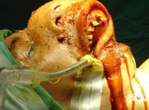
During the surgical procedure, the patient was hemodynamically stable with adequate ventilatory parameters:
At the end of surgery, extubation was conducted with the patient awake, without complications. Postoperatory evolution was adequate; thus, the patient was discharged from the hospital 3 days after the surgical procedure, has been followed-up to these days without complications or recurrence of the facial tumor.