|
#1
●
10-01-2022, 11:47 AM
| ||||||||
| My Rank: PRIVATE FIRST CLASS Poster Rank:4991 Female Join Date: Jan 2021 Posts: 54 Mentioned: 0 Post(s) Quoted: 21 Post(s)
| ||||||||
|
Treatment of Elephantiasis and Fournier's Gangrene of the Scrotum
A case of Fournier's gangrene complicated by massive lymphedema of the scrotum and right leg in a 43 year-old man from Panama, later confirmed to be filarial. A 43 year-old male from Panama presented to the emergency department with fever, tachycardia, and increasing swelling and drainage from his scrotum. His medical history was limited. He had not sought medical care in many years. Over the past THREE DECADES his scrotal swelling had gradually worsened. He relied on a walker for ambulation and his mother attended to most of his daily needs. He had no prior urologic history and only previous surgery was a diaphragmatic hernia repair as a child. On examination, there was massive scrotal edema with displacement past the level of his knees (Fig. 1). The scrotal skin was thickened and there were two open wounds with foul smelling, purulent fluid located at the bottom of the left hemi-scrotum. His right lower extremity had extensive edema and skin thickening of the upper leg. He was febrile to 102.2F with a pulse of 137 bpm, concerning for sepsis. 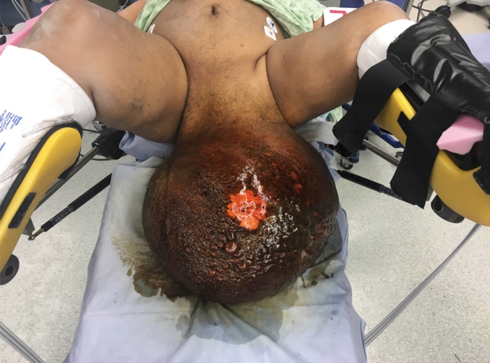 Fig. 1. Presentation of the patient with massive scrotal edema and open wounds over the left hemi-scrotum. Computed tomography (CT) scan (Fig. 2) of the abdomen and pelvis revealed a massive left inguinal hernia containing non-inflamed colon and a large subcutaneous abscess with gas measuring up to 12.8 cm with draining tract to the skin at the anterior aspect of the scrotum. Massive hydrocele (swelling around testicle) was noted in the left hemiscrotum, extensive scrotal wall thickening, soft tissue ulceration at the left posterior aspect of the scrotum, severe left kidney swelling, and extensive bilateral inguinal inflammation.  Fig. 2. CT imaging illustrating impressive scrotal edema and massive inguinal hernia. Due to the concern for Fournier's gangrene and patients declining clinical condition, he was taken to the operating room for emergent debridement of infected scrotal skin and subcutaneous tissue. Intra-operative findings demonstrated a large abscess and sinus tract toward the left inguinal region, with areas of skin necrosis. Initial pathology revealed scrotal skin with extensive deep dermal acute inflammation and liquefactive necrosis (liquified tissue). The next day the patient returned to the operating room for minimal secondary debridement. On hospital day 5, after several days of intravenous antibiotics his wound appeared to be improving but dressing changes and wound vacuum were too painful. A decision was made to proceed with multidisciplinary (urology, general surgery, plastic surgery), surgical treatment. The hernia repair was necessary to reduce the hernia contents. Next, the patient then underwent a scrotectomy (removal of the scrotum) and left orchiectomy (left testicles removal). On hospital day 8, the patient was taken back to the operating room by the plastic surgery team for a split thickness skin graft for the penis and primary closure of the perineum with advancement flap closures. He was discharged two days later to rehabilitation facility. He was seen four weeks postoperatively and was healing well with satisfactory cosmetic and functional outcomes (Fig. 3). 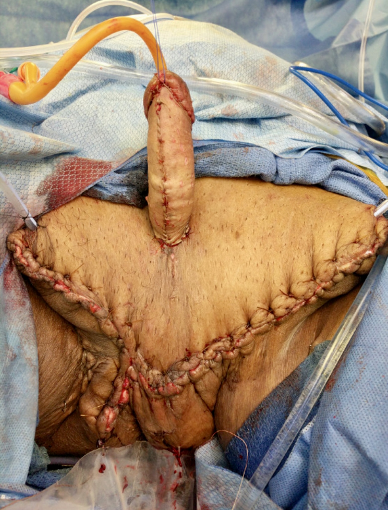 Fig. 3. Initial outcome following surgical debridement and multi-disciplinary staged repair. *I have an unreasonable fear of reposting, so sorry if it is. I did a search for relevant terms before prior. |
|
#2
●
10-01-2022, 12:07 PM
| ||||||||
| ★ Legacy Member ★ Poster Rank:247 So many choices now Join Date: Jul 2015 Posts: 5,548 Mentioned: 15 Post(s) Quoted: 2120 Post(s)
| ||||||||
|
Re: Treatment of Elephantiasis and Fournier's Gangrene of the Scrotum
Must have been hell to know there was a penis in there, somewhere. Every time you look down, you see what looks like the charred remains of the Death Star.
|
|
#3
●
10-01-2022, 05:50 PM
| ||||||||
| These are the rooms Poster Rank:25 of ruin. Join Date: Sep 2014 Posts: 54,011 Mentioned: 145 Post(s) Quoted: 30403 Post(s)
| ||||||||
|
Re: Treatment of Elephantiasis and Fournier's Gangrene of the Scrotum
They could have given him a bigger penis.
|
|
#5
●
10-02-2022, 01:41 AM
| ||||||||
| ✝Mudderator from Hell✝ Poster Rank:10 e-mail Join Date: Oct 2006 Posts: 94,979
Contributions: 817
Mentioned: 472 Post(s) Quoted: 10078 Post(s)
| ||||||||
|
Re: Treatment of Elephantiasis and Fournier's Gangrene of the Scrotum
after 3 decades imagine if he just waited another 2 decades or so and by then he would have looked like this.  |
|
#6
●
10-02-2022, 04:49 PM
| ||||||||
| My Rank: STAFF SERGEANT Poster Rank:877 Female Join Date: Feb 2010 Posts: 854 Mentioned: 1 Post(s) Quoted: 196 Post(s)
| ||||||||
|
Re: Treatment of Elephantiasis and Fournier's Gangrene of the Scrotum
Just like that episode of south park where all the men were bouncing on their big cancerous balls!
|
|
#7
●
10-02-2022, 07:57 PM
| ||||||||
| My Rank: CAPTAIN Poster Rank:208 Boob enthusiast Join Date: Jul 2017 Posts: 7,249 Mentioned: 8 Post(s) Quoted: 2713 Post(s)
| ||||||||
|
Re: Treatment of Elephantiasis and Fournier's Gangrene of the Scrotum
3 decades of suffering, that's some dedication right there He would wait a little more and his scrotum would fill the Panama channel |
|
#9
●
10-02-2022, 11:13 PM
| ||||||||
| My Rank: LANCE CORPORAL Poster Rank:2488 Join Date: Mar 2010 Posts: 175 Mentioned: 0 Post(s) Quoted: 44 Post(s)
| ||||||||
|
Re: Treatment of Elephantiasis and Fournier's Gangrene of the Scrotum
You would think his mom would eventually say, “Listen, I’m getting to old to take care of you, you’re going to HAVE to get these balls looked at.”
|