|
#1
●
10-08-2024, 05:31 PM
|
 These are the rooms Poster Rank:26 of ruin. Join Date: Sep 2014 Posts: 54,008 54,008Posts 87,658Thanked 30,403Quoted 145Mentioned 0Contrib. |
|
Subcutaneous Horns
An 84-year-old female presented to our orthopedic clinic after many years of suffering from large hard protrusions over her right sole. She claimed that it might have started after she stepped over a small piece of metal; months later, she noticed a small projection over a previous localized area of hard sole's skin. The growth of the protrusions was gradual in onset over the years and slowly progressed. With increasing pain and discomfort due to location and size, she could not sleep, and walking was made well nearly impossible. There was no history of discharge from the lesion or related lesions in the different parts of her body. She did not give any history of weight loss and loss of appetite. Her medical history was only significant for hypertension, which was well controlled. She did not give a history of similar lesions in any of her family members. On examination, the skin was found dry and crusted; there was a solitary, bony hard thickened keratinous mass with five projections arise from the surface of the sole [Figure - 1]. It was firmly adherent to the dermis, and its base measured 8.5 cm in length and 3 cm in width, and the height of the longest projection was 5.5 cm [Figure - 2]. The lesion at its base and the projections were not tender. There was no bleeding, weeping, or discharge from the lesion. Clinically, the skin surrounding the base of the lesion was normal in texture with no infiltrative palpable pathology. There was no associated lymphadenopathy. The foot neurovascular examinations were intact. The remainder of the patient's physical examination was unremarkable. The patient's laboratory tests were within the normal range. Radiography of the foot confirmed that the lesion was arising from the skin of the sole with no deep extension [Figure - 3]. We did not feel that the magnetic resonance image (MRI) study was warranted, based on its very superficial location. For such a large lesion, several punch biopsies of selected areas are appropriate for achieving a diagnosis. After consultation from a dermatologist, we decided to proceed with the excisional biopsy. With the patient under general anesthesia and a pneumatic tourniquet applied to her thigh, a marginal surgical excision was performed with 5 mm margins [Figure - 4], and the specimen was sent to the histopathology laboratory [Figure - 2]. The base of the lesion was cauterized to reduce the recurrence rate [Figure - 5]. A skin graft was not applied. We preferred the healing by secondary intention with this superficial excision in the sole. The histopathology result was consistent with a verruca vulgaris with clear surgical margins, and no evidence of malignancy was noted. Unfortunately, the patient missed the follow-up after discharge from the hospital 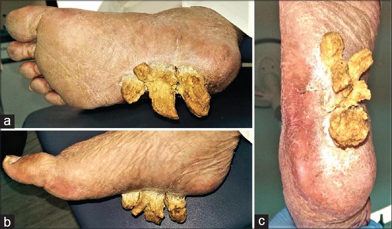 · 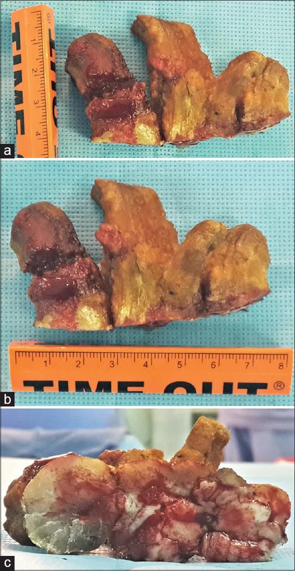 · 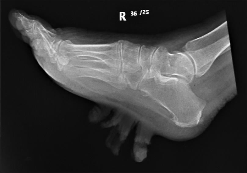 · 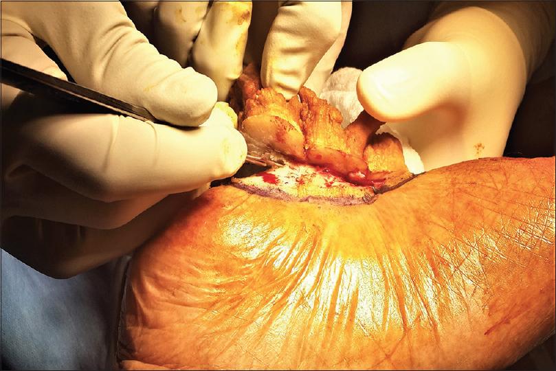 · 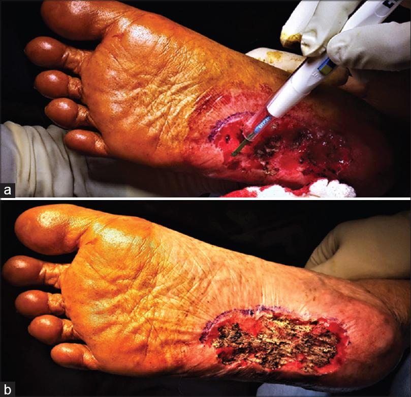 · |
|
#2
●
10-08-2024, 10:46 PM
|
 My Rank: MASTER GUNNERY SERGEANT Poster Rank:352 Manly Man Join Date: Sep 2017 Posts: 3,601 3,601Posts 12,427Thanked 700Quoted 0Mentioned 0Contrib. |
|
Re: Subcutaneous Horns
Horns? More like hooves.
|
|
#3
●
10-08-2024, 11:13 PM
|
 My Rank: CORPORAL Poster Rank:1269 female Join Date: May 2009 Posts: 489 489Posts 411Thanked 35Quoted 0Mentioned 0Contrib. |
|
Re: Subcutaneous Horns
You know her other leg and foot cut to shit . Try sleeping and not brush over skin with a dry heel .
|
|
#4
●
10-09-2024, 12:29 PM
|
 ★ Legacy Member ★ Poster Rank:110 Secret Agent Join Date: Dec 2009 Posts: 14,787 14,787Posts 49,109Thanked 3,050Quoted 6Mentioned 0Contrib. |
|
Re: Subcutaneous Horns
I was expecting some fool with metal implants under the skin. Was a bit shocked when seeing the first foot photo.   Yep, it would be a bit difficult to walk with that on your foot bottom. Once again another human anomaly to see.  Thanks for your posts friend! They’re always interesting and much appreciated! |
|
#5
●
10-09-2024, 12:46 PM
|
 ♚ Legacy Gold Member ♚ Poster Rank:268 ꓕSIꓤIꓕ∀S ϽINOᗡꓤ∀S Join Date: Feb 2010 Posts: 5,177
Contributions: 8
5,177Posts 31,152Thanked 1,441Quoted 45Mentioned 8Contrib. |
|
Re: Subcutaneous Horns
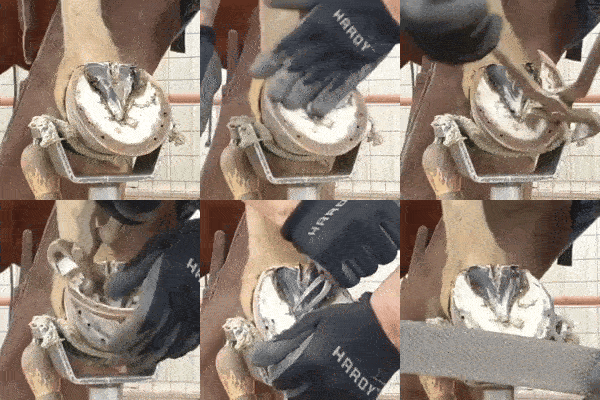
She just needed a farrier...
__________________ 200+ Additional Smilies Custom DR Search Engine ✦ Live life to it's fullest and leave a sexy corpse ✦ |