|
#1
●
07-26-2024, 04:12 PM
|
 These are the rooms Poster Rank:26 of ruin. Join Date: Sep 2014 Posts: 54,008 54,008Posts 87,658Thanked 30,403Quoted 145Mentioned 0Contrib. |
|
Spider Baby
A 32 year old gravida 3 para 2 living 1 death 1 [they are referring to her previous pregnancies and the outcomes, she has had three pregnancies and two living kids] patient came to the out-patient department of Cama and Albess hospital, Mumbai with 17 weeks gestation with a sonogram done at 14 weeks suggestive of spinal dysraphism opting for medical termination of pregnancy. A repeat Sonogram was done at our institute which revealed a fetus with severe spinal deformities. The fetus had lumbar meningomyelocele and spina bifida at multiple levels with lumbar lordosis and a protuberant abdomen. The patient was married since 8 years and had one living female child of 6 years of age followed by an intrauterine fetal demise at full term 2 years back. She had no significant obstetric complications or any major medical or surgical illness in the past. There was no family history of twinning in either parent. General and systemic examination was carried out. The patient was vitally stable with a 16 weeks sized uterus per abdomen and per vaginum and a patulous cervical os. After consultation with the pediatrician as well as the pediatric surgeon, the patient and her husband decided to opt for termination of pregnancy. The patient was admitted and all routine investigations were carried out. The patient's hemoglobin was 10.8 gm%, blood group was B positive and all other investigations were within normal limits. The patient was negative for HIV, hepatitis B, and VDRL. Medical Termination Pregnancy was done with detailed consent. Induction was done with dinoprostone gel followed by injection Pitocin augmentation. The patient aborted after 12 hours uneventfully and completely. Further inspection of the abortus revealed an apparently normal head with a thorax with two normally formed arms and an omphalocele. The abortus had two caudal ends each with a set of male genitilia and completely formed lower limbs arising laterally from either side of the thorax. One of the caudal ends also had spina bifida. There was yet another arm and a limb bud arising from a rudimentary head like structure at the other end of the fetus. An X-ray was done showing the normal skull of 1 fetus from which arose 1 set of ribs and thoracic vertebrae and two normally formed upper limbs. The lumbar vertebrae continued almost at perpendicular from the right side of the fetus with one set of pelvic bones and completely formed lower limbs with evidence of spina bifida. There was yet another set of lumbar vertebrae, pelvic bones and lower limbs which was fused to the left side of the fetus and one arm arising from the other end of the fetus. There was no evidence of another cranium or thoracic cage. There was a single placenta and cord which did not have any gross abnormalities. The abortus was sent for histopathology which revealed single liver and G-I tract that had herniated into an omphalocele. There was one set of lungs and heart. The parasitic fetus did not have a cranium or thorax and was fused at the epigastrium on left side of the normal fetus. The patient was stable post abortion and was managed with antibiotics. She and her husband refused any further investigations due to financial constraints and she was discharged from the hospital on the third day. 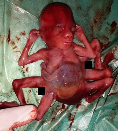 · 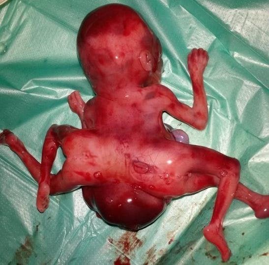 · 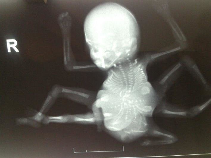 · 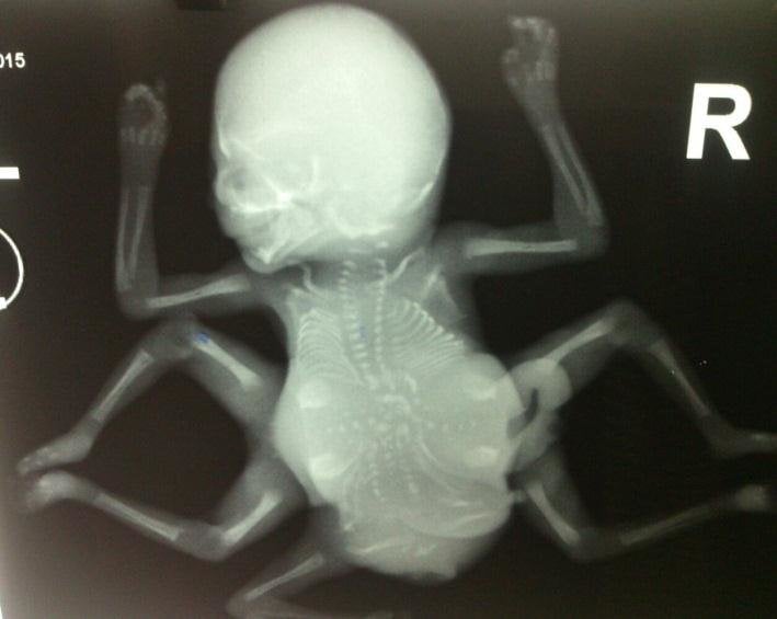 · 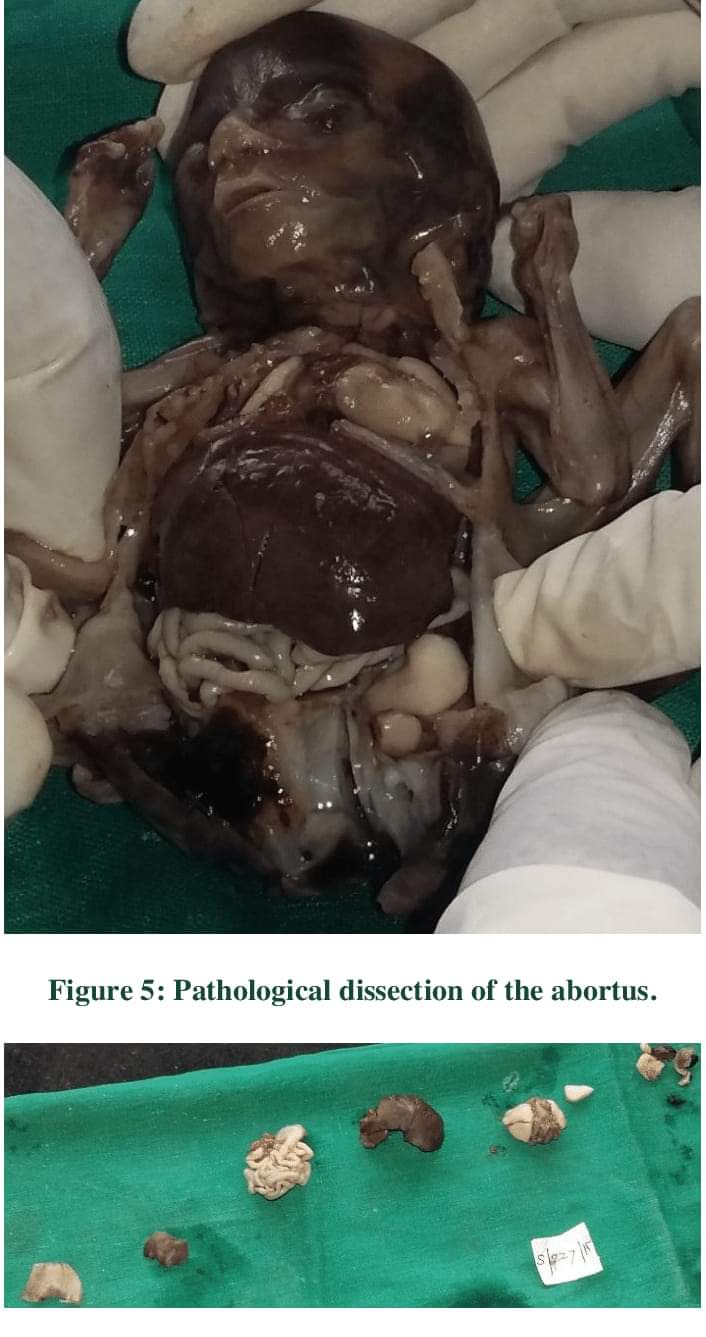 · |
|
#2
●
07-26-2024, 05:14 PM
|
 ♚ Legacy Gold Member ♚ Poster Rank:1095 ★☠ ★ ☠★ Join Date: Sep 2023 Posts: 607
Contributions: 1
607Posts 6,978Thanked 141Quoted 0Mentioned 1Contrib. |
|
Re: Spider Baby
wow that is so weird  |
|
#3
●
07-26-2024, 06:53 PM
|
 My Rank: GUNNERY SERGEANT Poster Rank:606 Join Date: Aug 2017 Posts: 1,542 1,542Posts 2,777Thanked 274Quoted 0Mentioned 0Contrib. |
|
Re: Spider Baby
I am going to have the Spider-Man song in my head now.  |
|
#4
●
07-26-2024, 09:14 PM
|
 ★ Legacy Member ★ Poster Rank:110 Secret Agent Join Date: Dec 2009 Posts: 14,839 14,839Posts 49,315Thanked 3,059Quoted 6Mentioned 0Contrib. |
|
Re: Spider Baby
It would have been even weirder if it was a living adult.   |
|
#5
●
07-26-2024, 10:06 PM
|
| ♚ Legacy Gold Member ♚ Poster Rank:92 Male Join Date: Nov 2009 Posts: 17,193 17,193Posts 44,961Thanked 4,709Quoted 8Mentioned 0Contrib. |
|
Re: Spider Baby
Her drinking water is probably heavily contaminated.  |
|
#6
●
07-27-2024, 09:29 AM
|
 ✝Mudderator from Hell✝ Poster Rank:10 e-mail Join Date: Oct 2006 Posts: 96,702
Contributions: 902
96,702Posts 543,770Thanked 10,372Quoted 479Mentioned 902Contrib. |
|
Re: Spider Baby
conjoined twinning  |
|
#9
●
08-02-2024, 04:40 AM
|
| My Rank: MASTER SERGEANT Poster Rank:538 Join Date: Oct 2008 Posts: 1,895 1,895Posts 4,653Thanked 318Quoted 1Mentioned 0Contrib. |
|
Re: Spider Baby
If it lived they would never catch it
|