|
#1
●
07-09-2014, 07:14 PM
|
 ✖ The Antique Christ ✖ Poster Rank:106 Gonzo Punk Join Date: Jan 2009 Posts: 15,478
Contributions: 62
15,478Posts 39,153Thanked 2,046Quoted 23Mentioned 62Contrib. |
|
Sledding Accident
A 7-year-old presented to the emergency department (ED) with complaints of right eye pain and double vision. The patient had been involved in a sledding accident 1 week before, in which he crashed into a fence. There was no loss of consciousness. On examination in the ED, the right eye was proptotic and dystopic (Figure 1), although with normal visual acuity and no deficits during visual field testing. The pupils were equal, round, and reactive to light, without evidence of an afferent pupillary defect. Dilated funduscopic examination was normal. Extraocular mobility demonstrated limitation of upward gaze and adduction of the right eye. Computed tomographic imaging of the head including facial cuts was performed (Figure 2, Figure 3). 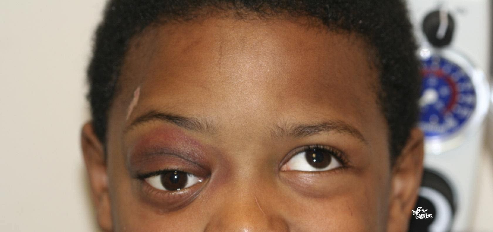 Patient with proptosis and orbital dystopia, with the right eye lower than the left. There is also limitation of upward gaze. 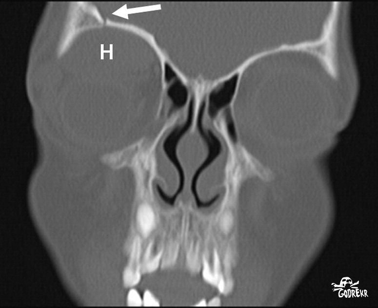 Coronal computed tomographic image. 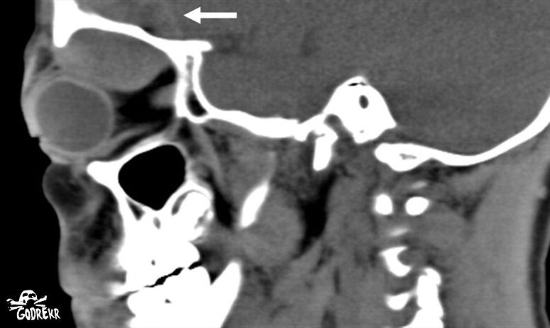 Sagittal computed tomographic image. Diagnosis Superior orbital wall fracture with retro-orbital hematoma. Limitation of upward gaze in the setting of trauma is most commonly associated with a fracture of the floor of the orbit, typically referred to as “blowout fracture.” Entrapment of the inferior rectus muscle within the orbital floor may lead to ophthalmoplegia and diplopia. In such cases, posterior displacement of the globe and prolapse of orbital contents into the maxillary sinus more commonly leads to enophthalmos (recession of the globe into the orbit), as opposed to proptosis. This patient had limitation of upward gaze, with proptosis and orbital dystopia with the right eye lower than the left (Figure 1). Here, limitation of upward gaze was due to a mass lesion (retro-orbital hematoma) impinging on the globe (Figure 2), rather than extraocular muscle dysfunction. Orbital roof fractures are rare, although they are more common in young children because of their high cranium-to-midface ratio. Such fractures have a high association with intracranial injury, most commonly epidural hematoma, as was seen in our patient (Figure 3). |
|
#2
●
07-09-2014, 08:31 PM
|
 My Rank: FIRST SERGEANT Poster Rank:440 Comrade Join Date: Aug 2013 Posts: 2,627 2,627Posts 6,239Thanked 1,167Quoted 14Mentioned 0Contrib. |
|
Re: Sledding Accident
Was that the before photo? The kid looks fine to me.  |
|
#5
●
07-10-2014, 07:50 AM
|
 ✝Mudderator from Hell✝ Poster Rank:10 e-mail Join Date: Oct 2006 Posts: 96,703
Contributions: 902
96,703Posts 543,835Thanked 10,374Quoted 479Mentioned 902Contrib. |
|
Re: Sledding Accident
don't give me the eye will you ?
|
|
#8
●
07-10-2014, 01:16 PM
|
| My Rank: FIRST SERGEANT Poster Rank:440 Comrade Join Date: Aug 2013 Posts: 2,627 2,627Posts 6,239Thanked 1,167Quoted 14Mentioned 0Contrib. |
|
Re: Sledding Accident
What you talk'n bout Willis?
|
|
#10
●
07-11-2014, 10:44 PM
|
 ★ Legacy Member ★ Poster Rank:53 Join Date: Mar 2013 Posts: 28,044
Contributions: 33
28,044Posts 47,276Thanked 11,875Quoted 175Mentioned 33Contrib. |
|
Re: Sledding Accident
His left eye looks wonky too...it's making me dizzy to look at him!  |