|
#1
●
09-03-2024, 07:55 PM
|
 These are the rooms Poster Rank:26 of ruin. Join Date: Sep 2014 Posts: 54,008 54,008Posts 87,658Thanked 30,403Quoted 145Mentioned 0Contrib. |
|
Scabies Complicated by Necrotizing Lymphocyte Vasculitis
Scabies is very common among children. It is often a harmless infectious disease, responding well to antiparasitic medication. Nevertheless, severe forms can occur in immunocompromised populations like newborns and infants. We report a unique case of scabies in a three-months-old infant, complicated by generalized cutaneous lymphocytic vasculitis and unilateral acral necroses. Lymphocytic vasculitis is a morphological term that includes clinically heterogenous diseases like connective tissue disease, infection, lichenoid diseases, drug reaction, Bechet's disease, superficial thrombophlebitis and leukemic vasculitis. Necrotizing forms are possible. Among infectious causes, our case is the first pediatric case where scabies is incriminated. Moreover, acral necroses occurred and imputation was necessary. A well-appearing three-month-old boy, with no past medical history, was admitted to the emergency department for an acute onset of ischemia of the left foot evolving for twelve hours. The mother reported a generalized rash evolving for one month. The boy was under no medication. Both parents suffered from generalized scratching. At the examination, the infant was afebrile and had severe itchiness and a pimple-like rash typical of scabies. The presence of pustules suggested additional bacterial infection. Swab sampling was performed and methicillin-sensitive Staphylococcus aureuswas isolated on culture. The child was put on oral antibiotics (amoxicillin + clavulanic-acid: 80 mg/kg/day, three times a day, for 10 days) and benzyl benzoate emulsion. Parents´ treatment, as well as decontamination of clothes and blankets were started. The boy had also necrosis of the third and fort toes of the left foot (Figure 1). Peripheral pulses of the four limbs were well perceived. Doppler of the left limb did not visualize any thrombus. A typical rush consisting of vasculitic elements was also found on skin examination. Biopsy and pathology examination found a lymphocytic vasculitis of the dermis and hypodermis. There was no IgG, IgM, IgA or C3 perivascular deposits on immunofluorescence. The next day after admission, the infant developed acrocyanosis of the left foot and necroses of all five toes (Figure 2). We started aspirin at 5mg/kg/day. Abdominal and lower limbs CT-angiography was normal. There was a mild inflammatory syndrome (CRP = 50.86mg/l, ESR = 34mm, ferritin=109ng/ml, ANC = 13230/mm3, platelets = 760000/mm3) that was most likely due to infection. Laboratory workup to look for thrombophilia and deep vein thrombosis was normal (PT = 100%, PTT = 32sec, Fibrinogen = 1.92g/l, D-Dimer = 0.66ng/ml, C protein = 72%, S protein = 70%, antithrombin III = 81%). Also, cryoglobulinemia blood essay was negative. Capillaroscopy found no abnormalities of the microcirculation in the nail folds of the right foot. We looked also for an underlying auto-immune condition. Plasma protein electrophoresis was normal and the coombs test was negative. Antinuclear antibodies were weakly positive and speckled. Anti-ds DNA antibodies, anti-Sm antibodies, anti-Ro/La antibodies, anti-RNP antibodies, anti-P-ANCA/C-ANCA antibodies, anti-cardiolipin and anti-beta-2 glycoprotein 1 antibodies were negative. No lupus anticoagulant was found. Evolution was marked by regression of acrocyanosis by the third day. Acral ischemia progression stabilized. Amputation of the five toes was performed by the seventh day. Meanwhile, scabies lesions have healed by the second week and CRP decreased to 17.1mg/l. After what, the boy was discharged home. He is now five-month-old and still healthy. 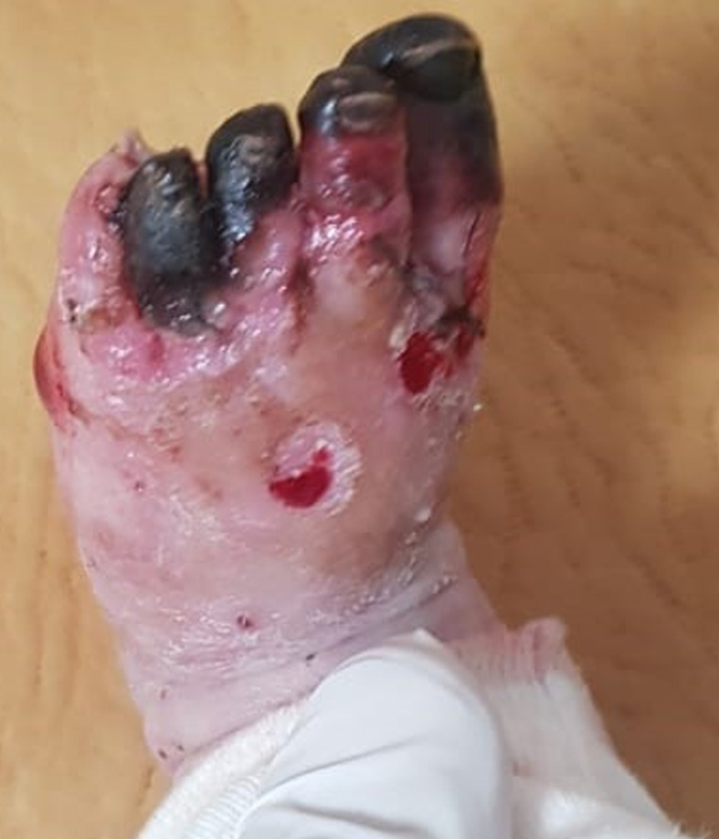 · 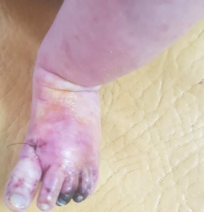 · |
|
#2
●
09-04-2024, 03:38 AM
|
 ✝Mudderator from Hell✝ Poster Rank:10 e-mail Join Date: Oct 2006 Posts: 96,660
Contributions: 901
96,660Posts 543,393Thanked 10,369Quoted 479Mentioned 901Contrib. |
|
Re: Scabies Complicated by Necrotizing Lymphocyte Vasculitis
What a way to start life.  The main cause of Lymphocyte Vasculitis in this infant is not yet clear. Hospital docs tend to believe it is staphylococcal infection rather than scabies. |