|
#1
●
03-20-2023, 09:31 PM
| ||||||||
| These are the rooms Poster Rank:25 of ruin. Join Date: Sep 2014 Posts: 54,011 Mentioned: 145 Post(s) Quoted: 30403 Post(s)
| ||||||||
|
Ring Finger Avulsion
Source: The patient is a 21-year-old right-hand-dominant male who presented to the emergency department one hour following injury to his left hand. He is an active-duty military male with no comorbidities who was running as part of a physical training exercise. During the run, he jumped and attempted to strike a stop sign with his left hand when his wedding band caught on the top of the sign causing a ring avulsion type injury to his left ring finger. His wound was dressed, and the amputated digit was wrapped in a latex glove and placed in a plastic bag of water and ice. Upon initial examination, the patient was noted to have sustained a complete amputation of his ring finger at the level of the middle phalanx proximal to the FDS insertion with significant circumferential soft tissue degloving (Figures 1-4). The patient reported intact sensation to both the radial and ulnar aspects of the finger proximal to the injury, and complete avulsion of the radial and ulnar proper digital neurovascular structures was noted. With the nearest replantation center approximately 100 miles away, a warm ischemia time of 2-3 hours from injury at point of contact, and the significant extent of soft tissue injury, it was determined that the likelihood of successful replantation was low, and revision amputation with primary closure or coverage was recommended to the patient. Consent was obtained for debridement and primary closure versus graft coverage. Local anesthesia was achieved with a digital block, and a thorough bedside irrigation and sharp debridement were performed under the supervision of the senior staff. The avulsed radial and ulnar arteries were tied off with 4-0 nylon suture. The middle phalanx, which had essentially been skeletonized by the injury, was shortened with a rongeur in an initial attempt to obtain soft tissue coverage. Adequate soft tissue coverage was still not possible despite step-wise shortening of the middle phalanx to the point of complete removal through the PIPJ. The decision was then made to take the extra time to create a full-thickness skin graft from the amputated digit. This would be performed in order to provide adequate soft tissue coverage to the underlying bone and neurovascular structures and increase the likelihood of graft uptake due to the use of autologous tissue, and with the hope of avoiding a future, scheduled trip to the operating room. Evaluation of the amputated digit revealed the soft tissue envelope to be intact; the flexor digitorum profundus tendon was also intact and had ruptured proximally from its myotendinous junction in the forearm while still maintaining its insertion at the base of the distal phalanx of the amputated part. The amputated portion of the digit was sterilized in a 10% betadine prep solution. Longitudinal incisions were made on the radial and ulnar sides and connected at the distal tip of the amputated finger to form a fishmouth-type incision. The incision was taken to the bone with a no. 15 blade, and the soft tissue was carefully dissected away from the bone and nail bed. The size of the required graft was then templated and the excess tissue was removed. Subcutaneous fat was excised to the level of the dermis as possible. The graft was secured with 4-0 Prolene suture (Ethicon, Somerville, NJ, USA) in a simple interrupted fashion (Figures 5, 6). A sterile dressing consisting of Adaptic™ non-adherent dressing (3M, St. Paul, MN, USA), 4x4 gauze, and loosely wrapped Kerlix™ (Cardinal Health, Dublin, OH, USA) was applied in addition to an AlumaFoam® splint (Hartmann USA, Inc., Rock Hill, SC, USA) secured with Coban™ wrap (3M) (Figure 7). The patient was discharged the same day from the emergency department with a prescription for double-strength Bactrim to be taken two times per day for 14 days. He was instructed to follow up in the clinic the following day for wound evaluation. At the initial follow-up, the patient maintained complete coverage of the injury without evidence of necrosis or drainage. He had intact sensation throughout the remaining finger. Nonoperative wound care and monitoring for incorporation of the graft was elected for further treatment. The patient was seen on a weekly basis for the following two weeks with non-weight-bearing restrictions. He continued to demonstrate signs of healing and graft incorporation, with definite signs of central perfusion and only minor ischemia medially and laterally. At his six-week follow-up, he was evaluated for suture removal and was noted to have developed a seroma. His sutures were removed and a small incision was made in the graft in the office to allow for fluid evacuation. He was allowed to return home with continued wound care and occupational therapy. At the final follow-up, the graft had fully healed with appropriate vascularization and full sensation over the radial and ulnar aspects . He was cleared to return to full active duty without restrictions. 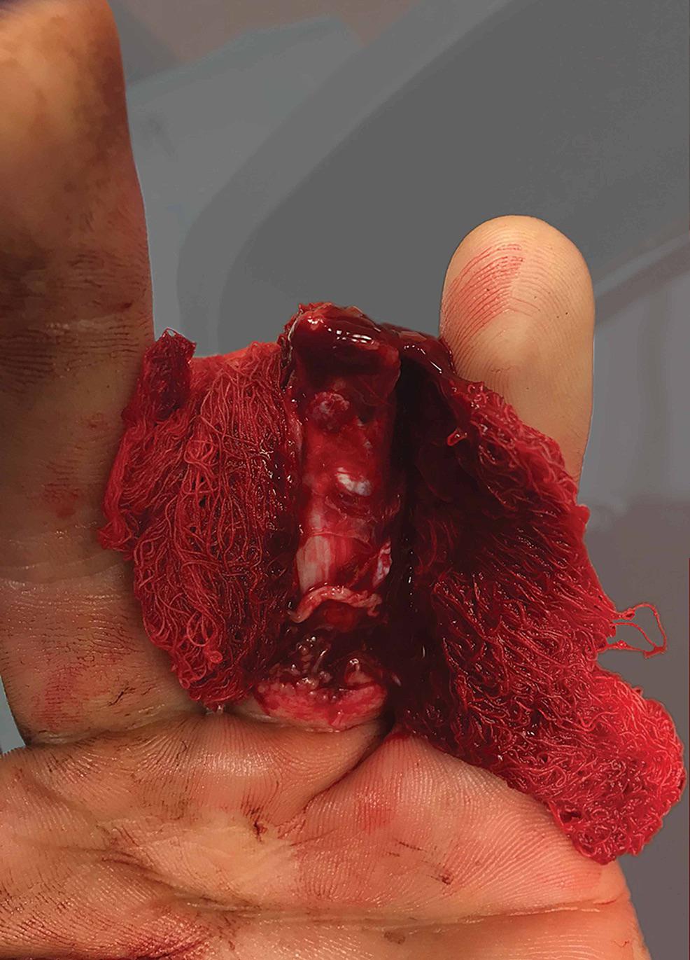 · 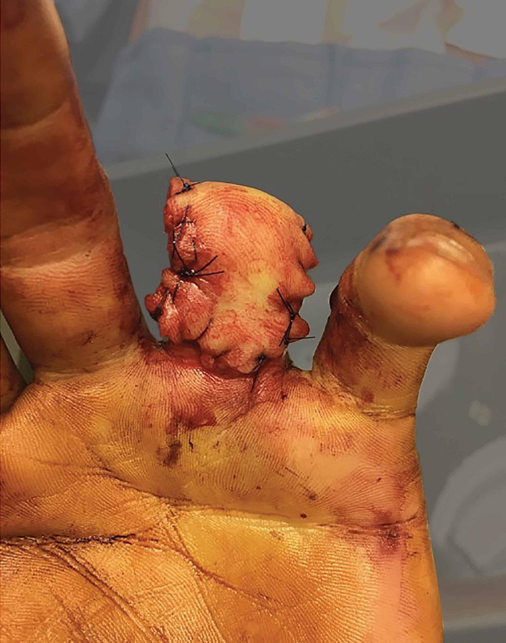 · 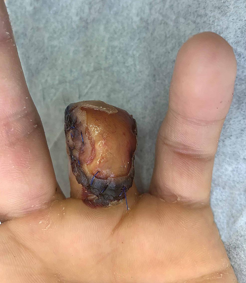 · 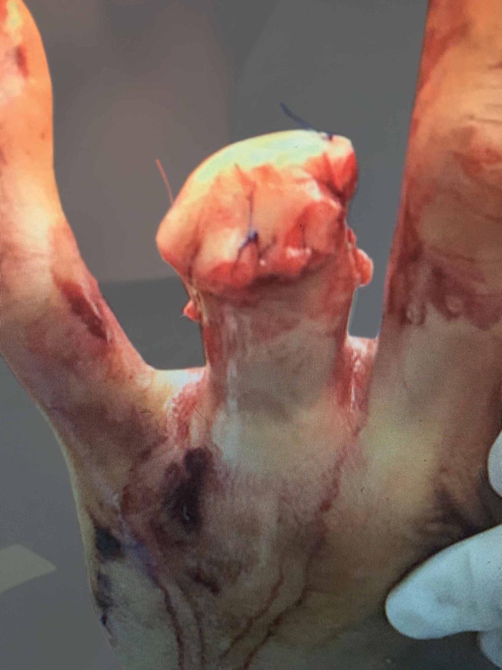 · 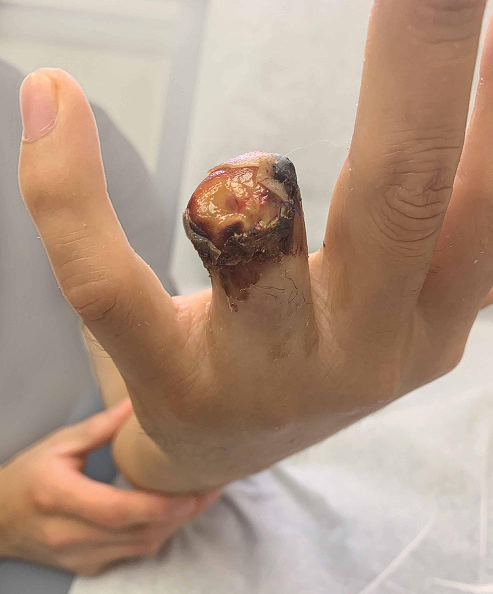 · 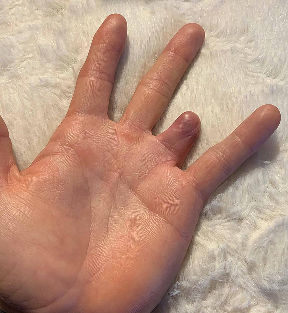 · 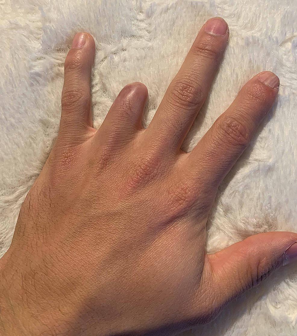 · |
|
#3
●
03-20-2023, 10:31 PM
| ||||||||
| ★ Legacy Member ★ Poster Rank:247 So many choices now Join Date: Jul 2015 Posts: 5,548 Mentioned: 15 Post(s) Quoted: 2120 Post(s)
| ||||||||
|
Re: Ring Finger Avulsion
He high-fives a fucking stop sign. Thats a class-C on-duty mishap. It'll be worth some government disability, even though it was his fault.  |
|
#4
●
03-21-2023, 12:34 AM
| ||||||||
| My Rank: MASTER GUNNERY SERGEANT Poster Rank:347 Manly Man Join Date: Sep 2017 Posts: 3,601 Mentioned: 0 Post(s) Quoted: 700 Post(s)
| ||||||||
|
Re: Ring Finger Avulsion
That's his fault for letting someone put a ring on that finger...
|
|
#5
●
03-21-2023, 12:11 PM
| ||||||||
| ★ Legacy Member ★ Poster Rank:119 Secret Agent Join Date: Dec 2009 Posts: 13,206 Mentioned: 6 Post(s) Quoted: 2787 Post(s)
| ||||||||
|
Re: Ring Finger Avulsion
The 5th photo is my favorite. Love him holding the bottom of his arm. The foreshortening makes it look huge and out of proportion. |
|
#6
●
03-21-2023, 05:46 PM
| ||||||||
| ✝Mudderator from Hell✝ Poster Rank:10 e-mail Join Date: Oct 2006 Posts: 94,983
Contributions: 817
Mentioned: 472 Post(s) Quoted: 10078 Post(s)
| ||||||||
|
Re: Ring Finger Avulsion
i would rather lose the entire digit than keeping a little stumpy
|