|
#1
●
01-09-2023, 09:41 PM
| ||||||||
| These are the rooms Poster Rank:25 of ruin. Join Date: Sep 2014 Posts: 54,011 Mentioned: 145 Post(s) Quoted: 30403 Post(s)
| ||||||||
|
Polyarteritis Nodosa in the Vulva of a Patient with Behçet's Disease
A 31-year-old single woman, of Mediterranean descent from Lebanon, presented with deep, painful genital ulcers and multiple oral aphthae for two weeks. Her condition was accompanied by arthralgia of the knees and elbows, light flashes, and blurry vision in both eyes. Her past history was significant for recurrent genital lesions and photosensitivity. She had no history of sexual activity or recent travel. The review of systems was otherwise unremarkable. Examination showed well-defined punched-out erosions over the buccal and gingival mucosa and multiple deep ulcers with overlying yellow exudate and black eschar involving two-thirds of both labia majora and minora sparing the clitoris and punched-out unremarkable non-violaceous borders with bilateral inguinal lymphadenopathy (Figure 1). Pathergy test was negative. Ophthalmological examination showed reduced visual acuity in both eyes; 20/60 in the right eye and 20/30 in the left eye with quiet anterior segments. Dilated fundus examination showed few cotton wool spots and intraretinal hemorrhage. No evidence of vitritis, retinitis, or vasculitis was found (Figure 2). Fundus fluorescein angiography showed multiple arteriolar infarctions involving the macula in both eyes, more in the right eye. The retinal venous system did not appear to be affected (Figure 3). Optical coherence tomography of the right and left eyes showing inner nuclear and outer plexiform layers hyper-reflectivity indicating ischemia (Figure 4). Her immunological workup showed negative antinuclear antibody ANA, double-Stranded DNA, antineutrophil cytoplasmic antibodies ANCA, and complement C3 & C4. Tests for herpes simplex virus (HSV) IgM 1 and 2, hepatitis B and C viruses, cytomegalovirus (CMV) IgM, human immunodeficiency virus (HIV), QuantiFERON-TB, and syphilis were negative. The HLA B-51 was positive. Microscopic examination of hematoxylin and eosin (H&E)-stained tissue sections from the vulvar biopsy revealed epidermal hyperplasia and dense dermal and subcutaneous inflammatory infiltrate with large numbers of neutrophils. A medium-sized subcutaneous artery was involved and showed neutrophilic infiltration of its wall. No granuloma was observed (Figure 5). Stain for elastic lamina showed medium-sized subcutaneous artery involvement (Figure 6). Considering the above, a final diagnosis of the coexistence of polyarteritis nodosa in Behçet's disease patient was made. She was admitted and started on pain management, intravenous methylprednisolone 1,000 mg for three days suggested by the ophthalmologist to suppress the ocular damage and to stop further mutilation of the genitalia followed by oral prednisolone 50 mg (1 mg/kg) with a slow taper of 5 mg weekly, oral colchicine 0.5 mg twice daily, and adalimumab 40 mg once every two weeks. On follow-up after four months, there was sizeable improvement of the ulcer and no new cutaneous or systemic complaints reported (Figures 7-8). 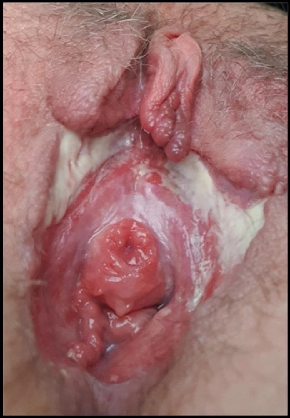 · 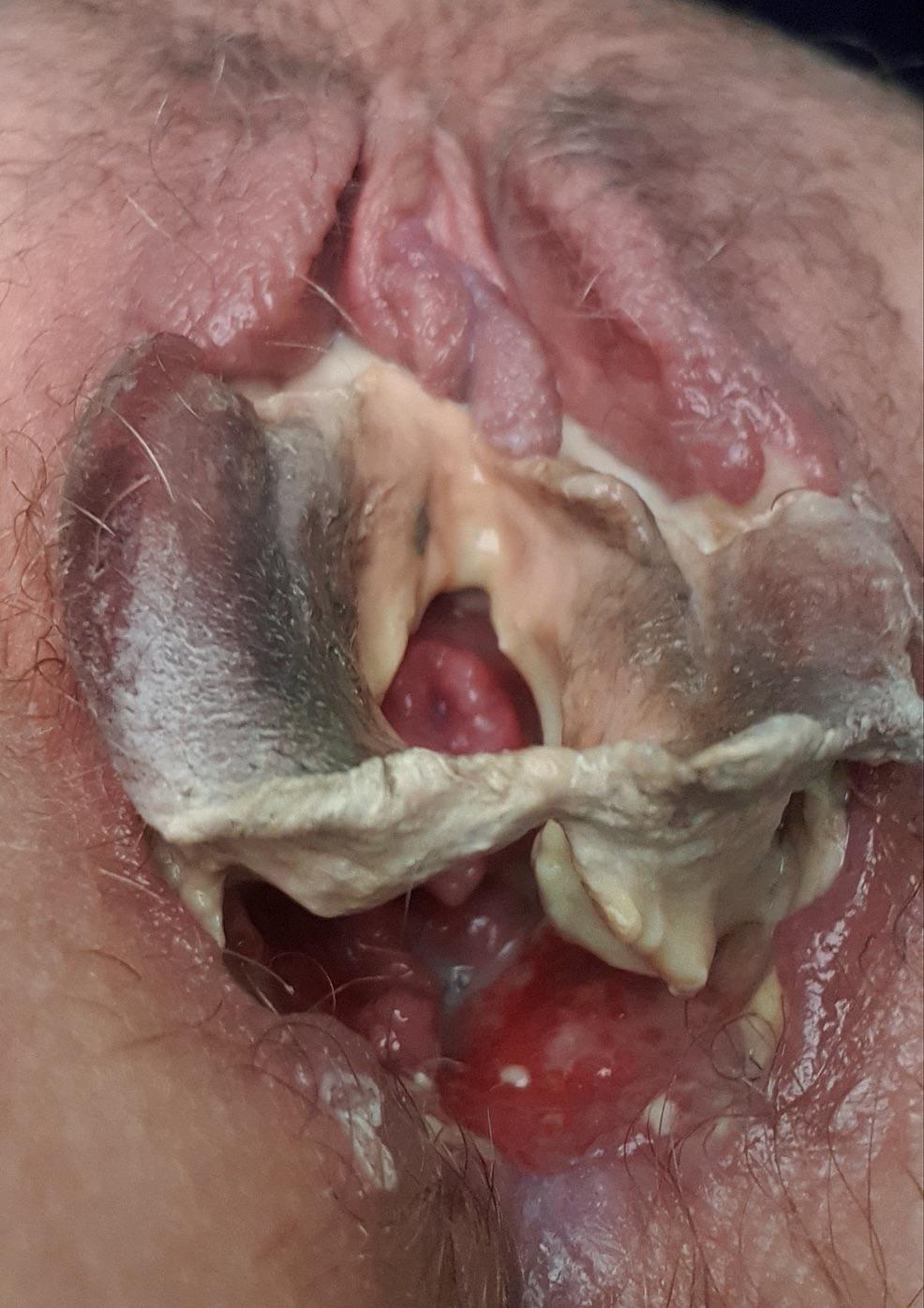 · 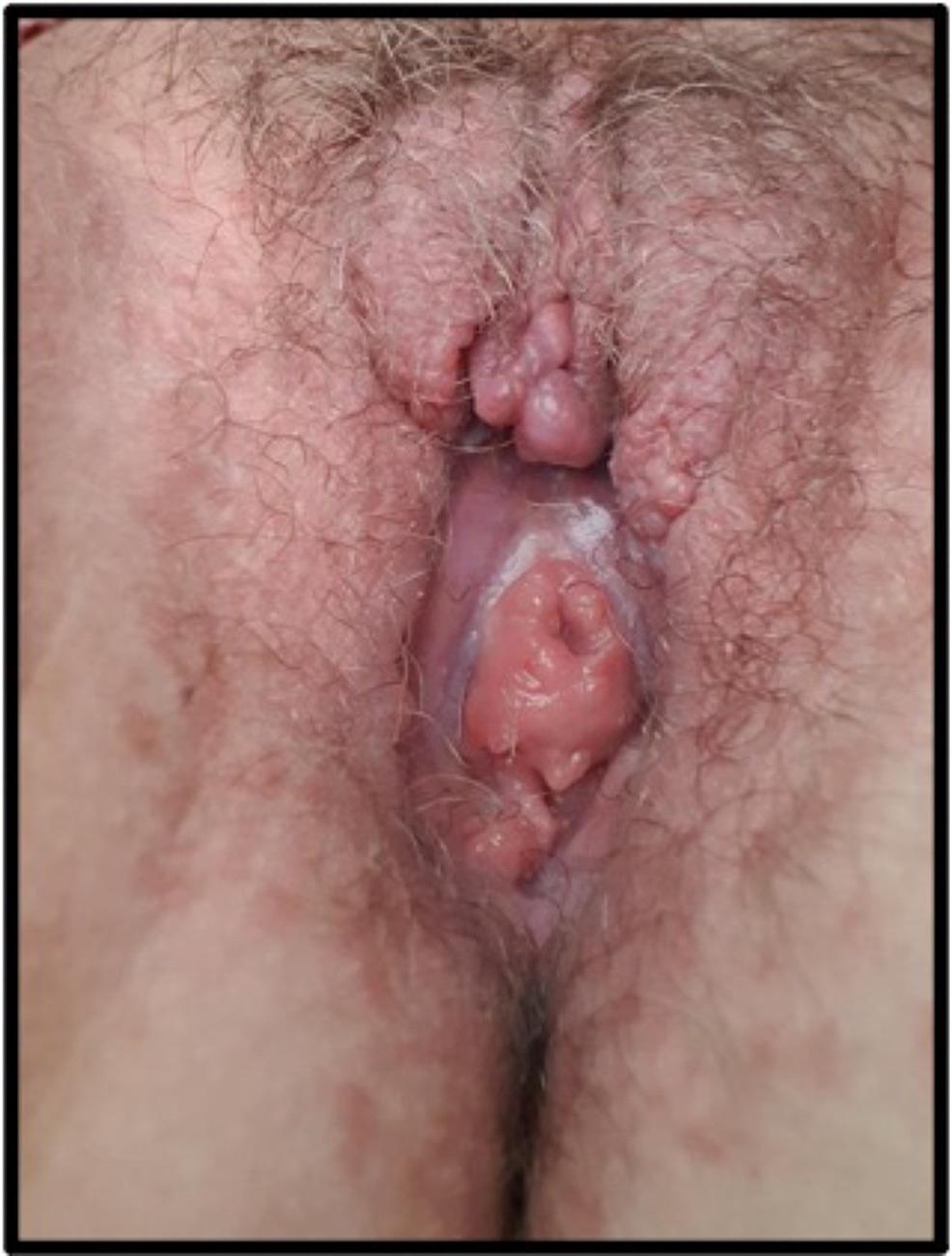 · |
|
#2
●
01-09-2023, 11:20 PM
| ||||||||
| ♚ Legacy Gold Member ♚ Poster Rank:202 Iceman Join Date: Aug 2017 Posts: 7,505 Mentioned: 19 Post(s) Quoted: 3629 Post(s)
| ||||||||
|
Re: Polyarteritis Nodosa in the Vulva of a Patient with Behçet's Disease
In that second picture it looks like a mushroom grew over her vagina
|
|
#3
●
01-09-2023, 11:51 PM
| ||||||||
| ★ Legacy Member ★ Poster Rank:247 So many choices now Join Date: Jul 2015 Posts: 5,548 Mentioned: 15 Post(s) Quoted: 2120 Post(s)
| ||||||||
|
Re: Polyarteritis Nodosa in the Vulva of a Patient with Behçet's Disease
The pee hole still looks a bit angry. Clitoris appears to be a bit easier to find, though. All in all, I guess its better than a bowling pin stuck in the ass.
|
|
#4
●
01-09-2023, 11:51 PM
| ||||||||
| These are the rooms Poster Rank:25 of ruin. Join Date: Sep 2014 Posts: 54,011 Mentioned: 145 Post(s) Quoted: 30403 Post(s)
| ||||||||
|
Re: Polyarteritis Nodosa in the Vulva of a Patient with Behçet's Disease
Yeah. That's gross but accurate. |
|
#5
●
01-10-2023, 02:11 AM
| ||||||||
| My Rank: STAFF SERGEANT Poster Rank:741 Female Join Date: Jun 2019 Posts: 1,095 Mentioned: 2 Post(s) Quoted: 510 Post(s)
| ||||||||
|
Re: Polyarteritis Nodosa in the Vulva of a Patient with Behçet's Disease
Christmas tree vagina... any takers |
|
#6
●
01-10-2023, 12:02 PM
| ||||||||
| Addict Of Misery Poster Rank:69 Join Date: May 2012 Posts: 22,176 Mentioned: 18 Post(s) Quoted: 9547 Post(s)
| ||||||||
|
Re: Polyarteritis Nodosa in the Vulva of a Patient with Behçet's Disease
Even without all the extra goo and such, that's one ugly snatch.
__________________ A deep well of despair I found, the day my dreams came true... |
|
#7
●
01-10-2023, 06:12 PM
| ||||||||
| My Rank: LANCE CORPORAL Poster Rank:3502 Join Date: Jun 2015 Posts: 100 Mentioned: 0 Post(s) Quoted: 27 Post(s)
| ||||||||
|
Re: Polyarteritis Nodosa in the Vulva of a Patient with Behçet's Disease
Did the lips rot off, is that what im seeing?
|
|
#9
●
01-11-2023, 05:04 AM
| ||||||||
| My Rank: MAJOR Poster Rank:92 Male Join Date: Aug 2009 Posts: 16,925 Mentioned: 9 Post(s) Quoted: 1885 Post(s)
| ||||||||
|
Re: Polyarteritis Nodosa in the Vulva of a Patient with Behçet's Disease
Well that's something I wish I could unsee.
|