|
#1
●
06-21-2024, 06:24 PM
| ||||||||
| These are the rooms Poster Rank:25 of ruin. Join Date: Sep 2014 Posts: 54,011 Mentioned: 145 Post(s) Quoted: 30403 Post(s)
| ||||||||
|
Parasitic Twin
Male newborn, monozygotic twin pregnancy, cesarean delivery at 37 weeks and 4 days and Apgar score 7/8. The parents were at the second decade of age and there was not a history of comorbidities or use of drugs or other substances during the pregnancy. It was the first pregnancy of the mother. At 34 weeks of gestation, she presented an echographic diagnosis of conjoined parasitic twin with shared liver (omphalopagus), without head pole, showing thoracic structures, limbs, kidney, bladder and umbilical hypertension. Parasitized twin with cardiomegaly, interventricular communication (IVC) and aortic coarctation. The baby can be visualized in Fig. 1 below. Free and informed consent was signed and the study was approved by the ethics committee of the institution. Multidisciplinary surgery was scheduled at 10 days of life. Preoperative ultrasound of the newborn with situs solitus in dextroposition, patent foramen ovale, intraventricular communication, patent ductus arteriosus, showing moderate mitral and tricuspid level. On computed tomography (Fig. 2), the fetus was parasitically connected anteriorly in the thoracoabdominal region, showing: kidney in the transition between the two fetuses, with no identification of the heart, in addition to partial herniation of the liver to the neck of connection between the twins. Regarding vascularization between the twins, it was possible to observe that two arteries irrigate the incomplete twin, both with a diameter of 2 mm, one of which is an extension of the right internal thoracic artery and the other possible persistence of the right umbilical artery. The resection was initiated at the base of the pedicles of the lower and upper limbs of the parasite, in which a urinary bladder, a structure suggestive of a kidney, was observed, in addition to loops of the small intestine and colon that were continuous and blind. The resected structures can be seen in Fig. 4 below. The newborn had viable intestinal loops, without malformations, liver with a bifurcated lobe, but completely belonging to the patient. No organ sharing between twins was identified. Resection of excess skin and synthesis was performed. The patient had renal insufficiency, with favorable evolution in the postoperative period. The surgery finished after 8 h and there was not important blood loss. Discharge occurred after 3 months of pediatric intensive care. There were not important complications in the immediate postoperative period. The follow up time is 24 months and the child is healthy. 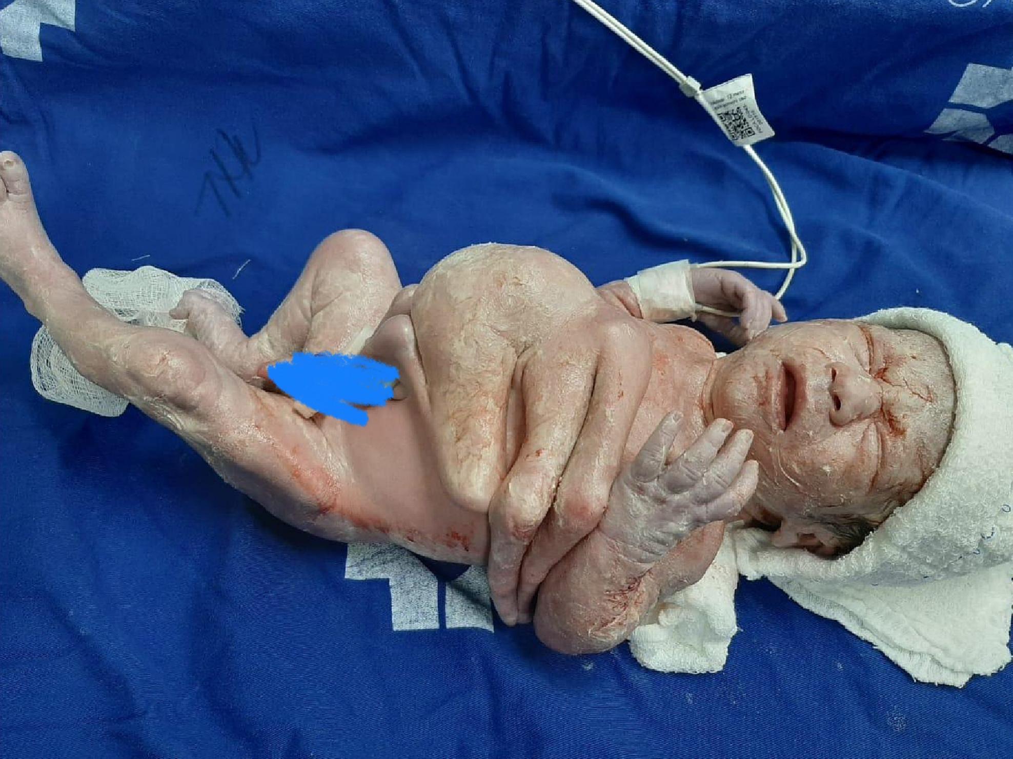 · 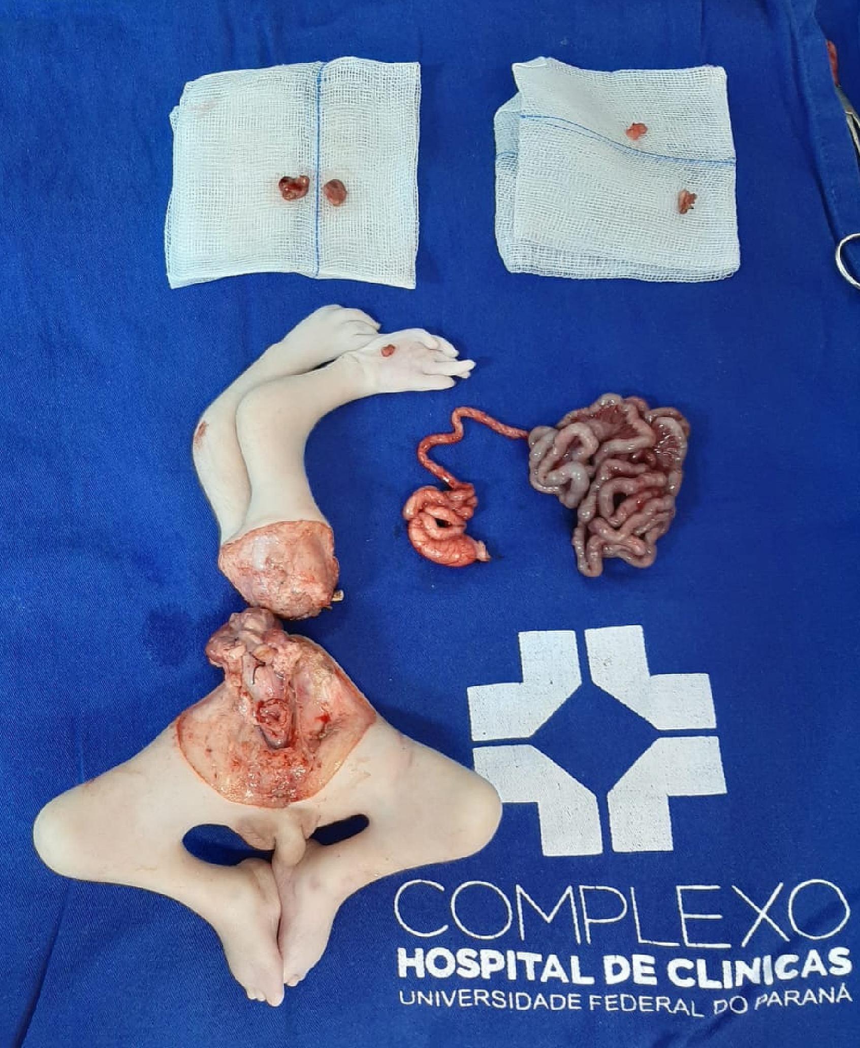 · |
|
#2
●
06-21-2024, 10:08 PM
| ||||||||
| ♚ Legacy Gold Member ♚ Poster Rank:130 Happy Happy Happy Join Date: Dec 2009 Posts: 12,406 Mentioned: 20 Post(s) Quoted: 4089 Post(s)
| ||||||||
|
Re: Parasitic Twin
Kill it with fire before they lay eggs
|
|
#4
●
06-22-2024, 03:46 AM
| ||||||||
| ✝Mudderator from Hell✝ Poster Rank:10 e-mail Join Date: Oct 2006 Posts: 94,990
Contributions: 817
Mentioned: 472 Post(s) Quoted: 10081 Post(s)
| ||||||||
|
Re: Parasitic Twin
reminded me a bit of the Bhagat's case where a man carried his twin brother for 36 years in his stomach without noticing
|