here's the story :
A 4-year-old boy presenting with generalized 'rash' since birth was misdiagnosed as having epidermolysis bullosa simplex (EBS).
The initial biopsy taken several years earlier by a non-dermatologist was signed out as EBS. We do not have access to the original pathology slides and were only able to gather information from the patient's records from an outside clinic.
These reports stated that the initial biopsy showed an "intraepidermal blister within the basal epidermal layer, along with clumping of the tonofilaments." This is a second hand report of the biopsy results and we are left to assume that clumping of the tonofilaments was determined by electron microscopy.
A second reading of the biopsy by an EBS expert noted no definite cleavage planes and normal type VII collagen. These findings excluded generalized recessive dystrophic EB and junctional EB, but left EBS in the differential.
Although his clinical appearance did not fit EBS, the patient was seen and misdiagnosed by various non-dermatologists in the US. Working diagnoses ranged from EBS to BCIE to severe seborrheic dermatitis, highlighting the importance of clinicopathologic correlation by a trained specialist in difficult-to-diagnose cases such as this.
His mother believed that flaring of the 'rash' was secondary to specific foods in his diet such as chicken, dairy products, eggs, and milk. However, the flares did not improve with elimination of these food items from his diet.
Furthermore, dietary restrictions may have resulted in failure to thrive and a subsequent intervention by social services. His developmental milestones were normal.
In addition, the patient had been hospitalized five times in the last eight months for methacillin-resistant Staph aureus (MRSA) infections. He had also lost six pounds over the last year, and weighed less than he did at his third birthday.
His mother had tried various treatment options including topical application of Aquaphor™ ointment, intensive moisturizing with Cetaphil™ lotion and oral acitretin, without significant improvement. There was no family history of dermatologic disease.
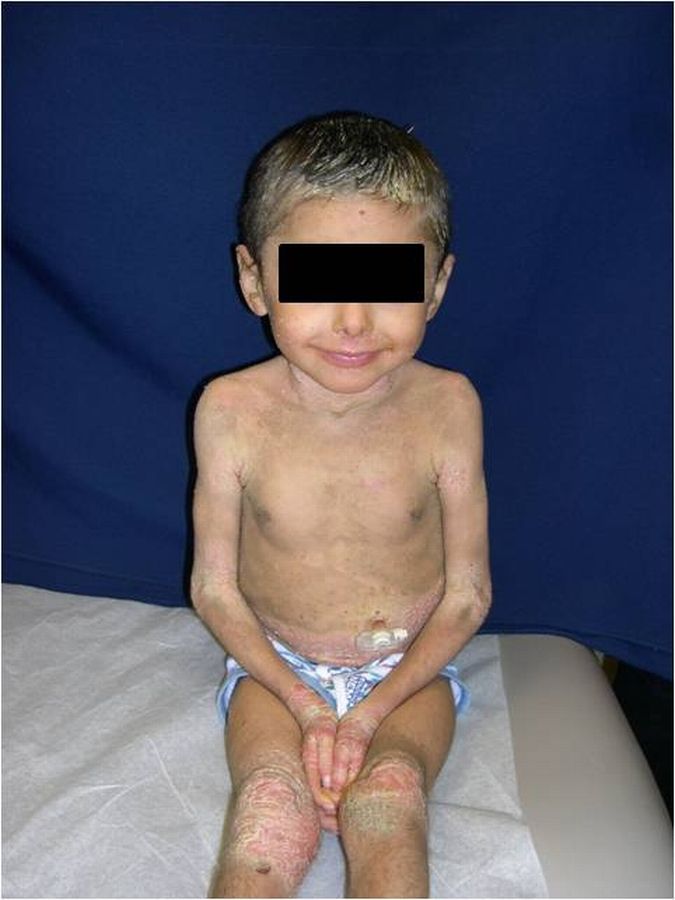
Physical examination revealed hyperpigmented, dirty-appearing scale with increased verrucous-warty linear plaques, especially in the flexural surfaces of the axillae, popliteal, and antecubital fossae.
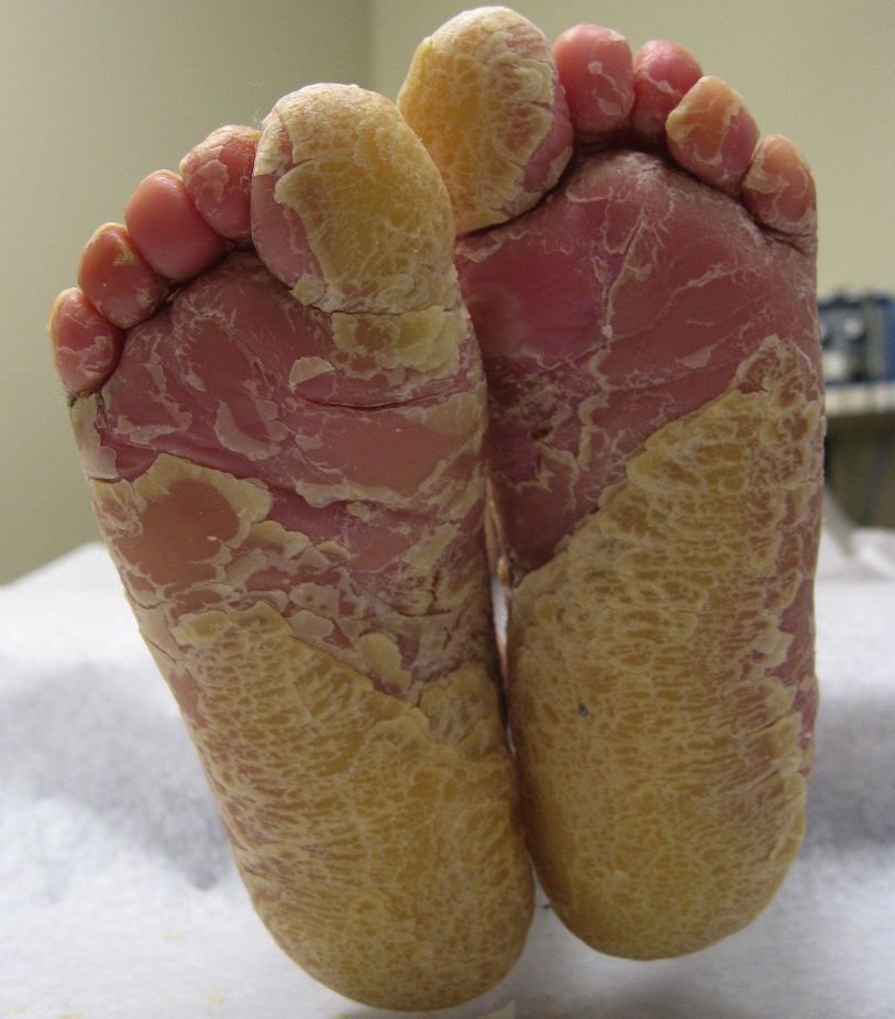
There was also thick yellow scale and palmoplantar keratoderma involving the hands and feet bilaterally and thick yellow-matted scale encompassing the scalp. Some degree of scale was present throughout all body surfaces, with approximately 95 percent involvement.
Fissuring was present on the hands, feet, and angles of the mouth. No blisters were noted and there was no involvement of the ocular mucosa. A generalized foul odor emanated from the child.
At this time, on the basis of clinical presentation alone, it was evident that EBS was not the correct diagnosis. The total body warty, porcupine quill-like hyperkeratosis, palmar hyperkeratosis, and thick scalp scale were characteristic of BCIE.
A repeat biopsy showed epidermal acanthosis, papillomatosis and hyperkeratosis. There was striking clumping and dissolution of the granular cell layer, both consistent with BCIE.
For treatment of his scalp, we prescribed olive oil to be generously applied under shower cap occlusion for approximately 2-3 hours prior to washing with T/Gel shampoo.
To his body, the patient was instructed to apply Vaseline™, Aquaphor™, and Crisco™ after bathing at a recommended twice daily frequency to improve moisturization.
Furthermore, a small quantity of liquid laundry bleach was added to the bath water (¼ to ½ cup per foot of water on side of tub) to kill and eliminate cutaneous bacteria responsible for the associated foul odor.
Oral anti-staphylococcal antibiotics were also used for purulent lesions. Oral retinoids, including isotretinoin and etretinate, are another treatment option that is relatively fast acting and effective for this condition; in many cases retinoids would provide benefits that would arguably be greater than any of the associated potential side effects (e.g. skeletal abnormalities such as cortical hyperostoses, diminished bone density and premature epiphyseal closure).
However, given that our patient was young, that his condition required long-term treatment and that he also suffered from baseline growth retardation, which had necessitated a percutaneous endoscopic gastrostomy (PEG) tube placement and overnight feeds, it was decided that we would first start treatment with topical therapy and oral antibiotics.
Retinoids could be considered at a later time if the patient's dermatologic condition failed to improve. Thus far, oral antibiotics along with the bleach baths have helped control and reduce skin infections and have consequently also helped the associated foul odor.
Furthermore, extensive use of emollients has helped soften and remove excess scale from his body and scalp.









 Gads. Poor little muffin.


