
|
#1
●
07-06-2024, 04:32 PM
| ||||||||
 These are the rooms Poster Rank:26 of ruin. Join Date: Sep 2014 Posts: 54,011 Mentioned: 145 Post(s) Quoted: 30403 Post(s)
| ||||||||
|
Nipple Lesion
A 40-year-old woman presented with a mass hanging from the tip of nipple of left breast for last 3 years (Figure 1). This lesion started as a small lesion of sub centimetric size. The lesion was painless and did not caused any symptoms. So, the patient ignored this lesion for long time. The lesion kept on growing slowly and silently to reach the present size. There was no history of lump in the breast and no discharge from nipple. She had three grown up children. The last pregnancy was 16 years back. Menstrual cycles were normal of 3–4 days/28 days. There was no history of any breast lesion in the family. On clinical examination, the mass shows a well circumscribed oval shape lesion of size 12x7 cm. The lesion was pedunculated and was having a long pedicle connected to nipple of left breast. The surface of this lesion was covered by thin skin all around which was glossy at certain places due to stretching. The surface of this lesion was uneven like cauliflower. There were no ulcers on its surface. This swelling was firm in consistency (Figure 2). The nipple areola complex and breast were normal. There was no axillary lymphadenopathy. The clinical diagnosis was of a benign polyp. The ultrasound of the bilateral pectoral breasts was normal. The mammography of both pectoral breasts was normal except showing a soft tissue mass with macro calcification outside the line of left pectoral breast (Figure 3). The patient was operated under local anesthesia. About 2 ml of 2% lignocaine was infiltrated under the skin of left nipple. An elliptical incision was given on the tip of nipple. The incision was deepened by 3mm and this wedge was excised along with long pedicle. There was a small feeder vessel which coagulated. The small elliptical wound was closed with 4–0 polypropylene suture. The patient was discharged after two hours on the same day as it was a day surgery procedure. There were no postoperative complications. The wound healed well and stitches were removed on 7th postoperative day. The excised specimen was sent for histopathological examination. The gross appearance of the excised specimen showed a 12x7 cm size, grey brown polypoidal mass with a long pedicle of about 5 cm (Figure 4). The cut section of specimen revealed white coloured, uniform smooth and whirly appearance. This polypoidal mass was covered with skin. It showed grey white areas infiltrating normal parenchyma. The microsection H&E stain 100 X showed lining of stratified epithelium with underlying subepithelial composed of loose collagenous tissue. Histopathological examination revealed it a fibroepithelial polyp as final diagnosis. The patient was followed at three months, the scar has healed well and no recurrence was observed. 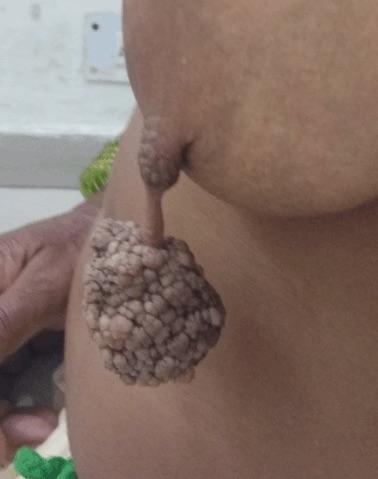 · 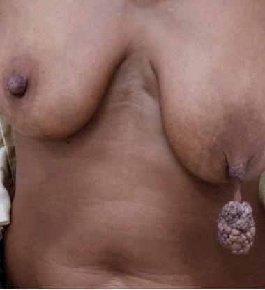 · 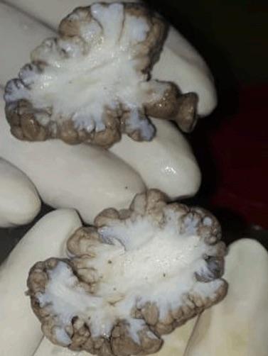 · 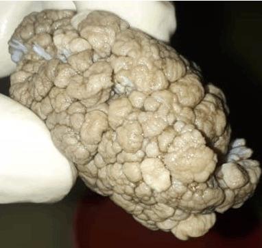 · |
|
#2
●
07-06-2024, 04:48 PM
| ||||||||
 ★ Legacy Member ★ Poster Rank:252 So many choices now Join Date: Jul 2015 Posts: 5,548 Mentioned: 15 Post(s) Quoted: 2120 Post(s)
| ||||||||
|
Re: Nipple Lesion
Any sort of craving I've ever had for cauliflower is now gone.
|
|
#7
●
07-07-2024, 05:15 AM
| ||||||||
 ✝Mudderator from Hell✝ Poster Rank:10 e-mail Join Date: Oct 2006 Posts: 96,550
Contributions: 817
Mentioned: 476 Post(s) Quoted: 10337 Post(s)
| ||||||||
|
Re: Nipple Lesion
How the hell can you ignore shit like this  |
|
#8
●
07-07-2024, 07:28 AM
| ||||||||
 My Rank: SERGEANT MAJOR Poster Rank:293 Join Date: Dec 2009 Posts: 4,484 Mentioned: 5 Post(s) Quoted: 874 Post(s)
| ||||||||
|
Re: Nipple Lesion
Clearly she never went to the gym. Jumping jacks would have been out of the question.
|
|
#9
●
07-08-2024, 10:18 AM
| ||||||||
 ♚ Legacy Gold Member ♚ Poster Rank:122 Happy Happy Happy Join Date: Dec 2009 Posts: 13,194 Mentioned: 23 Post(s) Quoted: 4312 Post(s)
| ||||||||
|
Re: Nipple Lesion
I never thought I'd see a titty that would make Cringe
|
|
#10
●
07-08-2024, 04:17 PM
| ||||||||
 ♚ Legacy Gold Member ♚ Poster Rank:268 ꓕSIꓤIꓕ∀S ϽINOᗡꓤ∀S Join Date: Feb 2010 Posts: 5,168
Contributions: 6
Mentioned: 45 Post(s) Quoted: 1438 Post(s)
| ||||||||
|
Re: Nipple Lesion
Very unique but festive Christmas ornament...
__________________  200+ Additional Smilies Custom DR Search Engine 200+ Additional Smilies Custom DR Search Engine ✦ Live life to it's fullest and leave a sexy corpse ✦ |








 I’ll bet she was a single ol lady…


