|
#1
●
03-21-2023, 09:44 PM
| ||||||||
 These are the rooms Poster Rank:26 of ruin. Join Date: Sep 2014 Posts: 54,011 Mentioned: 145 Post(s) Quoted: 30403 Post(s)
| ||||||||
|
Necrotizing Soft Tissue Secondary to Colon Cancer
Source: Our patient was an obese 54-year-old female who presented to the emergency department with a chief complaint of shortness of breath. She presented with a blood pressure of 176/80 mmHg, a pulse of 115 bpm, and a respiratory rate of 40 bpm. Labs on initial presentation showed a blood glucose of 653 mg/dL, leukocytosis of 17.5 K/μL, and an elevated lactic acid of 2.5 mmol/L. She had no previous diagnosis of diabetes mellitus. She was transferred to the ICU for management of diabetic ketoacidosis. After the patient was stabilized in the ICU, she started complaining of left abdominal pain. Physical exam showed a poorly defined erythema with underlying crepitus and a central stellate-shaped violaceous patch on the left flank and abdomen that was concerning for impending necrosis as shown in Figure 1. As shown in Figure 2, the CT of the abdomen and pelvis showed a large amount of subcutaneous emphysema within the left flank soft tissues that wrapped anteriorly along the left lower lateral abdominal wall into the anterior subcutaneous soft tissues of the left lower quadrant. There was also a small tract of air extending from the left retroperitoneum posterior to the left kidney and extending along the left iliopsoas muscle inferiorly. Mild soft tissue stranding and thickening adjacent to the proximal sigmoid colon in the region of the subcutaneous air was present as well. As a result of these findings, the patient was immediately taken to the operating room for management of a necrotizing soft tissue infection of the left hemi abdomen and left flank. Debridement of skin, soft tissue, fascia, and muscle was performed as shown in Figure 3. After further review of the initial CT on the following day, there was suspicion that there was a perforation secondary to a colonic malignancy as evidenced by soft tissue thickening along the wall of the sigmoid colon. Thus, a CT of the abdomen and pelvis with rectal contrast was ordered which showed intraabdominal abscesses and concerns for a tumor within the sigmoid to the descending colon, as seen in Figure 4. Further debridement was continued with the drainage of the intraabdominal abscesses along with a takedown of the splenic flexure, and an open left hemicolectomy with the creation of an end colostomy. During the operation, a perforation of the colon was identified. Two days later, a final debridement was completed with the placement of a wound VAC. Blood and specimen cultures grew Streptococcus anginosus (S. anginosus) and final pathology of the specimen demonstrated well-differentiated colonic adenocarcinoma measuring 6.5 cm in length and 1.1 cm in thickness, invading into the muscularis propria but not into the pericolonic fat or serosa. All fourteen lymph nodes were negative for malignancy, stage pT2N0M0 with all resection margins free of tumor. The surgical specimen is shown. Post-operatively, the patient was managed with broad-spectrum antibiotics and diabetes management with subcutaneous insulin. She was also followed up for surgical wound care and continuous antibiotics at a long-term acute care facility for two months. Follow-up wound healing at 2.5 months. 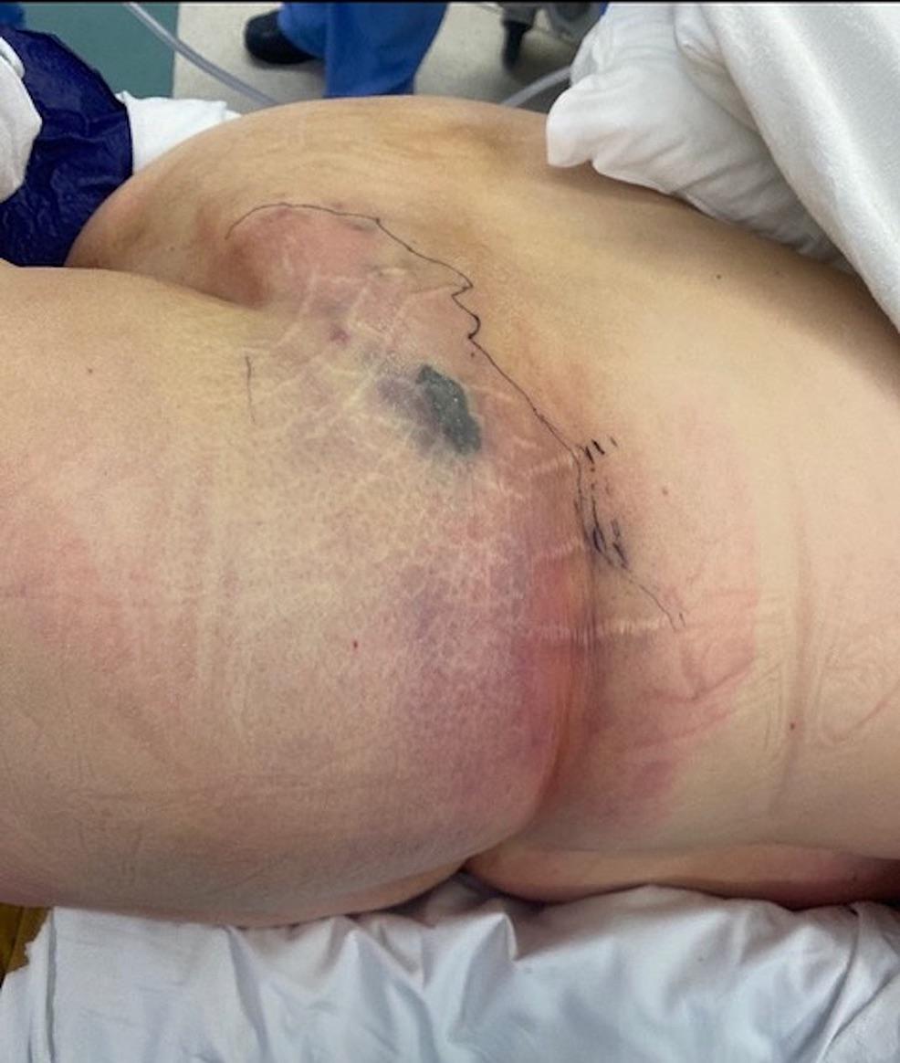 · 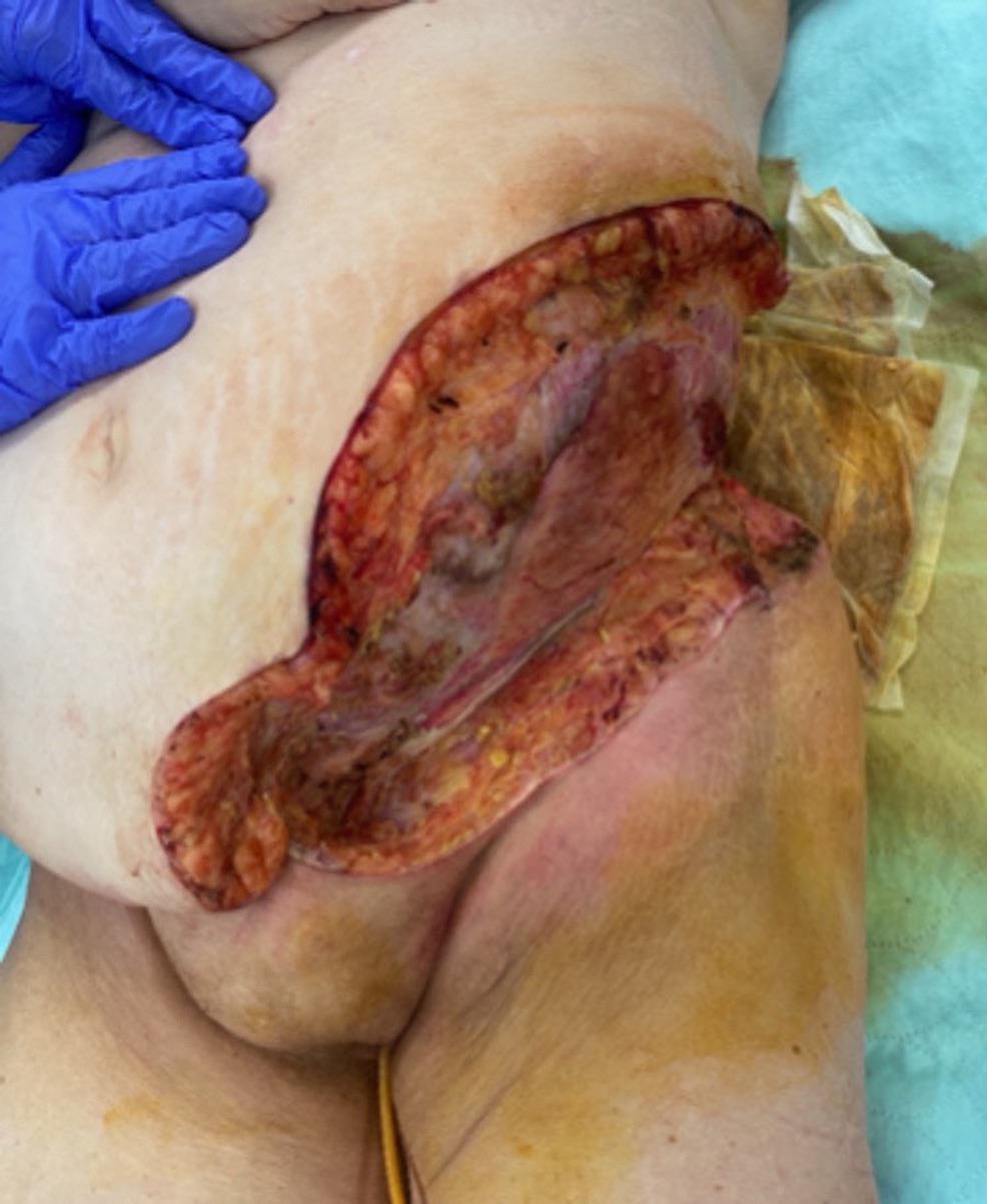 · 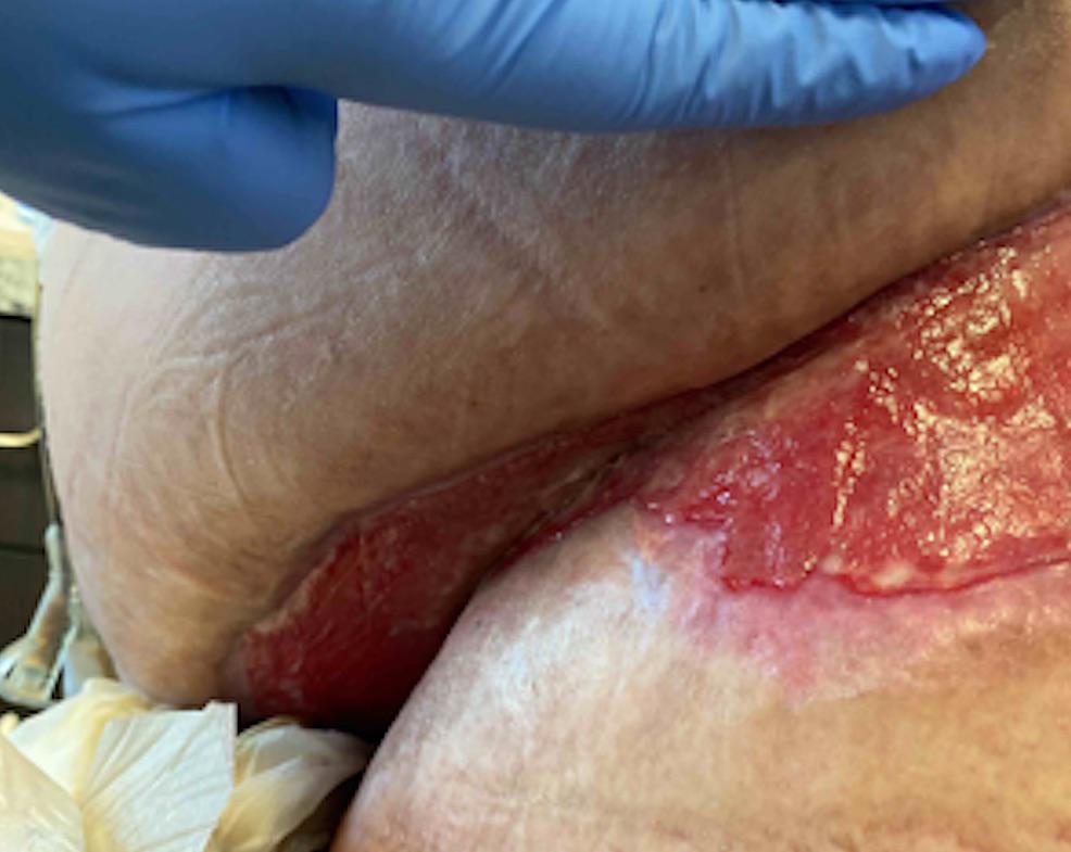 · 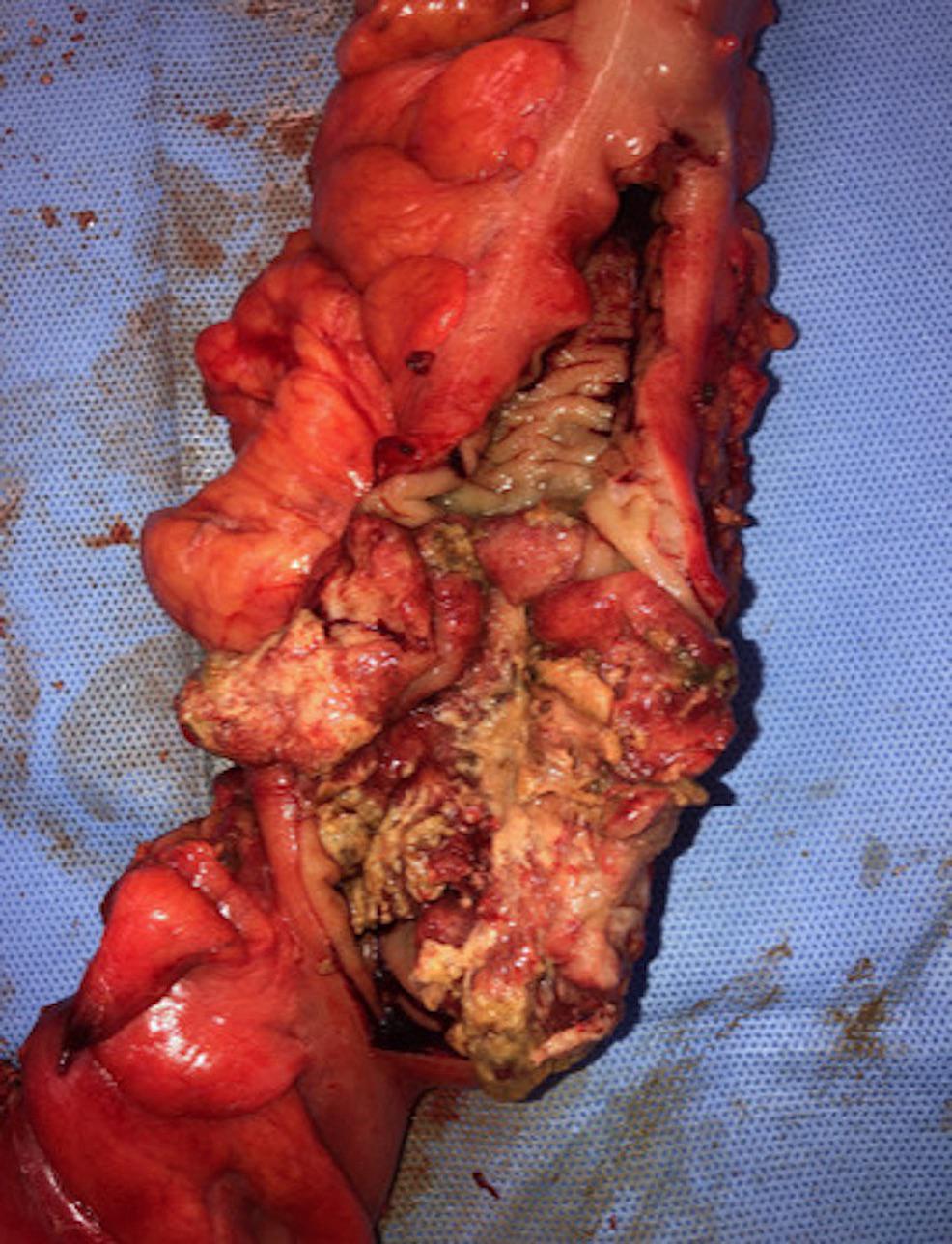 · |
|
#2
●
03-22-2023, 12:47 AM
| ||||||||
 My Rank: PRIVATE FIRST CLASS Poster Rank:3620 female Join Date: May 2015 Posts: 95 Mentioned: 0 Post(s) Quoted: 20 Post(s)
| ||||||||
|
Re: Necrotizing Soft Tissue Secondary to Colon Cancer
That’s insane!!
|