|
#1
●
03-14-2023, 10:42 PM
| ||||||||
 These are the rooms Poster Rank:26 of ruin. Join Date: Sep 2014 Posts: 54,011 Mentioned: 145 Post(s) Quoted: 30403 Post(s)
| ||||||||
|
Necrotizing Soft Tissue Infection: a Story in Pictures
Source: Our case is of a 48-year-old, African American female with a history of intravenous (IV) heroin abuse and multiple admissions for cellulitis and draining abscesses of the extremities in the past. She presented to the emergency room with 48 hours of body pain, aches, abdominal pain, and weakness. The patient endorsed using IV drugs earlier that day. She complained of severe pain in the right upper extremity. On examination, the patient was hypotensive to 80/60s mmHg and tachycardic to 120 beats/minute. The examination was also significant for multiple ulcerations on the bilateral upper and lower extremities. Most notably, two ulcers on the right forearm near the antecubital fossa were particularly tender (Figure 1). The right forearm was swollen compared to the left. There was no evidence of crepitus, fluctuance, or drainage. The compartments were soft and the pulses were palpable with a normal sensorimotor examination. Two sets of blood cultures were obtained. Further, fluid resuscitation with crystalloid and broad-spectrum antibiotics, vancomycin and piperacillin/tazobactam, were initiated immediately. Laboratory analysis revealed an anion gap metabolic acidosis with a pH of 7.19 and lactate of 6.9 mmol/L. She had an acute kidney injury (AKI) with a blood urea nitrogen (BUN) level of 77 mg/dL and creatinine of 6.5 mg/dL. The white blood cell (WBC) count was 1.67 × 10³/µL and the international normalized ratio (INR) was 1.63. She was admitted to the medical intensive care unit (ICU) for septic shock with multiorgan dysfunction. A surgical consult was obtained; however, due to the unclear nature of the disease and nonspecific symptoms, computed tomography (CT) without contrast of the right upper extremity was ordered. The CT showed nonspecific signs such as diffuse soft tissue swelling, fat stranding, and lymphadenopathy of the right medial upper extremity extending to the right axilla (Figure 2). On serial examination three hours later, the patient was noted to have a significant increase in pain to the right upper extremity out of proportion to the examination along with acute painful distress. Ecchymosis and purple skin changes had developed in the interval and were extending along the medial aspect of the upper extremity toward the axilla (Figure 3). She had developed a significant motor deficit in this interval, with 0/5 strength of the hand, forearm, and shoulder. She remained hypotensive and tachycardic and unresponsive to volume resuscitation and antibiotics, requiring norepinephrine (20 µg/kg/minute). With this change in the clinical picture, she was taken immediately to the operating room (OR) for radical debridement of the right upper extremity, with concern for an NSTI. An accurate LRINEC score could not be calculated at this time due to the lack of C-reactive protein (CRP) levels. Intraoperatively, the fascia was noted to be grossly healthy appearing, with no frank purulence noted. The findings were subtle, without obvious necrosis. The subcutaneous tissue and fat were very edematous with a slight gray tinge and easily separated with blunt dissection of a finger sweep. The muscles appeared grossly healthy, with edematous, albeit otherwise healthy-appearing fascia (Figure 4). The tissues were not bleeding normally, with multiple areas of venous thrombosis noted, and radical excision of the affected tissue was performed (Figure 5). Proximally, at the medial aspect of the upper arm, similar findings of the fat and subcutaneous tissues were noted and excised. The basilic vein was noted to be thrombosed. Distal pulses were successfully obtained with Doppler throughout the extremity. Fasciotomy of all muscle compartments of the forearm and upper arm was performed. After the initial radical debridement, the remaining tissues appeared healthy and bleeding (Figure 6). The patient was experiencing increasing vasopressor requirements despite two units of packed red blood cell (PRBC) transfusion and crystalloids; therefore, we dressed the wounds with betadine-soaked gauze and transferred her to the surgical intensive care unit (SICU) for further resuscitation. Over the next 16 hours, the patient briefly improved with decreasing pressor requirements and a downtrend of lactate to 3.5 mmol/L. Shortly after, she began to deteriorate again with increased pressor requirements and increased lactate to 5.9 mmol/L. The right upper extremity muscles and the neurovascular bundle was notably dusky and edematous. There were now skin changes extending from the axillary region onto the lateral chest wall (Figures 7, 8). She was immediately returned to the OR for further debridement. The shoulder skin and subcutaneous tissues were debrided and incisions extended to the lateral chest wall. The tissues were nonviable nonbleeding, and, subsequently, the lateral torso soft tissues were excised to the level of the iliac crest. Only at this point were the tissues firmly adherent, healthy-appearing, and bleeding. The rapid spread of the infection with subsequent involvement of the axilla and neurovascular bundle was thought to have compromised the entire right arm. The arm was frankly necrotic with multiple areas of venous thrombosis. The family was informed of the necessity for amputation as a life-saving procedure, and she agreed. A right shoulder disarticulation was performed (Figure 9). The left forearm was explored with two incisions, volar and dorsal, to explore ecchymosis that had developed in the interim, ruling out an additional source of infection. The tissues were noted to be healthy and bleeding. A diagnostic laparoscopy was performed to rule out any intraabdominal source as a CT abdomen/pelvis performed on admission showed nonspecific dilated loops of bowel with a possible transition point. There was no significant pathology or mechanical obstruction noted upon diagnostic laparoscopy, attributed to likely ileus. She was returned to the SICU for continued resuscitation. The pH, lactate, and vasopressor requirements began to improve. Blood cultures and surgical swab cultures returned positive for group A, beta-hemolytic S. pyogenes, indicative of a type 2 NSTI. 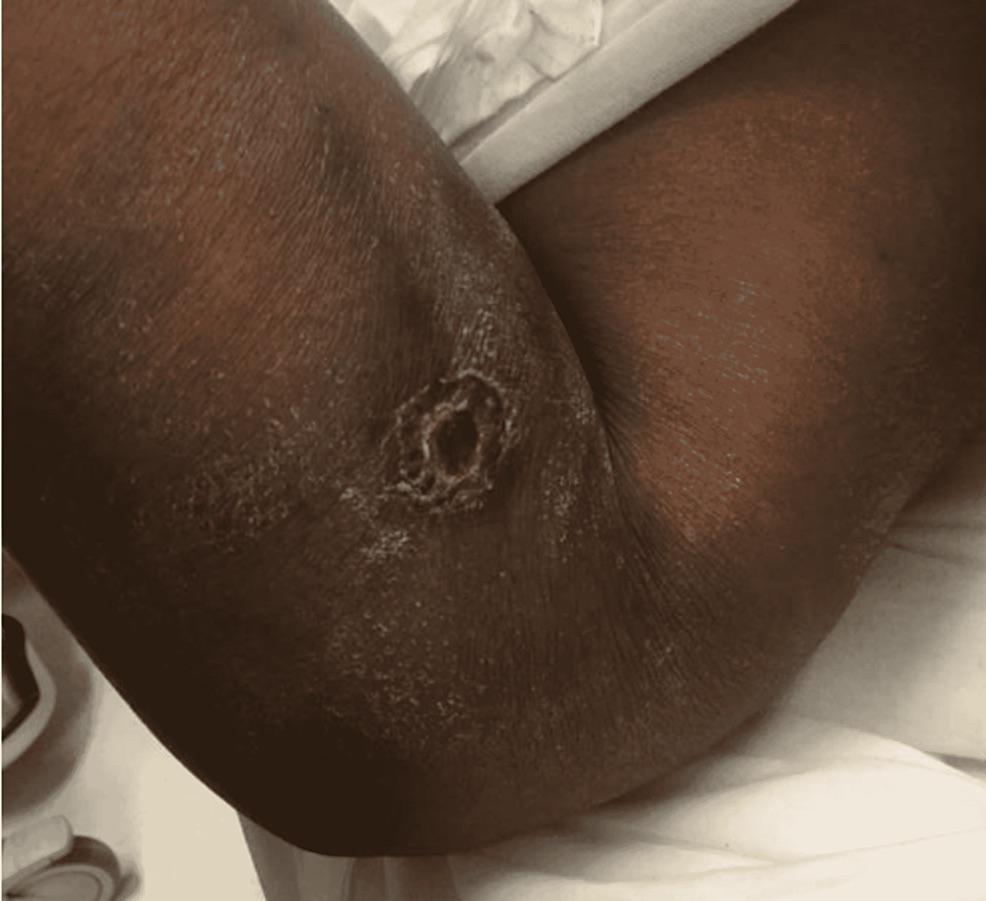 · 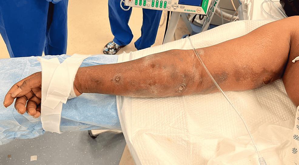 · 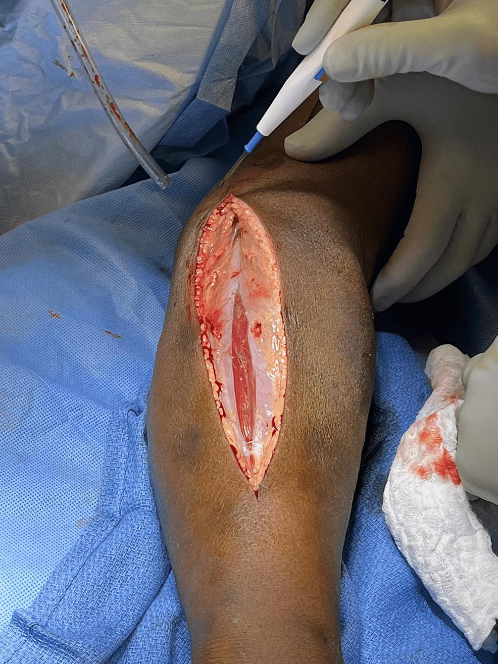 · 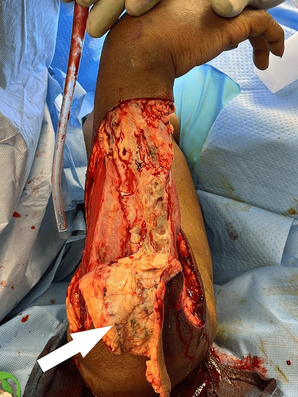 · 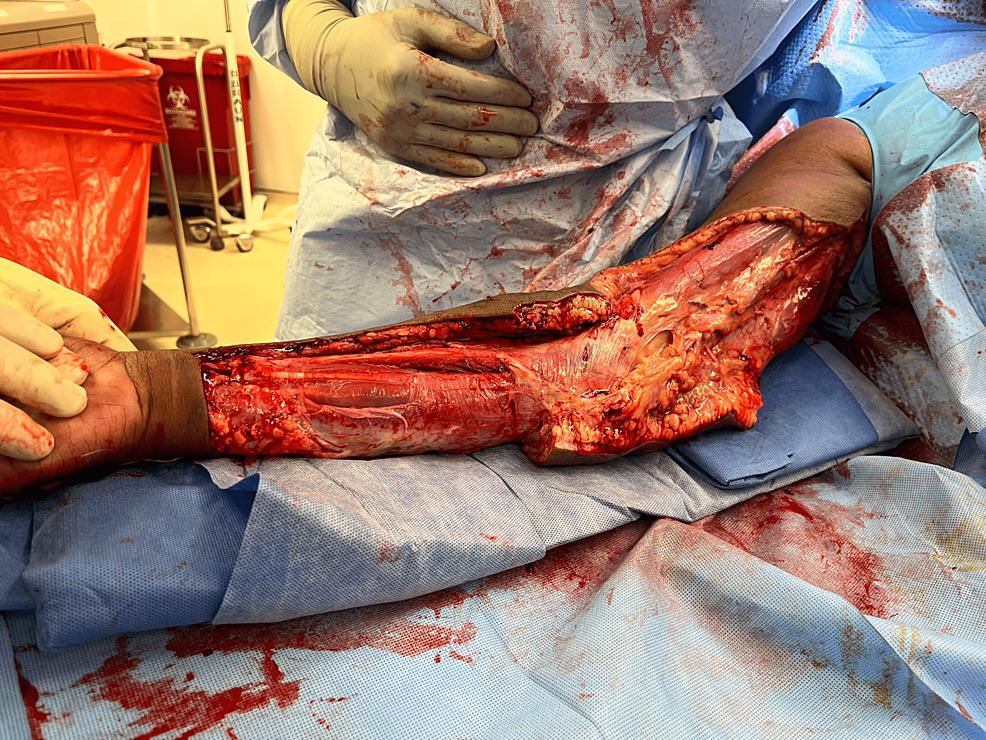 · 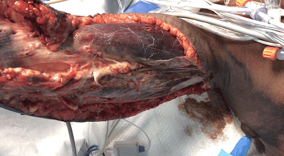 · 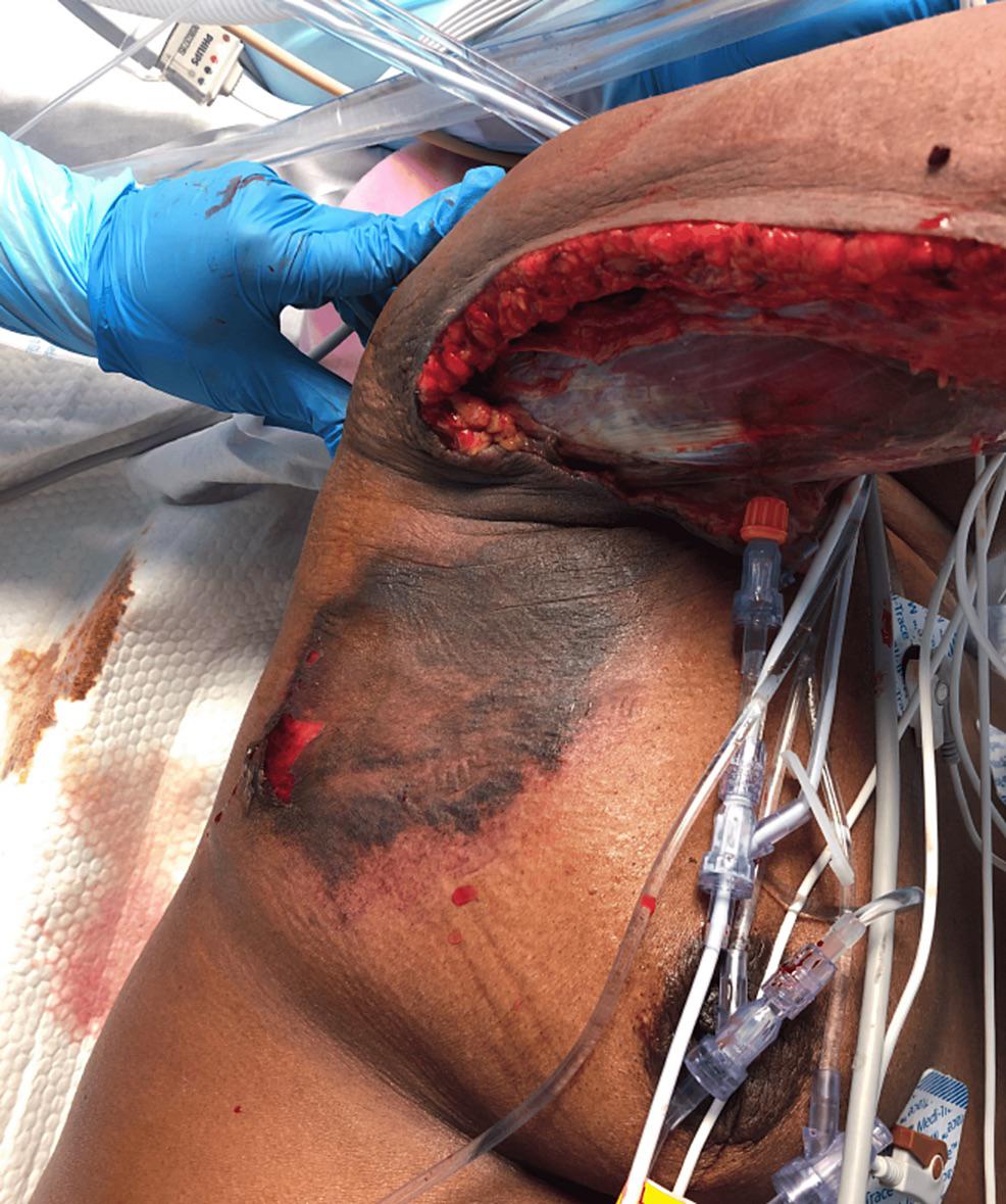 · 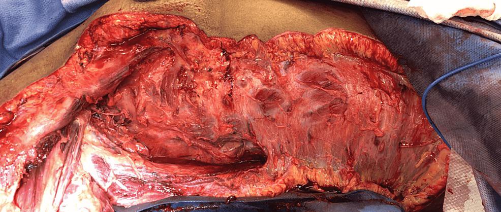 · 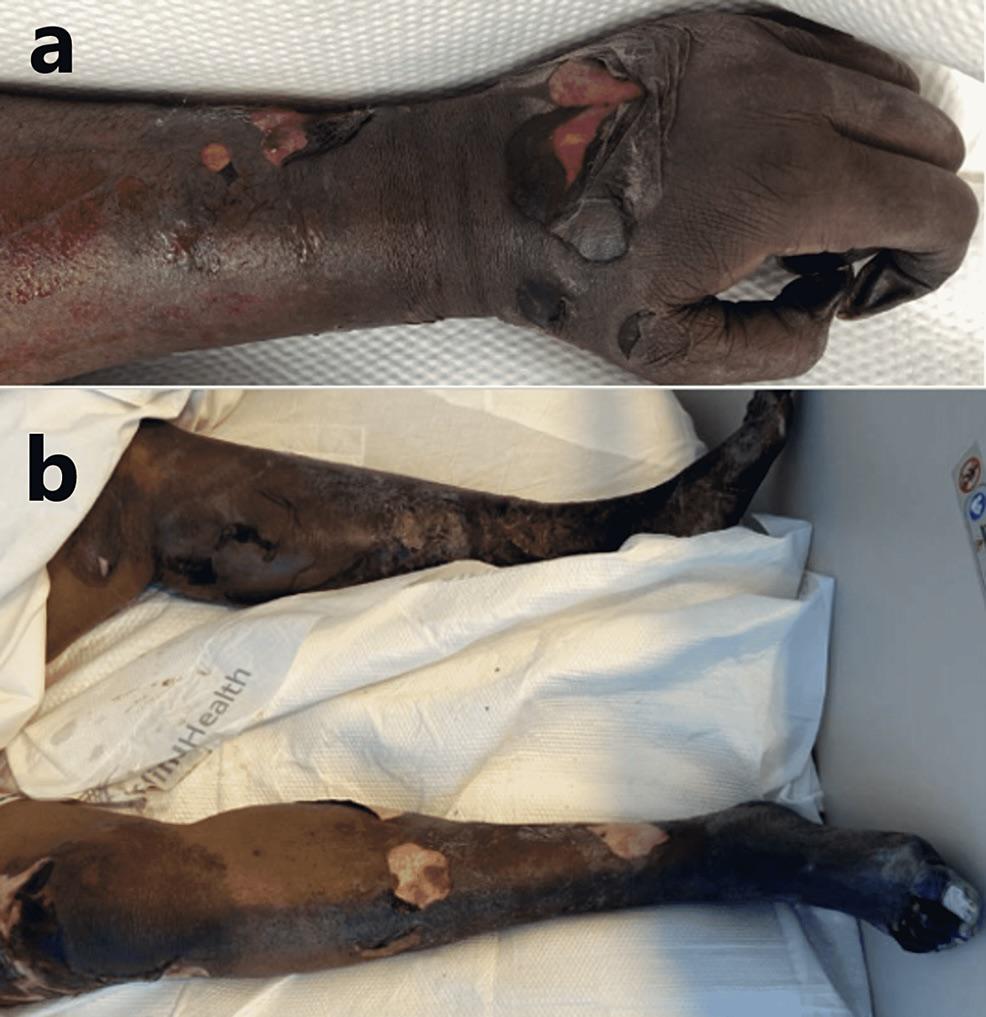 · 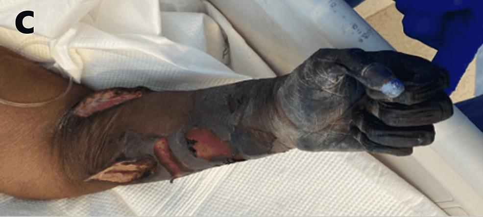 · 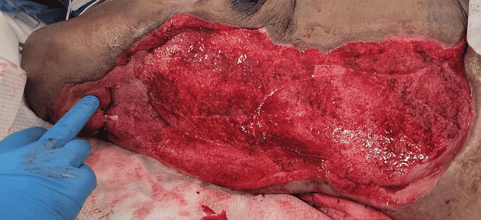 · 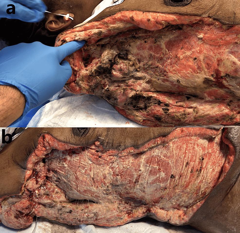 · |
|
#2
●
03-15-2023, 02:27 AM
| ||||||||
 ★ Legacy Member ★ Poster Rank:252 So many choices now Join Date: Jul 2015 Posts: 5,548 Mentioned: 15 Post(s) Quoted: 2120 Post(s)
| ||||||||
|
Re: Necrotizing Soft Tissue Infection: a Story in Pictures
There had to be a point at which she said to herself “Yup, I think I might have let this go a bit too long”.
|
|
#3
●
03-15-2023, 06:19 AM
| ||||||||
 The Devil's Left Hand Poster Rank:70 Male Join Date: May 2012 Posts: 22,207 Mentioned: 21 Post(s) Quoted: 9551 Post(s)
| ||||||||
|
Re: Necrotizing Soft Tissue Infection: a Story in Pictures
I wish my school library had picture books like this when I was a child.
__________________ Why so sad? Your tears are no match for mine. |
|
#4
●
03-15-2023, 06:29 PM
| ||||||||
| These are the rooms Poster Rank:26 of ruin. Join Date: Sep 2014 Posts: 54,011 Mentioned: 145 Post(s) Quoted: 30403 Post(s)
| ||||||||
|
Re: Necrotizing Soft Tissue Infection: a Story in Pictures
I always wonder because I know if I have a sore that never heals, I'm going to the doc.
|
|
#5
●
03-15-2023, 06:30 PM
| ||||||||
| These are the rooms Poster Rank:26 of ruin. Join Date: Sep 2014 Posts: 54,011 Mentioned: 145 Post(s) Quoted: 30403 Post(s)
| ||||||||
|
Re: Necrotizing Soft Tissue Infection: a Story in Pictures
We definitely should see shit like this at an appropriate stage.
|