|
#1
●
05-18-2024, 06:11 PM
| ||||||||
 These are the rooms Poster Rank:24 of ruin. Join Date: Sep 2014 Posts: 54,011 Mentioned: 145 Post(s) Quoted: 30403 Post(s)
| ||||||||
|
Necrotizing Fasciitis
>A 62-year-old female presented to the emergency department with complaints of abdominal pain and a decrease in urinary output. She has a past medical history of remote colon and endometrial cancers, chronic obstructions, colocutaneous fistulas, diabetes, hypertension, and atrial fibrillation (a-fib). Her surgical history consists of colostomy, right hemicolectomy, hysterectomy, and wound exploration related to the colocutaneous fistula. >>Upon assessment, she had ventral hernias with a leakage of foul-smelling fluid around her colostomy site. She presented with cellulitis of the entire pannus with some superficial epidermal blistering and necrosis, but the subcutaneous tissue appeared viable, as seen in Figure 1. The computed tomography (CT) scan revealed skin thickening and subcutaneous soft tissue stranding consistent with panniculitis/cellulitis with no drainable abscess. She was started on intravenous vancomycin and cefepime for the treatment of the cellulitis. >>Within 24 hours, her abdominal cellulitis had worsened despite receiving broad-spectrum antibiotics. The patient had increased tenderness with palpation to this area. Her white blood cell count (WBC) count increased from 10.2 on admission to 13.4. There was concern for a necrotizing soft tissue infection. She was taken to the operating room that day for emergent debridement. During the debridement, the skin around her hernias was left intact. The surrounding necrotic skin and fat were debrided to the fascia. Her peritoneum was not opened. Tissue samples were obtained, revealing Klebsiella pneumoniae, few Enterococcus faecalis, and Staphylococcus. After debridement, the wound was dressed with betadine-soaked gauze and sterile dressings. >>On post-op day (POD) one, the dressings were removed by the wound, ostomy, continence, nurse (WOCN) team. Upon removal of the dressings, an exposed bowel was noted (Figure 2). There was necrosis noted to the lateral aspects of the wound. The wound was packed with saline-moistened Kerlix and covered in abdominal pads (ABDs). An ostomy appliance was placed over the fistula. The patient was scheduled for further debridement the next day. >>On POD two, general surgery performed a wide debridement of the abdominal wall. The enterocutaneous fistula was taken down. >>On POD three, she returned to the operating room for another washout and an open abdomen negative pressure therapy (ABThera) was placed. General surgery placed an ABThera with each washout from POD three until POD seven. >>On POD seven, a washout was performed and vicryl mesh was placed over the exposed bowel. General surgery applied negative pressure wound therapy (NPWT) using white foam over the mesh followed by black foam. >>On POD nine, the patient was taken to the operating room for wound debridement of the skin and subcutaneous fat (Figure 3). After the procedure was completed, an NPWTi-d device was placed in the operating room, as seen in Figure 4. Four barrier rings were placed at the inferior and lateral wound edges to facilitate a good seal and prevent maceration. Four white foam pieces were placed to protect the area with mesh. A total of four kits consisting of both NPWTi-d and ROCF-CC were utilized. The instillation ports were Y connected to the trac pads and applied to four areas in the wound to improve instillation coverage and prevent leakage in the lateral aspects. The NPWTi-d settings were 130 milliliters (ml) of normal saline solution (NSS) to soak for 10 minutes every three and a half hours. Normal saline solution was chosen because it has been shown that despite its direct antimicrobial activity, it can be as effective as other solutions when used with NPWTi-d [6]. The settings for the device were based on the parameters set forth by the review of evidence and recommendations for a cycle frequency of two-four hours at -125 mmHg and a dwell time of 10-20 minutes [7]. >>On POD 11, the patient returned to the operating room for a washout and dressing change. The NPWTi-d with ROCF-CC was removed. General surgery placed NPWT utilizing white foam over the mesh and covered then with black foam. >>On POD 15, the first NPWTi-d change was performed at the bedside. The wound base had 40% pink tissue, 20% red tissue, and 40% slough, as seen in Figure 5. White foam was placed over the vicryl mesh. NPWTi-d was placed to the central portion. ROCF-CC was applied to the lateral aspects to assist in the removal of the non-viable tissue, as seen in Figure 6. The goal of therapy was to improve granulation tissue and prevent repeated trips to the operating room. >>The patient underwent four dressing changes at the bedside, utilizing NPWTi-d and ROCF-CC. On POD 22, ROCF-CC was discontinued. There was a decrease in devitalized tissue to the lateral aspects. New granulation tissue was present where the macrocolumns are located as seen in Figure 7. White foam was placed over the area with mesh followed by NPWTi-d. >>On POD 29, the final dressing change was performed utilizing NPWTi-d. The patient was deemed medically stable for discharge. The receiving facility was unable to accept the patient with NPWTi-d in place. She was transitioned to NPWT, utilizing white foam over the bowel and one NPWT machine. >>The patient spent a total of 31 days in the hospital. During this time, she had three ABThera applications, five applications utilizing NPWTi-d, and two applications with NPWT. There was a decrease in devitalized tissue and the wound edges were noted to have contracted. The patient’s measurements reduced from 32 x 54 x 5 cm (8,640 cmᵌ) to 19.5 x 53 x5 cm (5,167 cmᵌ). >>The patient was readmitted to our facility six days after her discharge for altered mentation and possible wound infection. There was concern for purulent drainage noted in the cannister. The dressing was changed the next day in coordination with the surgery and infectious disease teams. Once the dressing was removed, there were no obvious signs of infection. The wound base had 85% red tissue and 15% tan slough, measuring 16 x 54 x 3.5 cm (3,024 cmᵌ) (Figure 8). The patient underwent three dressing changes throughout her readmission. She was discharged to a rehabilitation center where they continued NPWT. 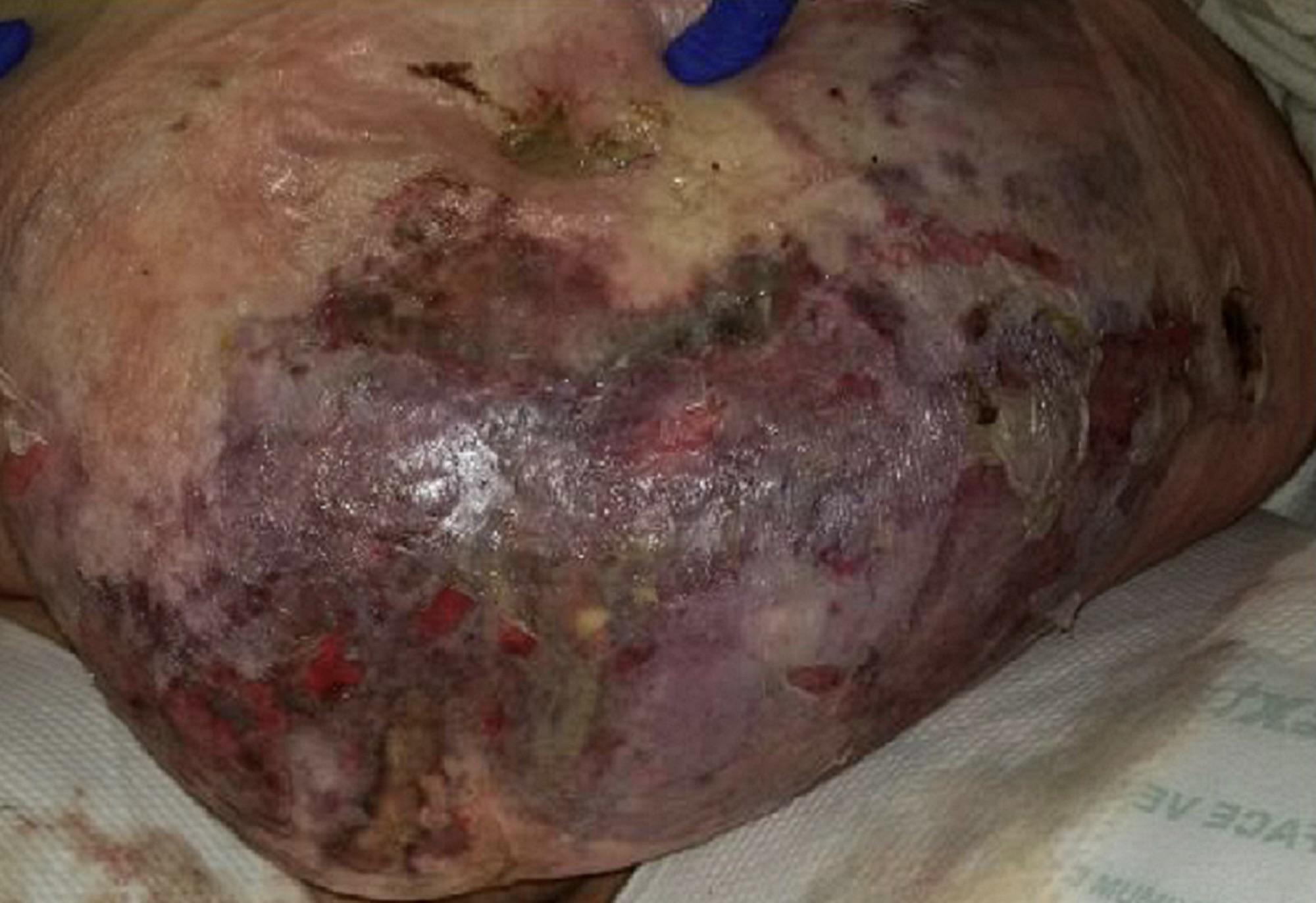 · 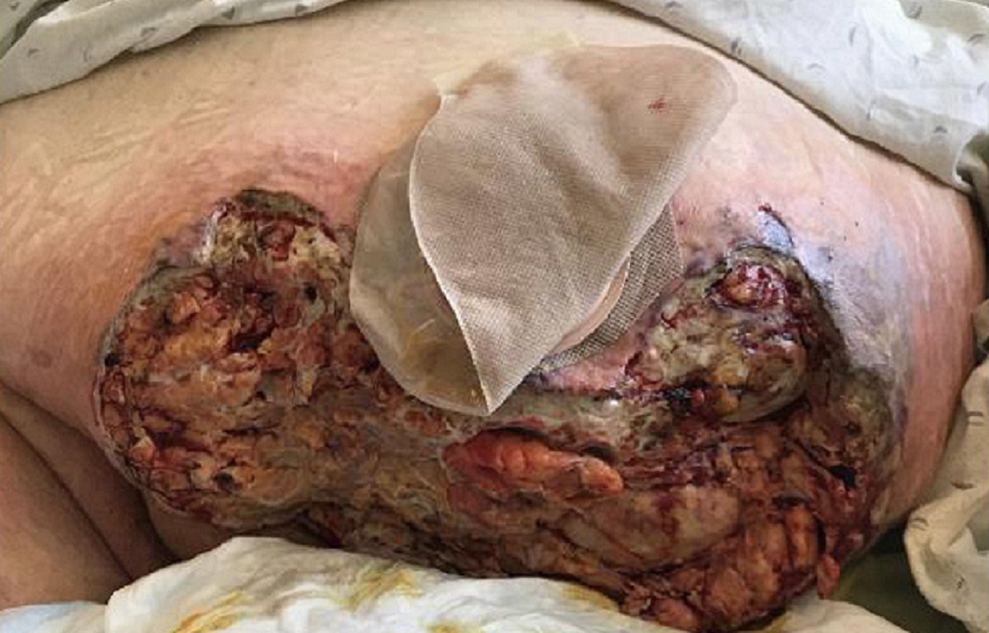 · 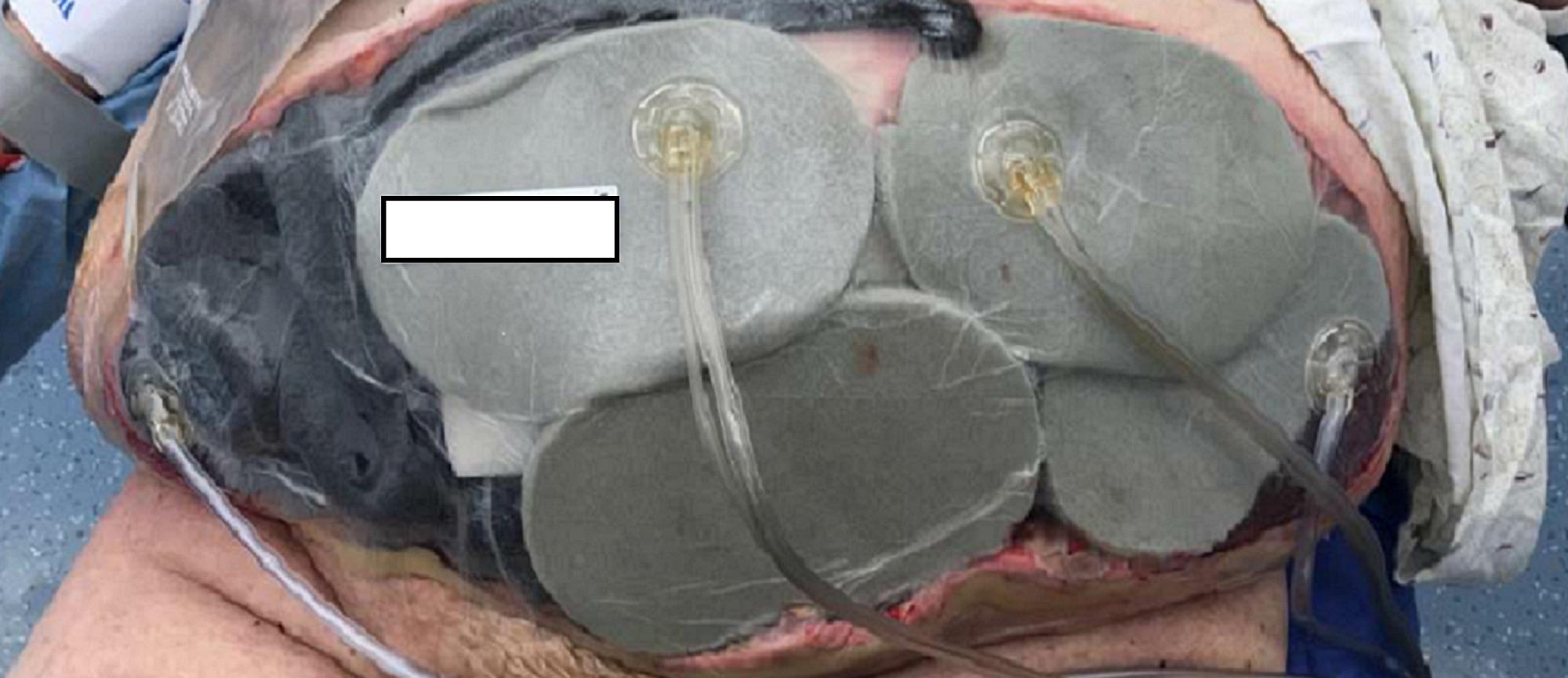 · 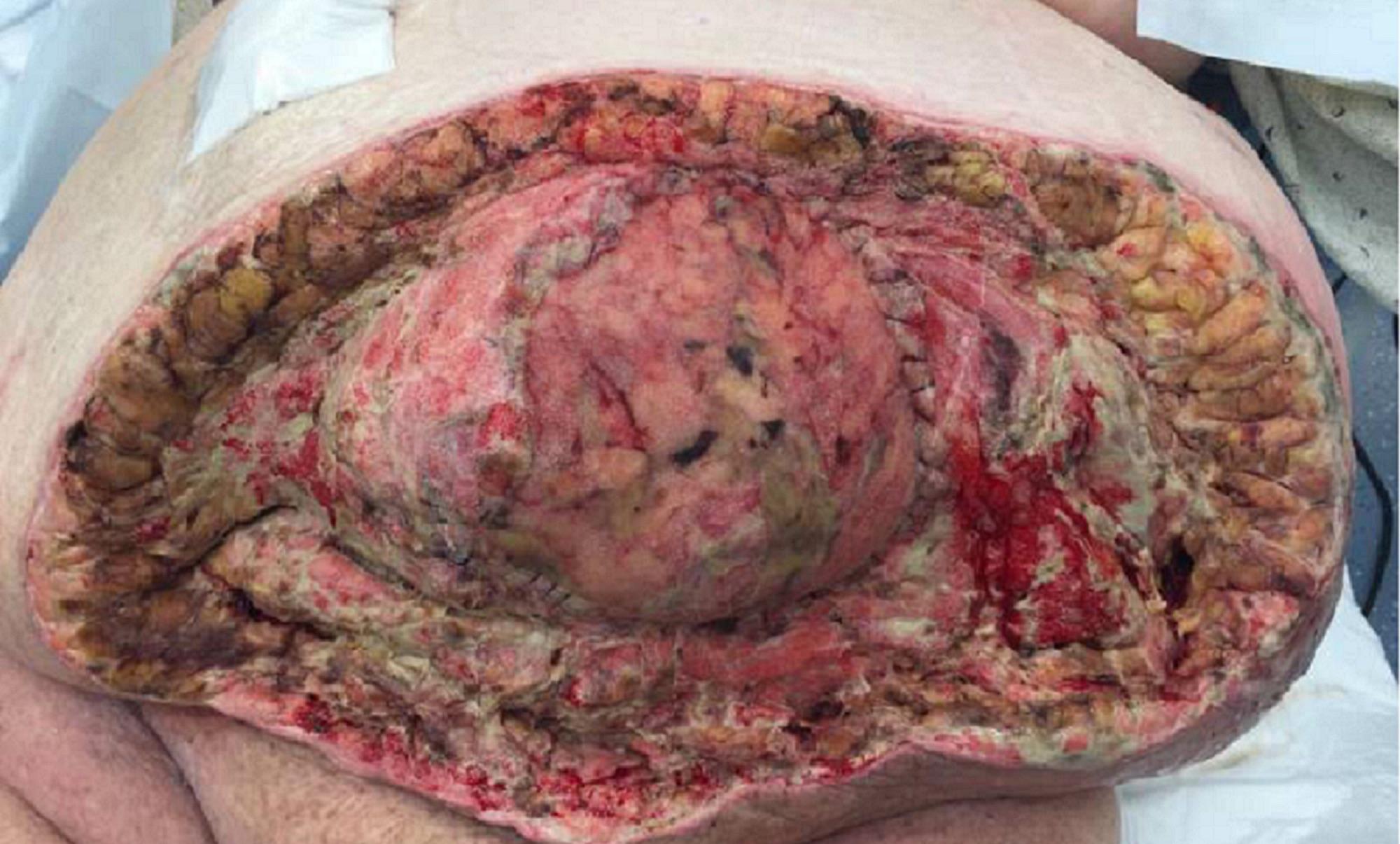 · 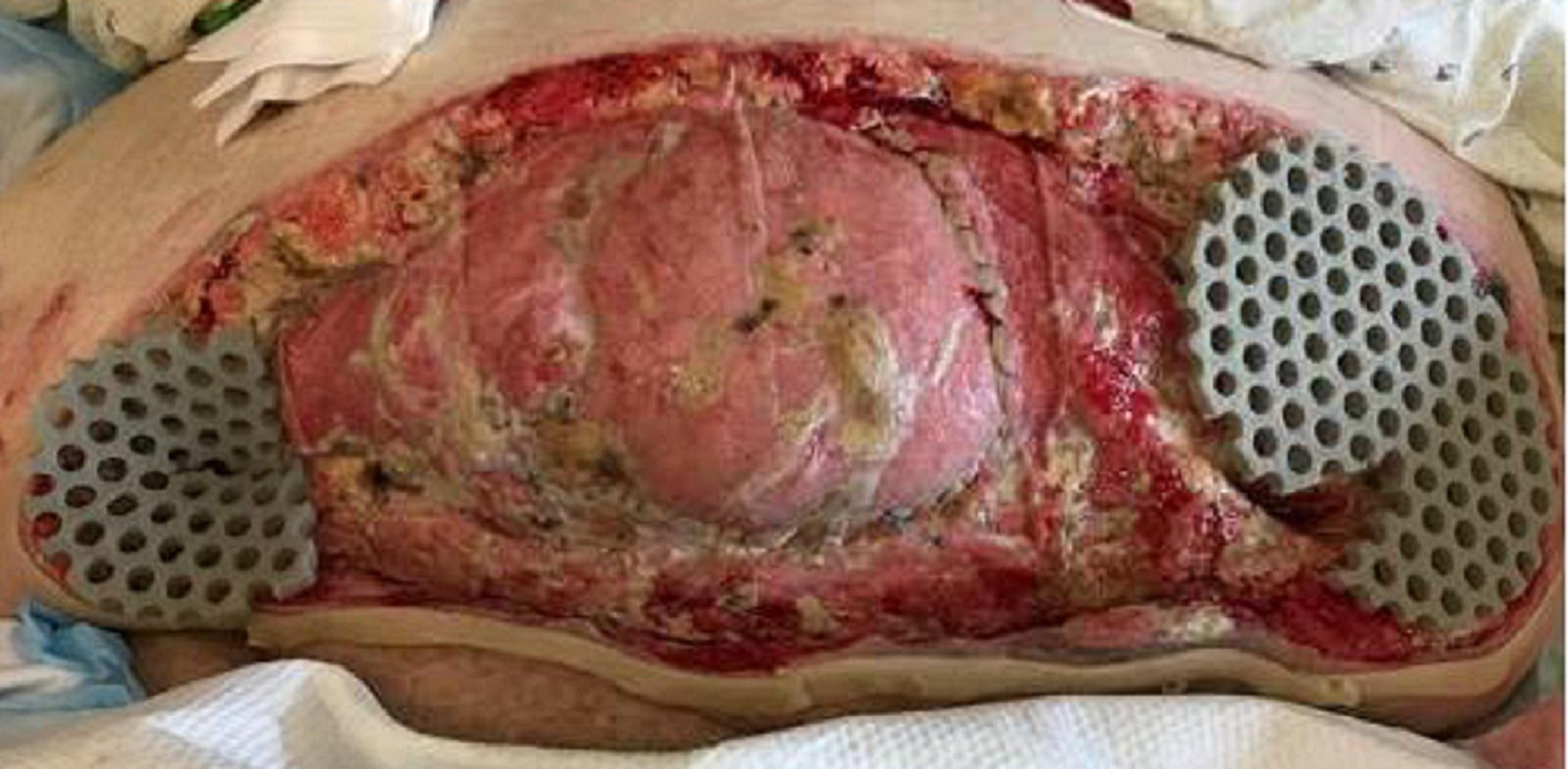 · 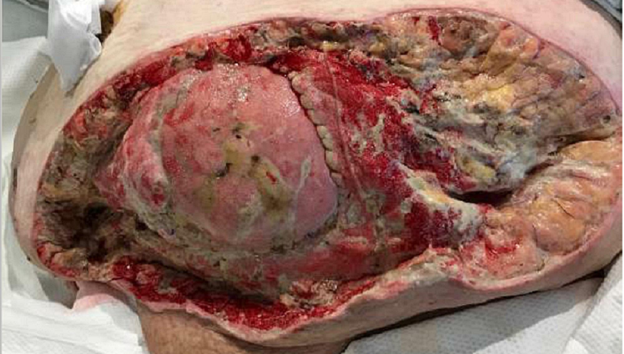 · 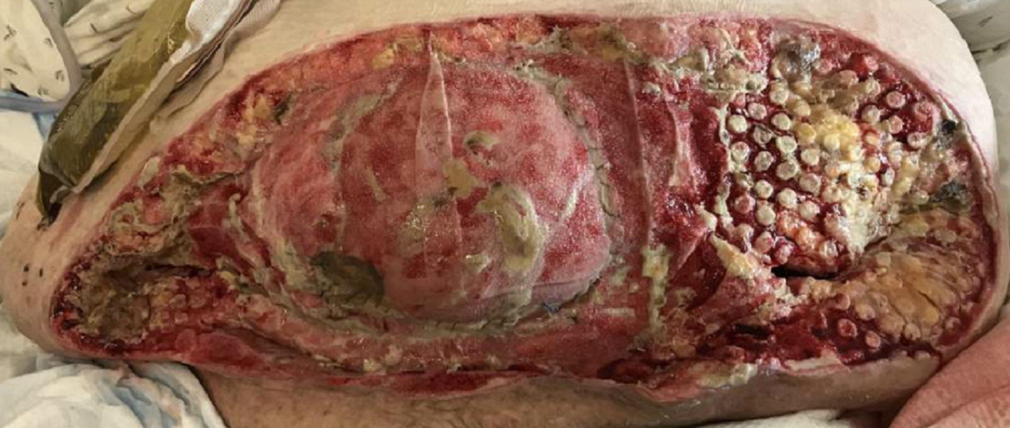 · 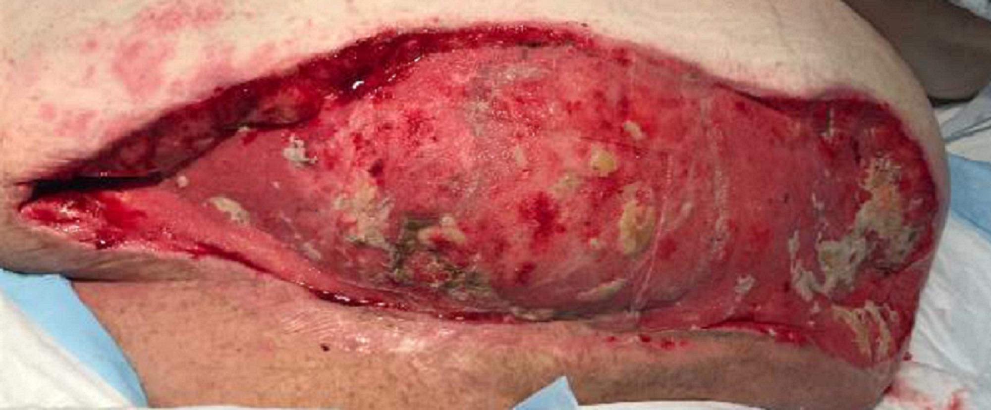 · |
|
#2
●
05-18-2024, 09:24 PM
| ||||||||
 ★ Legacy Member ★ Poster Rank:248 So many choices now Join Date: Jul 2015 Posts: 5,548 Mentioned: 15 Post(s) Quoted: 2120 Post(s)
| ||||||||
|
Re: Necrotizing Fasciitis
The addition of Air Filters (photo 5), obviously a performance upgrade, are a nice touch.
|