|
#1
●
10-28-2024, 07:56 PM
|
 These are the rooms Poster Rank:26 of ruin. Join Date: Sep 2014 Posts: 54,008 54,008Posts 87,658Thanked 30,403Quoted 145Mentioned 0Contrib. |
|
Nasty Pressure Wound
This happened in Italy: A 59‐year‐old Italian man, suffering from congenital paraplegia, reported the existence of a pressure ulcer in his left gluteal region for nearly 25 years. Four years prior to presentation at our outpatient clinic, he noticed the onset of an exuberant vegetating, smelly, and painful tumour close to the ulcer, with progressive growth since then (Figure 1). A skin biopsy showed an ulcerated squamous cell carcinoma. Computed tomography (CT) showed a wide involvement of the subcutaneous tissue, bilateral inguinal lymph nodes, perianal anatomical structures, anal sphincter muscles, coxofemoral joint, and bone. Surgery was excluded as well as radiotherapy as the presence of fistulae between urethra and adductor muscles. Indeed, the patient was initiated in February 2018 to a palliative chemotherapy (capecitabine 500 mg twice a day and vinorelbine 30 mg thrice a week). The patient is currently on therapy with partial control of the disease. A foam wound dressing is applied daily in order to better allow skin tissue restoration. Marjolin's ulcer is a cutaneous malignancy that arises in previously chronically inflamed skin or scar tissue.1 It may also develop from pressure ulcers.2 Marjolin's ulcer may be distinguished into acute and chronic forms. The term acute ulcer refers to malignant transformation occurring within 12 months from skin injury. Nevertheless, the average duration of the cancerous change growth, from the time of skin damage to malignant transformation, may last more than 30 years. The most common histological tumour type is squamous cell carcinoma, especially during diagnoses of scars and chronic wounds. Furthermore, in acute conditions, basal cell carcinoma is more common. Pressure ulcers frequently occur in paraplegic subjects: this condition may lead to the development of an immunocompromised district, which is a skin area more vulnerable than the rest of the body for genetic or acquired reasons, prone to developing opportunistic infections, tumours, or dysimmune reactions (often of granulomatous type).3 Sacral and iliac areas are frequently affected by pressure ulcers in paraplegic patients. These regions have extensive lymphatic drainage into the pelvis, which explains the frequent local and remote metastases. In the case of suspicions, multiple tissue specimens should be collected from the ulcer and its margin, minimising false negative results of histopathological examination.4 Magnetic resonance imaging and/or CT can assess the level and extent of destruction as well as the inflammation of tissues.5 In order to avoid local recurrence, it is necessary to perform wide local excision with a 2–5 cm margin of healthy tissue, with primary or delayed skin graft.5 Nevertheless, sometimes, the disease's extension does not allow surgery intervention; therefore, radiotherapy as well as chemotherapy may be useful in order to reduce the tumour size or to improve quality of life in patients with short life expectancy.1 The sequential events that have occurred to our patient may well be explained by the concept of immunocompromised district: paraplegia has favoured the induction of pressure ulcers, and these have favoured the development of the cancer. 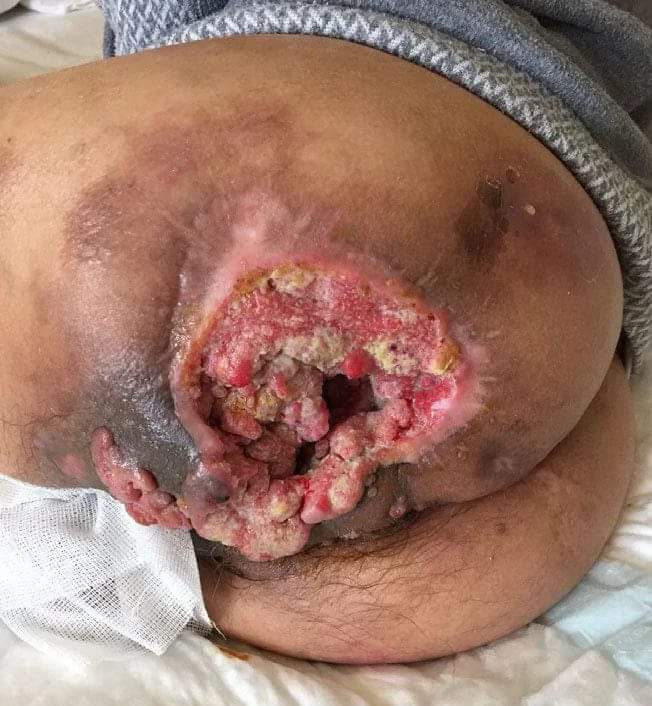 · |