She was aged 22 years. She had regular menstrual cycles of 3-4 days every 28-30 days. She conceived in 4 months of marriage and had first record of pregnancy with positive urine test for hCG at 42 days after the last menstruation. She first reported at 11 weeks 4 days of pregnancy with vaginal bleeding of 15 days duration and lower abdominal pain. Her blood pressure was 140/80 mm Hg and the uterus was palpable in hypogastrium to be of 14 weeks size. Abdominal ultrasound done at that time showed three gestational sacs and only one with fetal pole.
Estimation of beta human chorionic gonadotrophin (hCG) done was 1, 81,759 mIU/mL. Even after 3 weeks of observation on progesterone support with micronised progesterone and folic acid supplementation, vaginal bleeding persisted. At 14 weeks 2 days she noticed and complained of passing ‘grape like vesicles'. The uterus was enlarged to 18-20 weeks size; the blood pressure was 110/70 mm Hg. Repeat ultrasound examination revealed only two gestational sacs, the posterior one with 14 weeks live fetus and the other having vesicular mole (Fig.2). The serum beta hCG was 5, 21,698 mIU/mL.
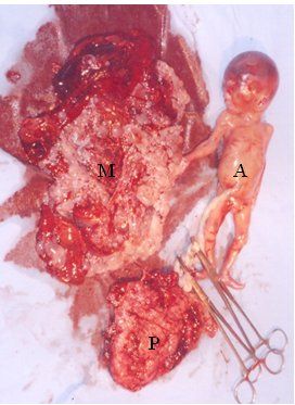
At 16 weeks 2 days, she had a miscarraige. First the molar pregnancy was expelled which weighed 500g. It was followed 20 minutes later by a live male fetus with no grossly detectable anomalies, weighing 250g and a placenta of 100g.(Fig.3).Serum beta hCG at this time was 3, 98, 100 mIU/mL and repeat estimate after a week was 3, 10, 500 mIU/mL.