
|
#1
●
08-14-2011, 04:37 PM
| ||||||||
 My Rank: MAJOR Poster Rank:12 Join Date: Jun 2009 Posts: 93,128
Contributions: 226
Mentioned: 91 Post(s) Quoted: 2009 Post(s)
| ||||||||
|
Large Teratoma of the Placenta
A 26-year-old Malay female at 31 weeks of pregnancy with abdominal pain after riding on a motorcycle. However, there was no uterine contraction, decreased fetal movement or per vaginal bleeding. There was no relevant past medical or surgical history. She weighed 44.5 kg (non-pregnant) and 56 kg (pregnant). Her blood pressure was 110/74 mmHg. Ultrasound examination of the abdomen revealed a solid cystic mass attached to the placenta but separated from the fetus. The mass measured 7.8 x 3.8 cm. At 39 weeks of gestation, she gave birth vaginally via vacuum assistance to a healthy baby boy weighing 2500 grams. The Apgar scores were 8 and 9 at 1 and 5 minutes. Gross examination showed a mass attached to the placenta which was arising within the fetal membrane. The mass measured 8.5 x 7.5 x 5 cm and has a cleavage at the center separating it into 2 ovoid structures. It was covered by well-formed skin with presence of hair. Cut section showed cystic and solid areas. The cystic spaces were filled with clear fluid and the wall thickness measured 0.1 to 0.2 cm. The cut surface of solid area was yellowish and glistening. The placenta appeared unremarkable with presence of an umbilical cord. 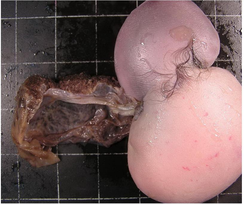  Microscopic examination revealed cyst wall which was lined by stratified squamous epithelium with underlying dermis and skin adnexal like sebaceous glands and hair follicles. The solid mass was composed of myxoid dermis, adipose tissue, nerve bundles and blood vessels. There was no organised bony skeleton, lung, brain, liver, intestine and genitourinary structures. The placenta was histologically unremarkable. 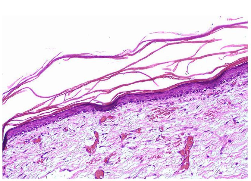 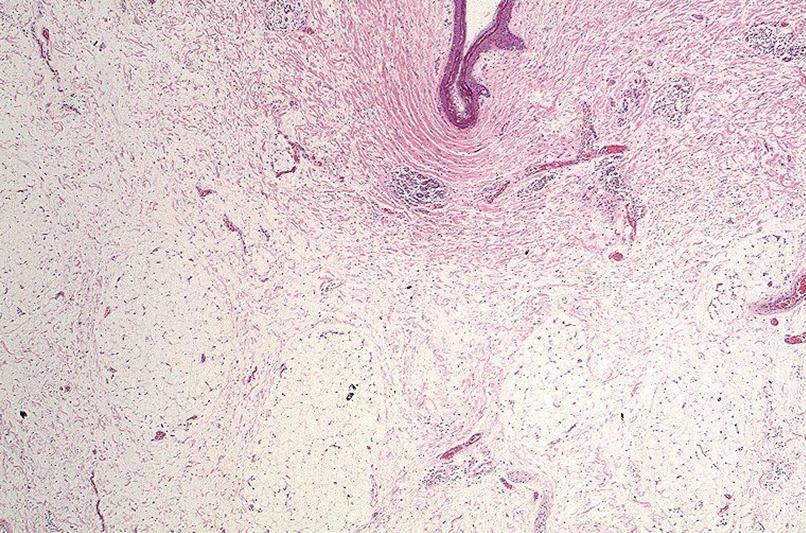 |





 Was that an unformed baby? The hair! Great post


