|
#1
●
01-26-2023, 02:11 AM
| ||||||||
 These are the rooms Poster Rank:26 of ruin. Join Date: Sep 2014 Posts: 54,011 Mentioned: 145 Post(s) Quoted: 30403 Post(s)
| ||||||||
|
Inflammatory Breast Cancer
Source. This case is out of Miami, Florida: A 60-year-old female presented to the emergency department with right-sided hip pain. Physical examination demonstrated bilateral, asymmetric, tender, immobile breast masses with nipple retraction, peau d'orange appearance, skin hyperpigmentation, and frank ulceration (Figure 1). Bloody nipple discharge was present on the left breast. Palpable nodules were present in the bilateral axillary region. In addition, innumerable superficial verruca-like skin nodules were found involving both breasts, extending to the supraclavicular region and to the periumbilical region in the mid-abdominal wall (not shown). Initial pelvic X-ray demonstrated multiple lytic lesions. This prompted further investigation with contrast-enhanced computed tomography (CECT) imaging. CECT of the chest revealed bilateral severe diffuse thickening of the skin overlying both breasts associated with extensive infiltrative fibroglandular densities and nipple retraction, findings supportive of diffuse inflammatory breast disease. Pathologically enlarged metastatic left-sided axillary lymphadenopathy and multiple small scattered breast masses in the upper inner quadrant of the left breast were also observed. CECT of the pelvis demonstrated multiple lytic lesions in the bony pelvis and the right femur. Multiple mixed lytic and blastic lesions to the thoracolumbar spine were also observed. A positron emission tomography-computed tomography (PET-CT) demonstrated infiltrative bilateral breast masses, axillary adenopathy, and diffuse bony metastasis, supporting the previous CECT findings. The PET-CT revealed multiple fluorodeoxyglucose (FDG)-avid bone lesions present throughout the spine, pelvis, sacrum, and proximal appendicular skeleton. Metastatic adenopathy was identified in the left supraclavicular and bilateral axillary lymph nodes. Bilateral breast pathology revealed triple-positive infiltrating ductal carcinoma with dermal lymphatic involvement. After a review of the clinical, radiological, and histological evidence, a diagnosis of stage IV bilateral inflammatory breast cancer was established. 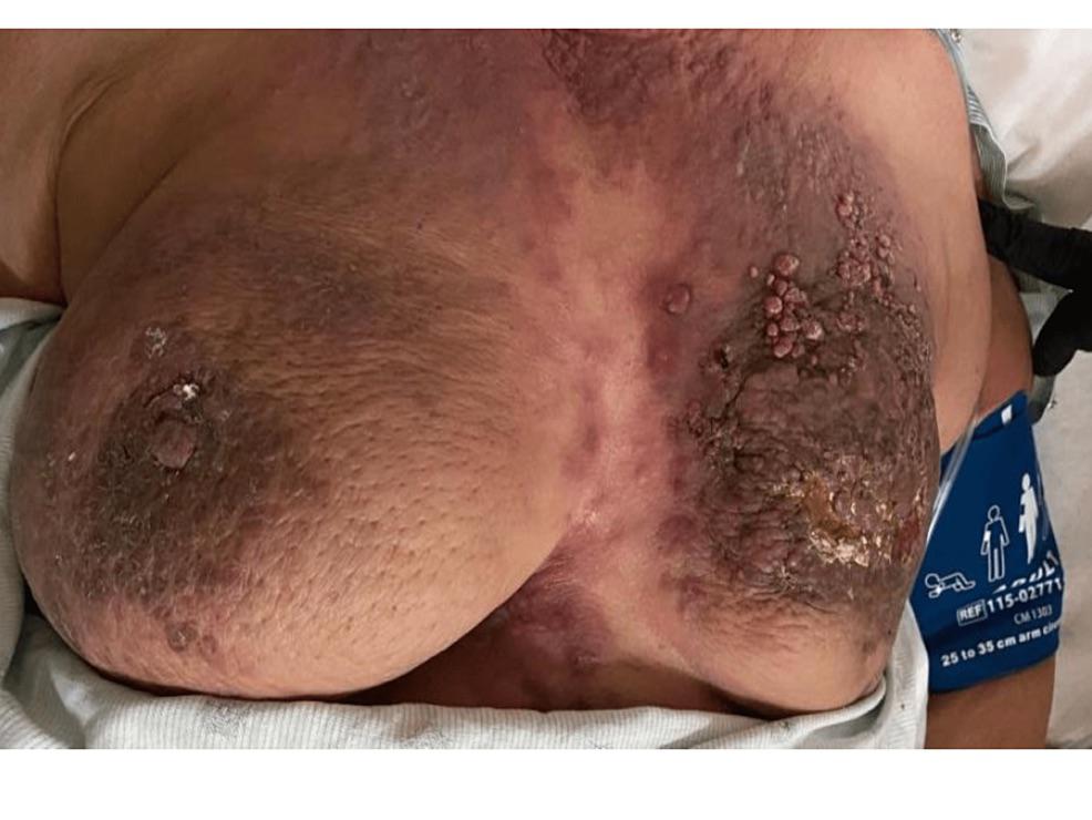 · 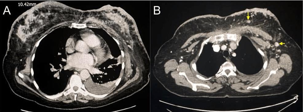 · |
|
#2
●
01-26-2023, 06:57 AM
| ||||||||
 The Devil's Left Hand Poster Rank:70 Male Join Date: May 2012 Posts: 22,207 Mentioned: 21 Post(s) Quoted: 9551 Post(s)
| ||||||||
|
Re: Inflammatory Breast Cancer
Looks like grandma should have used a bit more sunscreen during all those years spent on the topless beach.
__________________ Why so sad? Your tears are no match for mine. |
|
#3
●
01-26-2023, 03:34 PM
| ||||||||
 My Rank: SERGEANT Poster Rank:1028 Male Join Date: Jan 2010 Posts: 670
Contributions: 1
Mentioned: 1 Post(s) Quoted: 148 Post(s)
| ||||||||
|
Re: Inflammatory Breast Cancer
I don't understand how someone can let something get this bad before presenting to A&E?  |