|
#1
●
07-10-2010, 11:56 PM
| ||||||||
 My Rank: CORPORAL Poster Rank:1631 Male Join Date: Mar 2010 Posts: 340 Mentioned: 0 Post(s) Quoted: 0 Post(s)
| ||||||||
|
Human Botfly In Eye!
Could be a repost and i bet a ton of people knew about this, but i just found about it after i saw the botfly removal vid and this was fucking crazy. 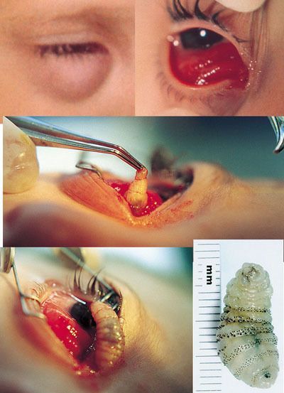 "Myiasis" is the medical term for a maggot (fly larva) infestation of a living body. In this case, the patient was a 5-year-old boy treated by U.S. Air Force surgeons in a rural area of the Republic of Honduras. "The respiratory pore of a late-stage larva of the human botfly (Dermatobia hominis) was located in the anterior orbit," says the article abstract. "The larva was gently removed under general anesthesia through a small incision in the conjunctiva." The patient was apparently none the worse for wear. According to entomologists, the human botfly lays its eggs on the bodies of other insects (such as mosquitoes), which then transfer the eggs to animal or human hosts by direct contact. When a botfly egg hatches, the larva burrows into the host's skin head-first and begins feeding. This nasty creature is found mainly in Central and South America, but there are other species of flies known to responsible for cases of myiasis in North America, mainly blowflies. According to an epidemiological study conducted in 2000, most instances of myiasis acquired in the United States are the result of blowflies laying their eggs in pre-existing wounds. Capt Randall L. Goodman, USAF, MC; Col Michael A. Montalvo, USAF, MC; Maj J. Brian Reed, USAF, MC; LTC Frank W. Scribbick, USA, MC; Chad P. McHugh, MPH, PhD; Randall L. Beatty, MD; Ricardo Aviles, MD A 5-YEAR-OLD boy with inferior orbital swelling and an erythematous mass arising from the inferior cul-de-sac of his right eye (Figure 1 and Figure 2 to the right) was seen by an Air Force Mobile Ophthalmic Surgical Team working in a rural area of the Republic of Honduras. The respiratory pore of a late-stage larva of the human botfly (Dermatobia hominis) was located in the anterior orbit. The larva was gently removed under general anesthesia through a small incision in the conjunctiva (Figure 3, Figure 4, and Figure 5 to the right). Figure 1. Five-year-old Honduran boy with right inferior orbital swelling and erythema. Figure 2. Examination under anesthesia, demonstrating right eye chemosis and inferior cul-de-sac mass. Figure 3. A large white larva being removed from the right anterior orbit through a conjunctival incision. Figure 4. Complete late-stage human botfly larva immediately after removal. Figure 5. Based on the morphology of the anterior and posterior spiracles and the exterior spines, the larva was determined to be a mature larva of the human botfly (Dermatobia hominis). |
|
#2
●
07-11-2010, 01:15 AM
| ||||||||
 My Rank: SERGEANT Poster Rank:990 transylvanian concubine Join Date: Dec 2009 Posts: 717 Mentioned: 0 Post(s) Quoted: 4 Post(s)
| ||||||||
|
Re: Human Botfly In Eye!
My ex used to be an army medic and was blessed to be the one to get to remove one of those things from some guy's back. G-ross.
|