|
#1
●
07-14-2024, 05:24 PM
|
 These are the rooms Poster Rank:26 of ruin. Join Date: Sep 2014 Posts: 54,008 54,008Posts 87,658Thanked 30,403Quoted 145Mentioned 0Contrib. |
|
Hole in the Face
The second case involves the orbit and the middle face, diplopia, erosion of the medial wall and orbital floor, bare medial and inferior extraocular muscles, loss of the conjunctiva, lagophthalmos, epiphora, and enophthalmos. A 43-year-old female was referred to the Nephrology Department with the diagnosis of GPA. She had a history of left mastectomy due to breast cancer, followed by chemotherapy and radiotherapy. She still takes tamoxifen. Subsequently, she had a lung tumor resection with wound healing difficulties. The biopsy of the wound tissues led to the diagnosis of GPA. The activity of the disease encompassed sinus involvement, palate–sinus fistula, conjunctivitis, inflammation of the left orbital tissue, hearing loss in the left ear, and an inflammatory tumor of the left lung. Additional sinus biopsy confirmed typical granulomas with PR3-ANCA-positive serology. The destruction of the left side of the nasal cavity was present at the initial presentation, along with necrotizing tissues covering the margins of the healthy tissues (Figure 6). The medial wall of the orbit was partially preserved. Imaging confirmed the involvement of the nasal and orbital tissues with significant destruction of bony structures as well as a large tumor (90 × 47 × 42 mm) with multiple smaller ones in the right lung. The lesions in the middle face were extremely painful for the patient. The treatment with CYC was initiated, with i.v. administration once a month. After the fifth dose, we still observed progression, so a decision to introduce an additional agent was made. Computed Tomography (CT) of the orbit (Figure 7) revealed the destruction of the medial orbital wall with an inflammatory involvement of the medial rectus and inferior rectus muscles with the limitation of fat tissue in the orbit. The sinuses were open, communicating with the nasal cavity. From the clinical point of view, the structures of the orbit were seen through the nose, the muscles were bare, and the medial part of the orbit was not covered with any tissue; no conjunctiva was present (Figure 8 and Figure 9). Moreover, the middle and lower parts of the respiratory tract were involved (lesions confirmed by bronchoscopy), and the patient presented dyspnea and chronic cough. The progression was confirmed clinically and with multiple imaging modalities, despite the intensive immunosuppressive treatment. After six months of CYC, RTX, a biologic agent, was added to the standard scheme of therapy inducing remission, administered in a weekly dose during a 1-day inpatient clinic stay (four doses). Following this therapy, she was reassessed by an ophthalmologist. She had diplopia when looking up, tearing, redness, and sometimes purulent discharge. Her visual acuity was logMar 0.0 in the RE and logMar 0.2 in the LE, which was involved. The left eyeball was situated slightly lower than the RE, and enophthalmos was diagnosed due to massive destruction of the medial wall and the orbital floor. No conjunctiva was present near the foci of necrosis. The sensation was impaired in the area of the lower eyelid. Bare medial and inferior rectus muscles were seen through the nasal cavity (Figure 8). The eyeball itself was well preserved but required intensive use of artificial tears. CT re-examination of facial structures revealed a severe progression in bony destruction. MTX was administered. Until now, the patient has been constantly monitored. Achieving remission is very difficult in such a severe case. As soon as remission is established, surgical reconstruction of the middle face will be performed. The outcome of the initial surgery is presented in Figure 10. It should be noted that the eyeball lost its bony support, and hypotropia and enophthalmos of 4 mm are present. Also, the lower eyelid does not have enough support, contributing to lagophthalmos and its negative consequences for the surface of the eye. From the ophthalmic point of view, the progression of the malposition of the eye and the eyelid was observed in the follow up. There is a relatively high risk of displacement of the affected eye into the left orbit. 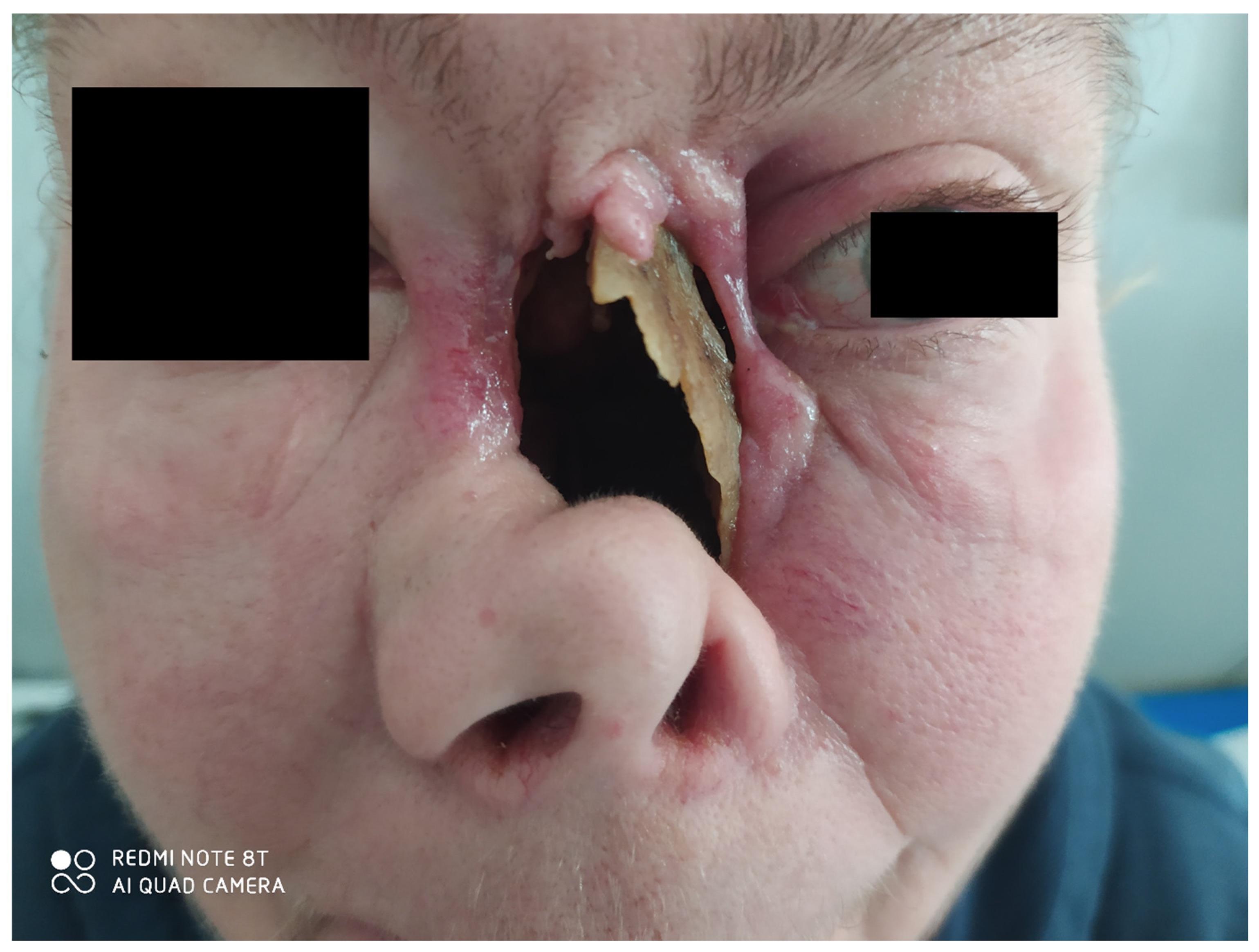 · 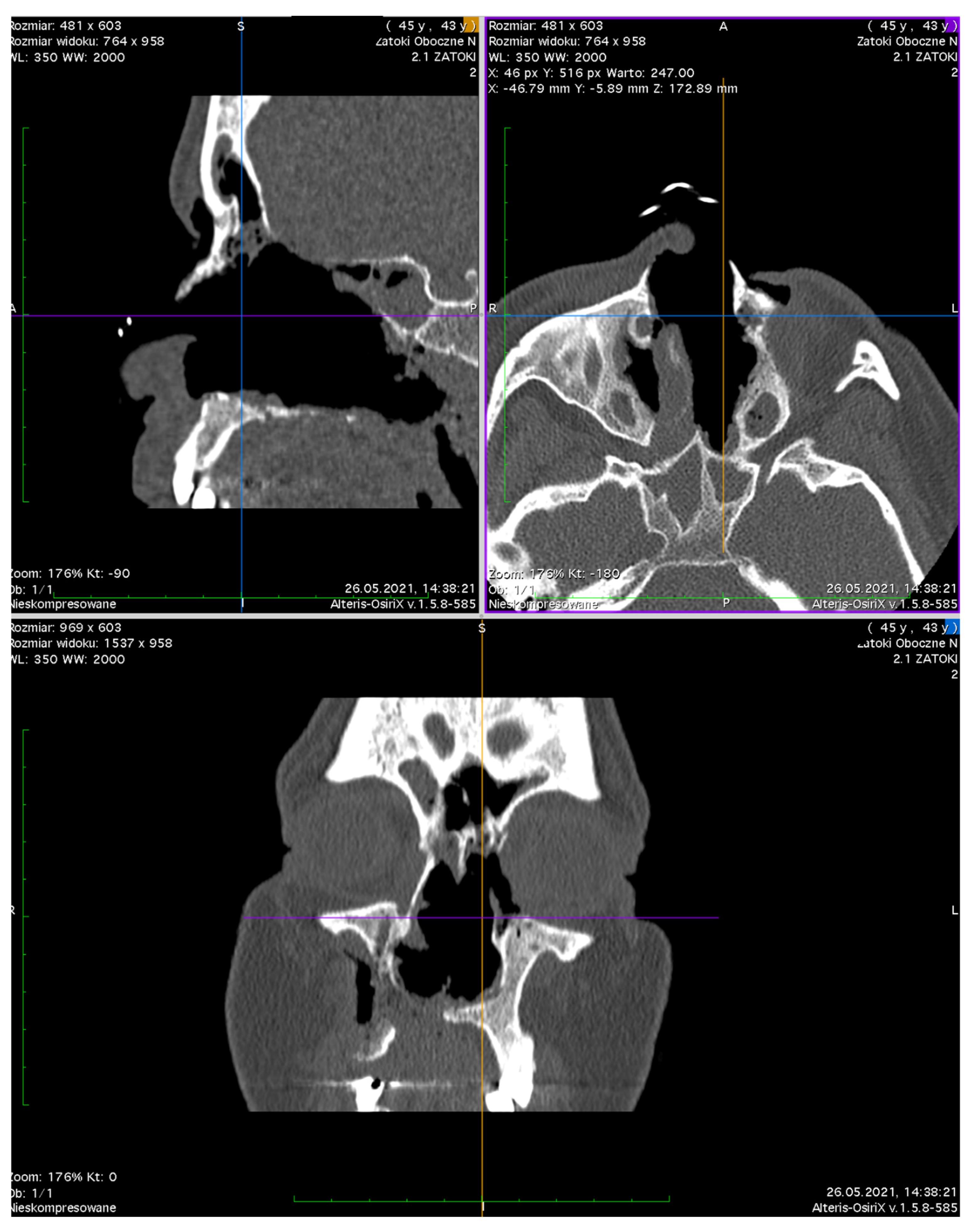 · 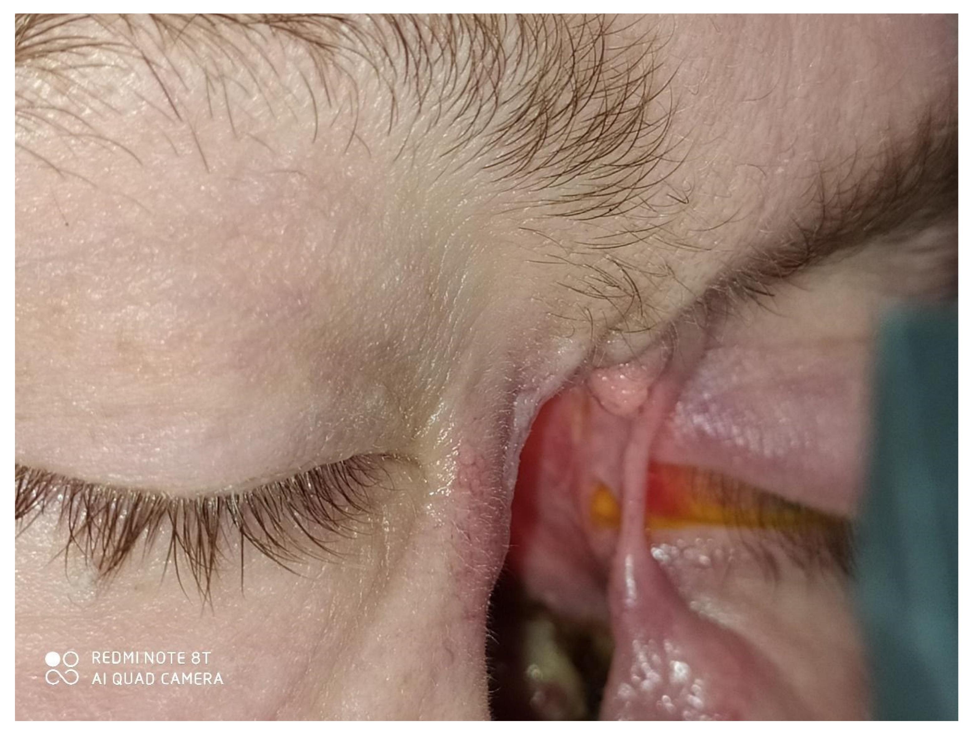 · 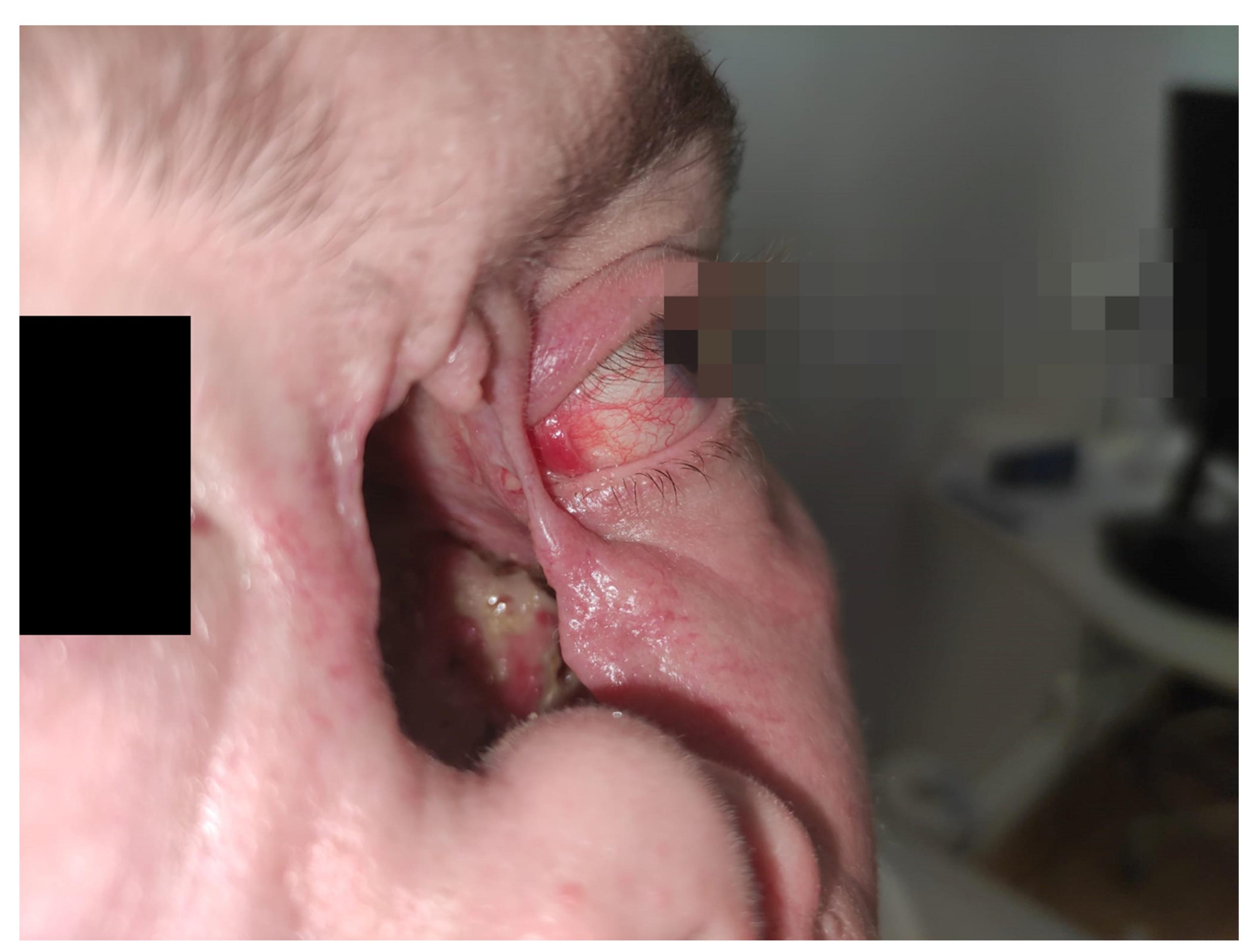 · 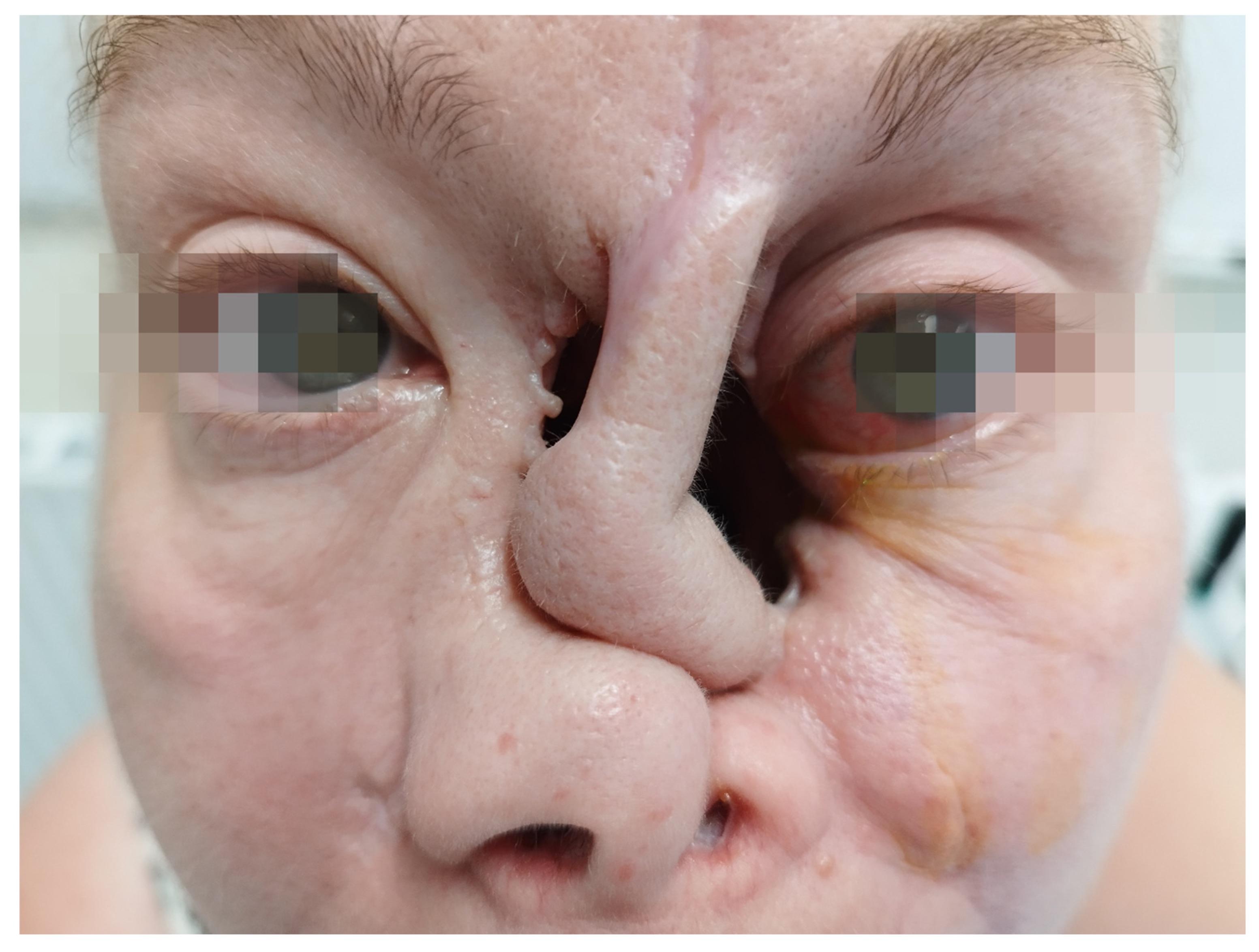 · |
|
#2
●
07-15-2024, 12:39 AM
|
 My Rank: MAJOR Poster Rank:120 Male Join Date: Feb 2010 Posts: 13,414 13,414Posts 15,836Thanked 3,535Quoted 15Mentioned 0Contrib. |
|
Re: Hole in the Face
Very interesting as usual! But imagine having to suffer like this, poor woman.
|
|
#3
●
07-15-2024, 01:16 AM
|
| My Rank: LANCE CORPORAL Poster Rank:3095 Join Date: May 2024 Posts: 125 125Posts 274Thanked 41Quoted 1Mentioned 0Contrib. |
|
Re: Hole in the Face
My friend's grandfather looked like this from some injury or disease. Scary looking dude
|
|
#4
●
07-15-2024, 01:34 AM
|
 ♚ Legacy Gold Member ♚ Poster Rank:537 your supreme lord & master Join Date: Apr 2017 Posts: 1,901 1,901Posts 3,392Thanked 376Quoted 2Mentioned 0Contrib. |
|
Re: Hole in the Face
Nice clit. I’d hit that.
|
|
#5
●
07-15-2024, 01:47 PM
|
 ♚ Legacy Gold Member ♚ Poster Rank:1299 Femme fatale Join Date: May 2024 Posts: 474 474Posts 825Thanked 191Quoted 1Mentioned 0Contrib. |
|
Re: Hole in the Face
I'd be constantly worrying my left eye would just drop into the caved in hole in the face. Or even worse flies getting inside and maggots residing inside, I'd also wear a mesh bag with a drawstring over my head to prevent that. Maybe hang some sticky fly paper down from the forehead.
|
|
#6
●
07-16-2024, 06:13 AM
|
| My Rank: PRIVATE Poster Rank:9917 Join Date: Oct 2021 Posts: 15 15Posts 68Thanked 6Quoted 0Mentioned 0Contrib. |
|
Re: Hole in the Face
Good thing their eyes are blurred now nobody will ever be able to recognize them.
|
|
#7
●
07-17-2024, 07:05 AM
|
 ]~Resident Asshole~[ Poster Rank:301 Stands to Pee Join Date: Aug 2009 Posts: 4,330 4,330Posts 6,564Thanked 2,203Quoted 12Mentioned 0Contrib. |
|
Re: Hole in the Face
Some Navy guys pay extra to hit those kinds of holes.
|