|
#1
●
01-05-2023, 07:21 PM
| ||||||||
 These are the rooms Poster Rank:26 of ruin. Join Date: Sep 2014 Posts: 54,011 Mentioned: 145 Post(s) Quoted: 30403 Post(s)
| ||||||||
|
Fungating Leiomyosarcoma of the Breast
Source: A 39-year-old female patient was referred to our hospital due to severe anorexia, weight loss (20 kg in one year), weakness, and prostration. After careful interrogation, she revealed the presence of a bulky, fast-growing breast mass with six-month evolution. Upon physical examination, the patient presented with paleness, severe cachexia, and hypotension upon admission. During clinical breast examination, we identified a 20 x 20cm fungating mass in the right breast with massive ulceration of the skin and small skin lesions compatible with cutaneous metastasis in the contralateral breast (Figure 1, 2). Laboratory tests showed severe anemia (5,1 g/dL), leukocytosis (60,700/mm3), elevated levels of C-reactive protein (331 mg/L), procalcitonin (3,52 ng/mL) and altered International Normalized Ratio (INR) of 2,1. The chest radiogram revealed a right pleural effusion with multiple bilateral pulmonary nodules and pleural retraction in the upper third of the right lung (Figure 3). The patient was admitted for a definite diagnosis, staging, and treatment of bacterial over-infection of the fungating breast neoplasm. The patient was given broad-spectrum antibiotics and transfused packed red cells. We began dressing care of the fungating wound with topical metronidazole. A contrast-enhanced computed tomographic (CT) of the thorax, abdomen, and pelvis was performed for staging, which demonstrated a large neoformative lesion in the right breast (with approximately 15 cm), ulcerated, with air-fluid levels inside and enlarged ipsilateral axillary lymph nodes (Figure 4). A loculated right pleural effusion with circumferential irregular pleural thickening due to pleural carcinomatosis and multiple pulmonary nodular lesions with spiculated contours in favor of multiple pulmonary metastases (Figure 5) were identified. The CT scan showed bone metastases in the pelvis. An ultrasound-guided core needle biopsy of the breast mass was conducted for a definite diagnosis. The histopathological examination revealed a spindle-cell neoplasm with moderated nuclear atypia and rare mitosis figures. Tumor cells were strongly positive for smooth muscle actin and vimentin and lacked expression of cytokeratin 7, CD34, Cam 5.2, and protein S-100. The ultrasound of the contralateral breast was clear, except for localized skin thickening suggestive of cutaneous skin metastasis. According to the histopathological and immunohistochemical findings, the tumor was classified as a leiomyosarcoma of the breast. Staging revealed massive dissemination of the leiomyosarcoma with lung, pleural, cutaneous, and bone metastasis. After a discussion of the case with a tertiary center with a Sarcoma Treatment Unit, the patient was referred to palliative care due to the extent of the disease and died one month after the diagnosis. 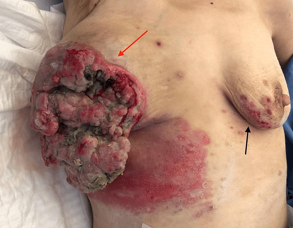 · 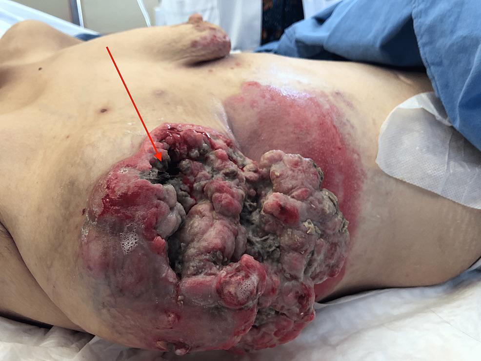 · |
|
#4
●
01-06-2023, 11:50 AM
| ||||||||
 The Devil's Left Hand Poster Rank:70 Male Join Date: May 2012 Posts: 22,207 Mentioned: 21 Post(s) Quoted: 9551 Post(s)
| ||||||||
|
Re: Fungating Leiomyosarcoma of the Breast
Mom?! 
__________________ Why so sad? Your tears are no match for mine. |
|
#5
●
01-06-2023, 05:31 PM
| ||||||||
 ★ Legacy Member ★ Poster Rank:252 So many choices now Join Date: Jul 2015 Posts: 5,548 Mentioned: 15 Post(s) Quoted: 2120 Post(s)
| ||||||||
|
Re: Fungating Leiomyosarcoma of the Breast
Imagine trying to stuff all that crap in a bra every day until you give up from the pain.
|
|
#6
●
01-07-2023, 04:05 AM
| ||||||||
 ✝Mudderator from Hell✝ Poster Rank:10 e-mail Join Date: Oct 2006 Posts: 96,642
Contributions: 817
Mentioned: 476 Post(s) Quoted: 10343 Post(s)
| ||||||||
|
Re: Fungating Leiomyosarcoma of the Breast
Can't imagine she didn't visit a doc earlier  |
|
#7
●
01-07-2023, 04:26 AM
| ||||||||
 My Rank: FIRST SERGEANT Poster Rank:399 Join Date: Apr 2011 Posts: 3,054 Mentioned: 2 Post(s) Quoted: 276 Post(s)
| ||||||||
|
Re: Fungating Leiomyosarcoma of the Breast
That's it. My boobs ran for the hills  |
|
#8
●
01-07-2023, 01:57 PM
| ||||||||
 ★ Legacy Member ★ Poster Rank:110 Secret Agent Join Date: Dec 2009 Posts: 14,660 Mentioned: 6 Post(s) Quoted: 3021 Post(s)
| ||||||||
|
Re: Fungating Leiomyosarcoma of the Breast
I don’t understand how a person can neglect themselves to that level of disfigurement. At the first sign of trouble, like shown in the left breast I’d be at the doctors asking WTF is up with my body…  I just don’t understand how you could let that go for the length of time it would take for that to happen to your own body.. Wouldn’t her bed sheets be covered in muck every morning with that kind of living wound? Clothes? Undergarments? Bath towels? I’m asking WTF!?!?! how how the fuck you can let yourself go like that…  |
|
#9
●
01-08-2023, 08:10 PM
| ||||||||
| My Rank: LANCE CORPORAL Poster Rank:2454 Male Join Date: Nov 2012 Posts: 181
Contributions: 3
Mentioned: 1 Post(s) Quoted: 33 Post(s)
| ||||||||
|
Re: Fungating Leiomyosarcoma of the Breast
Lack of education in the early stages, lack of access to healthcare, "alternative" remedies tried first, distrust of healthcare practitioners, fear...
|
|
#10
●
01-09-2023, 12:31 AM
| ||||||||
| ★ Legacy Member ★ Poster Rank:110 Secret Agent Join Date: Dec 2009 Posts: 14,660 Mentioned: 6 Post(s) Quoted: 3021 Post(s)
| ||||||||
|
Re: Fungating Leiomyosarcoma of the Breast
Thanks for helping me get a clear understanding. Much appreciated.  I’ve always been a believer in “modern medicine” as a survivor of a skydiving accident. Without it I most certainly wouldn’t be where I am today. |