|
#1
●
10-08-2024, 10:38 PM
|
 These are the rooms Poster Rank:26 of ruin. Join Date: Sep 2014 Posts: 54,008 54,008Posts 87,658Thanked 30,403Quoted 145Mentioned 0Contrib. |
|
Forequarter Amputation for Synovial Sarcoma
We present the case of a patient, ML, aged 33, who was admitted in our clinic for an arm and axilla radionecrotic tumour. The tumour was first seen in the patient 9 months ago. It presented as a round mass, with 0.75 cm diameter. The tumour had an explosive growing in the past 3 months. She was investigated in an Orthopaedic Clinic, where the diagnostic imaging included MRI, and which revealed a heterogeneous tumour in close contact with the axilla vascular and nervous bundle, invading the rhomboids and deltoid. The differential diagnosis included chondrosarcoma, liposarcoma, malignant fibrous histiocytoma, musculoskeletal tumors (percutaneous needle biopsy), osteochondroma and osteochondromatosis, osteosarcoma and synovial osteochondromatosis, etc. Further, a tumour biopsy was performed–tissue fragments representing malignant tumour proliferation with small, round cells, having a reduced cytoplasm, hyperchromatic nuclei with a compact disposition and large areas of tumoral necrosis. These hystopathologic aspects are synonymous with a Ewing PNET tumour, but do not exclude a synovial sarcoma or a rabdomiosarcoma Immunohistochemical studies revealed: EMA: positive in rare tumour cells; CK cocktail positive in tumour cells; S 100 negative in tumour cells; CD57 negative; CD 34 negative in tumour cells and positive in vessels; CLA negative in tumour cells and positive in small lymphocytes. Thus, these tests are compatible with a low’grade synovial sarcoma diagnostic. The tumour had a substantial growth from the day of biopsy. Further, due to the tumour’s large volume, surgical intervention was preceded by radiotherapy and chemotherapy (ifosfamide and doxorubicine). The tumour regressed a lot. When the patient was admitted in our clinic, she presented a 10/10 cm mass, involving the posterior left arm and axilla, fixed on the deep planes. The tumour was heterogeneous, ulcerated, with radiation necrosis, redness, oedema and tissue debris. Diagnostic methods included; blood tests, ultrasonography and pulmonary radiology that revealed no metastases. Local infection (staphylococcus aureus and Raoultella (K) ornithinolytica) control was made possible by pre and postoperative administration of medaxone and gentamicyn. Radical surgical procedure was imposed by: 1) the tumour’s large dimension, 2) shoulder girdle muscle invasion, 3) close contact and invasion of axilla vascular and nervous bundle and 4) histopathological form–low–grade sarcoma–more aggressive and a higher rate of metastases[5,6,7,8]. Forequarter amputation was imposed as a life saving measure. Surgical intervention was carefully planned so as to allow long time cover of the amputation stump. We decided to perform an anterior approach, the patient being placed in a right lateral position. The surgical technique consisted of an incision on the preoperative markings. These were deepened to the musculofascial layers. Platysma and supraclavicular nerves were cut. The clavicle was exposed and cut laterally to the sternocleidomastoidian with a Gigli saw. The tendons of pectoralis major, coracobrachialis and short head of the biceps were cut. Then, the subclavicular vessels were ligated. Surgical technique: After the subclavicular vessel ligature, brachial plexus branches were isolated, severed and let to retract. Scapula, trapezius, rhomboids, latissimus dorsi and seratus anterior were removed as the dissection progressed in the posterior plane [9]. The histopathology revealed a sarcoma–like tumour proliferation, with small, round cells presenting radiation necrosis changes. Axillary lymph nodes presented degenerative changes with massive reactive sinus histiocytosis 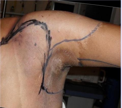 · 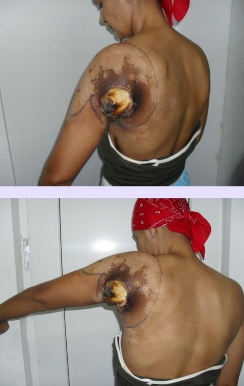 · 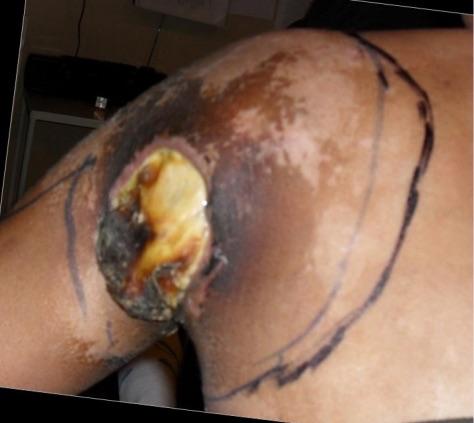 · 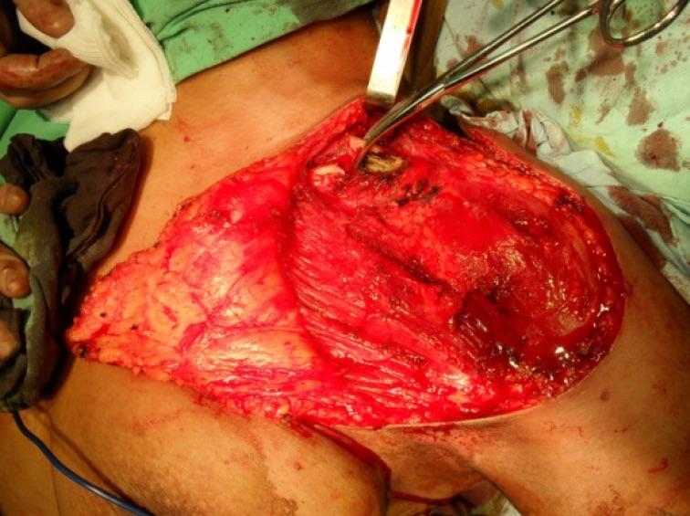 · 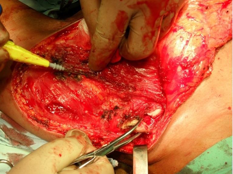 · 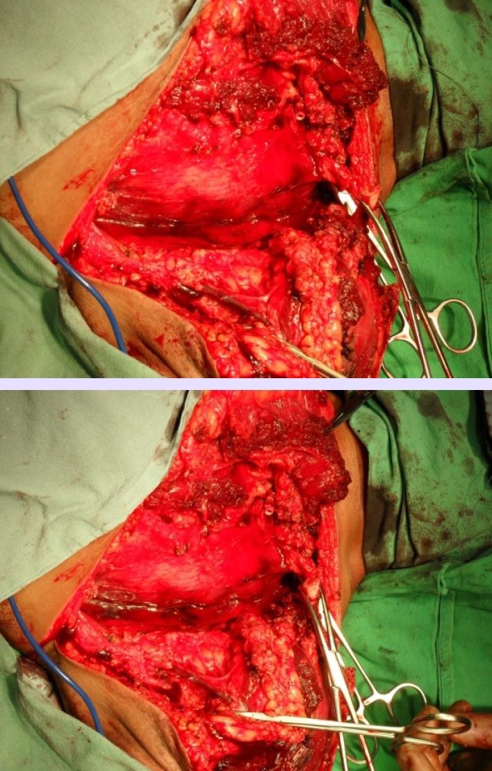 · 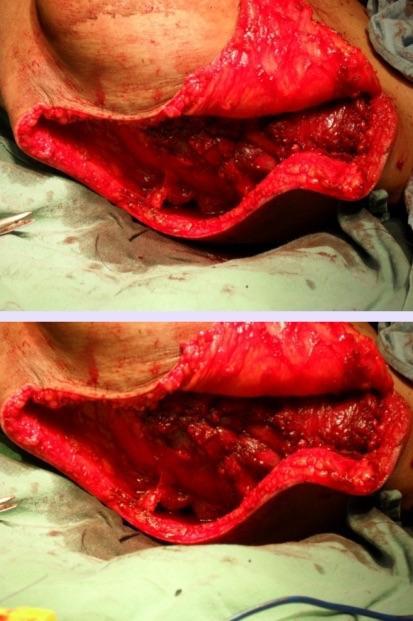 · 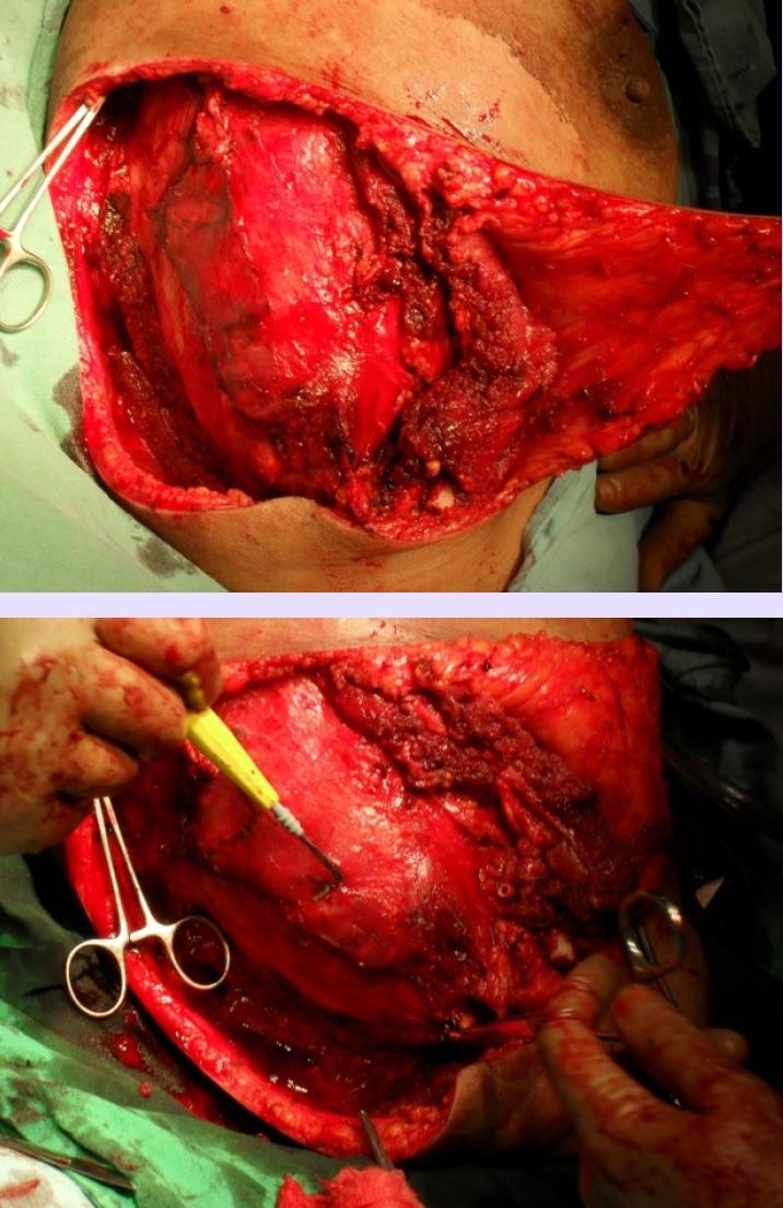 · 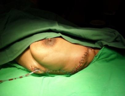 · 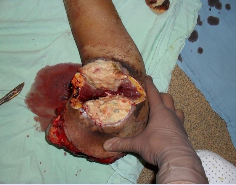 · 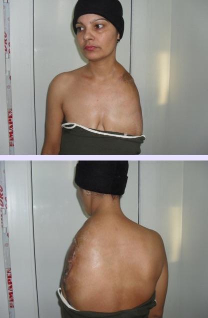 · 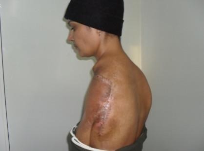 · |
|
#2
●
10-08-2024, 11:14 PM
|
 My Rank: CORPORAL Poster Rank:1269 female Join Date: May 2009 Posts: 489 489Posts 411Thanked 35Quoted 0Mentioned 0Contrib. |
|
Re: Forequarter Amputation for Synovial Sarcoma
I was not expecting that much .
|
|
#3
●
10-09-2024, 12:22 PM
|
 ★ Legacy Member ★ Poster Rank:110 Secret Agent Join Date: Dec 2009 Posts: 14,787 14,787Posts 49,104Thanked 3,050Quoted 6Mentioned 0Contrib. |
|
Re: Forequarter Amputation for Synovial Sarcoma
Scary tumor. Pretty amazing. She’s alive and got to keep her left breast.  |
|
#4
●
10-09-2024, 12:36 PM
|
 ♚ Legacy Gold Member ♚ Poster Rank:268 ꓕSIꓤIꓕ∀S ϽINOᗡꓤ∀S Join Date: Feb 2010 Posts: 5,177
Contributions: 8
5,177Posts 31,152Thanked 1,441Quoted 45Mentioned 8Contrib. |
|
Re: Forequarter Amputation for Synovial Sarcoma
Too bad this wasn't treated more aggressively when it was just 0.75cm. Ironically, the term "Gigli saw" always makes me giggly...
__________________ 200+ Additional Smilies Custom DR Search Engine ✦ Live life to it's fullest and leave a sexy corpse ✦ |
|
#5
●
10-09-2024, 09:56 PM
|
 ♚ Legacy Gold Member ♚ Poster Rank:537 your supreme lord & master Join Date: Apr 2017 Posts: 1,900 1,900Posts 3,385Thanked 375Quoted 2Mentioned 0Contrib. |
|
Re: Forequarter Amputation for Synovial Sarcoma
It was invented by Dr. Giggles. Bonus points to the surgeon for the titty picts.
|
|
#6
●
10-11-2024, 03:53 PM
|
 ✝Mudderator from Hell✝ Poster Rank:10 e-mail Join Date: Oct 2006 Posts: 96,654
Contributions: 901
96,654Posts 543,138Thanked 10,365Quoted 479Mentioned 901Contrib. |
|
Re: Forequarter Amputation for Synovial Sarcoma
So they waited for 9 months to let it grow from 0.75 cm to what it shows in the pic. Also:The tumour had a substantial growth from the day of biopsy. That seems to happen more. https://www.ncbi.nlm.nih.gov/pmc/articles/PMC10772461/ |