|
#1
●
01-22-2023, 04:42 AM
| ||||||||
 These are the rooms Poster Rank:26 of ruin. Join Date: Sep 2014 Posts: 54,011 Mentioned: 145 Post(s) Quoted: 30403 Post(s)
| ||||||||
|
Extensive Acral Melanoma of the Foot.
Extensive acral melanoma of the foot. The patient tried to “self-medicate holistically” for two years. An 82-year-old Samoan man, skin phototype V, presented with left heel pain due to a large, exophytic, hyperpigmented, polypoid mass. He had initially presented to the dermatology clinic 2 years previously with a small, dark macule, but had decided to self-medicate holistically on his home island until his heel pain prompted his return. Physical examination revealed a lesion consisting of black, necrotic nodules and friable granulation tissue along with ulcerations and purulent/serosanguinous discharge. At the base of the lesion was a hyperpigmented, asymmetrical macule with nodularity (figure 1). The patient had no other associated symptoms and no lymphadenopathy. An MRI demonstrated a 9×7 cm lobulated, exophytic left heel soft tissue mass with invasion into the fat pad, but no other tissue invasion or bone erosion. Biopsies from the medial and plantar surfaces of the macular lesion showed marked pigment deposition throughout all levels of the epidermis and in the superficial dermis (figure 2). The histopathological findings also revealed lentiginous proliferation of atypical melanocytes associated with irregular epidermal acanthosis along with prominent pagetoid spread of atypical intraepidermal melanocytes . Atypical melanocytes were noted at dermal papillae tips, but it was difficult to discern any definitive invasion into the dermis. These initial findings suggested a diagnosis of acral lentiginous malignant melanoma in situ with a vertical growth phase resulting in an extensive nodule. Although definitive features of invasive melanoma were not identified, given the patient's history, the small sample size of tissue, and the lack of biopsies from the fungating mass, it was felt that tumour excision was the most appropriate treatment. A sentinel node biopsy was negative so a below-the-knee amputation was carried out. Gross examination of the prosection revealed clear margins and findings of a variably pigmented (white to black) lesion with a focal area of fungating tumour measuring about 9×7×3 cm. Adjacent to this fungating mass, covering approximately half of the lateral aspect of the foot, was an ill-defined, grey-blue discolouration measuring 11×8 cm. Finally, located 0.4 cm medially from the variably pigmented lesion and grossly separate from the main lesion, was a well-demarcated, slightly firm, black lesion measuring 0.5×0.5×0.2 cm. Microscopic findings of the fungating mass revealed superficial ulceration and an underlying multinodular, heavily pigmented mass with gross extension into the adjacent adipose tissue that measured 3.2 cm Breslow depth. Additional histopathological findings of the mass and lesions were consistent with biopsy results and also demonstrated invasion into the dermis and subcutaneous tissues. Midway through the dermis, prominent regression was indicated by tumoural melanosis and fibrosis. Deeper to this, the tumour extended into the subcutaneous tissue without pigmentation, which was supported by findings of marked cytological atypia that included pleomorphism, abnormal chromatin patterns and atypical mitotic figures. The mitotic count was 15/mm2 with the hot spot method. No definitive lymphovascular invasion or perineural invasion was identified. Acral lentiginous melanoma, stage T4b was the final diagnosis . 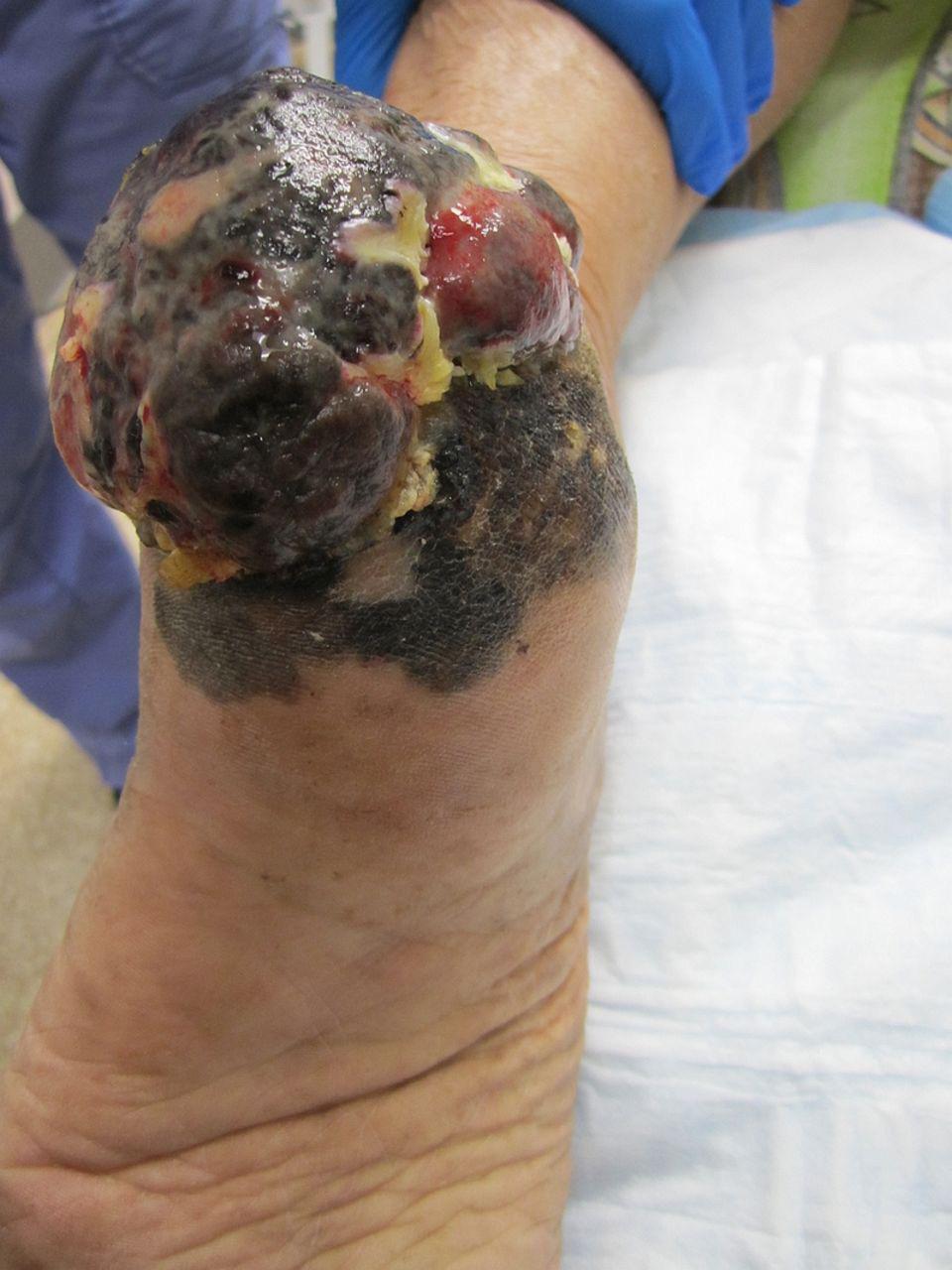 · 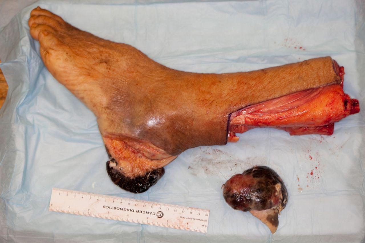 · |
|
#2
●
01-22-2023, 12:37 PM
| ||||||||
 ★ Legacy Member ★ Poster Rank:110 Secret Agent Join Date: Dec 2009 Posts: 14,666 Mentioned: 6 Post(s) Quoted: 3023 Post(s)
| ||||||||
|
Re: Extensive Acral Melanoma of the Foot.
They removed his foot so they could work on it for a while before they give it back to him…  Or it was so infected they had to amputate it to save his leg…  |
|
#3
●
01-22-2023, 02:08 PM
| ||||||||
 ★ Legacy Member ★ Poster Rank:252 So many choices now Join Date: Jul 2015 Posts: 5,548 Mentioned: 15 Post(s) Quoted: 2120 Post(s)
| ||||||||
|
Re: Extensive Acral Melanoma of the Foot.
Maybe tried the wrong fragrance of essential oils?
|
|
#4
●
01-22-2023, 10:31 PM
| ||||||||
 My Rank: MASTER GUNNERY SERGEANT Poster Rank:352 Manly Man Join Date: Sep 2017 Posts: 3,601 Mentioned: 0 Post(s) Quoted: 700 Post(s)
| ||||||||
|
Re: Extensive Acral Melanoma of the Foot.
They're going to send it back for warranty.
|
|
#5
●
01-22-2023, 11:01 PM
| ||||||||
| ★ Legacy Member ★ Poster Rank:110 Secret Agent Join Date: Dec 2009 Posts: 14,666 Mentioned: 6 Post(s) Quoted: 3023 Post(s)
| ||||||||
|
Re: Extensive Acral Melanoma of the Foot.
It’s good the other didn’t fail as well. The “body” shop would be installing two instead of just one. Money saved!  |
|
#9
●
01-25-2023, 12:36 AM
| ||||||||
| My Rank: MASTER GUNNERY SERGEANT Poster Rank:352 Manly Man Join Date: Sep 2017 Posts: 3,601 Mentioned: 0 Post(s) Quoted: 700 Post(s)
| ||||||||
|
Re: Extensive Acral Melanoma of the Foot.
You suck at keeping secrets. |