|
#1
●
03-30-2023, 09:26 PM
| ||||||||
 These are the rooms Poster Rank:26 of ruin. Join Date: Sep 2014 Posts: 54,011 Mentioned: 145 Post(s) Quoted: 30403 Post(s)
| ||||||||
|
Enormous Gossypiboma
Source: An 85-year-old male was sent to the Oral and Maxillofacial Unit for an enormous tumor of the right paranasal sinus. His medical history included the following: high blood pressure controlled by medication, treatment by aspirin 150 mg/day for 6 years, complete exeresis of a right paranasal basal cell carcinoma of the skin (1.2 cm in diameter) eleven years before, and some repetitive episodes of epistaxis that usually benefitted from a bilateral nasal packing using gauzes provided with fatty ointment Biogaze® (Bottu Pharmaceuticals, Casablanca, Morocco). The patient presented with a facial CT scan that showed a large osteolytic tumor of the right maxillary sinus (Fig. 1). Thoraco-abdominal CT scan was normal. Biopsies were performed by the ENT team through nasal endoscopy. The samples’ histology examination revealed necrosis and inflammatory tissues. New sets of biopsies were performed using a Caldwell–Luc approach. The histological findings showed no different features. Although no histological signs of malignancy were reported, the tumor showed a very aggressive behavior. It spread rapidly through the site of the meatotomy and the vestibular incision. Within a few days, the tumor had invaded the right nasal fossa and a large part of the oral cavity, inducing a destruction of 1/3 of the oral cavity's roof in its lateral part (hard palate; Fig. 2). The histological tests raised suspicion regarding the presence of Candida Albicans and Actinomyces, but specific cultures were all negative. The patient, however, was put under antifungal and antibiotic treatments without any improvement. During the screening time, bleeding from the nose and the oral cavity was accompanying the growing of the tumor. The patient suffered some episodes of severe anemia (hemoglobin concentration: 5–6 g/dL) that necessitated blood transfusion. The growth of the tumor toward the pharynx was so impressive (Fig. 3) that the patient showed rapidly increasing signs of respiratory discomfort and frequent episodes of coughing. Considering the imminent risk of asphyxiation, the patient was admitted to the operating theater where he underwent an oral and pharyngeal tumor removal under local anesthesia. The specimen (Fig. 4) was then sent to the laboratory. The histological examination found necrosis, inflammatory tissue, and fabric fibers suggesting the presence of the remains of gauze (Gossypiboma; Fig. 5). The patient was then prepared for a second surgery under general anesthesia 2 weeks later. A right paranasal approach with a back cut in the right lower eyelid was performed. The infraorbital nerve was exposed and protected. The gossypiboma partially destroyed the anterior and the medial paranasal sinus bony walls. The floor of the sinus was completely destroyed, and only the orbital floor's periosteum was left intact. The complete removal of the tumor was performed using a submucoperiosteal dissection (Fig. 6A and B). A trilayer reconstruction of the defect of the lateral one-third of the hard palate was immediately performed using the following: a flap of the remaining nasal wall mucosa to create the upper layer (sinusal floor mucosa), a flap of the buccal fat pad to create the intermediate layer, and a mucosal cheek flap harvested above the level of the parotid duct to create the lower layer (palatal mucosa; Fig. 7A–E). Saline solution spray was indicated for nasal and sinusal cleaning. Nose blowing was prohibited for 15 days. The immediate follow-up was uneventful, and the patient was discharged 5 days after surgery. A monthly follow-up was scheduled, including clinical examinations and plain X-rays. No complication was noticed. Six months after the surgery, the oral cavity's roof is still completely closed (Fig. 8A and B). An MRI showed clear right paranasal sinus and nasal fossa with a large communication between them. There are no signs of recurrence. There is still persistent thickening of the sphenoidal sinus mucosa (Fig. 9). 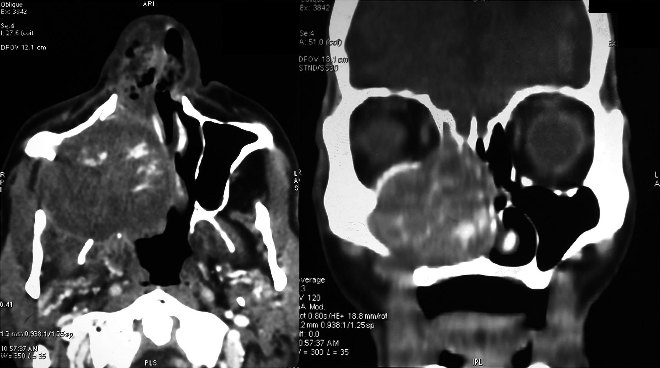 · 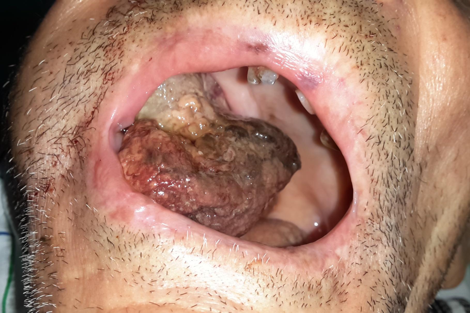 · 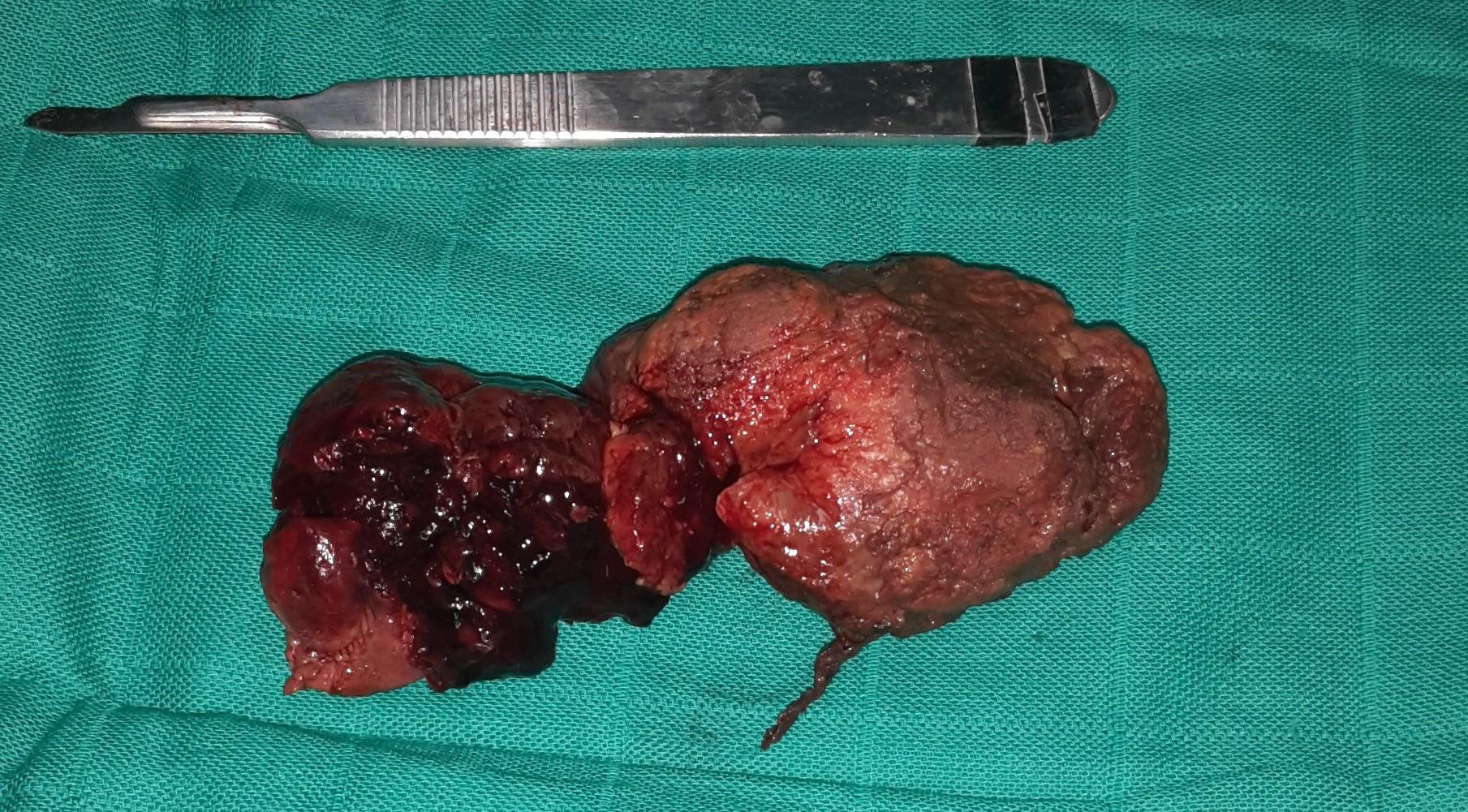 · 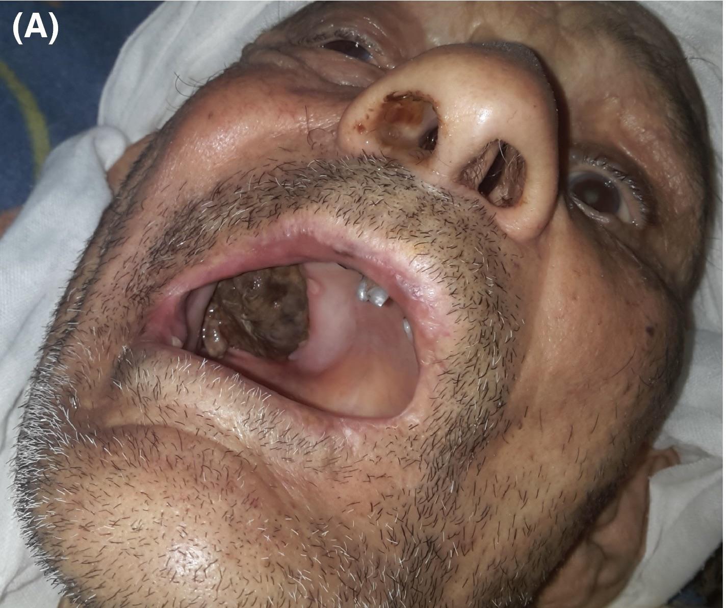 · 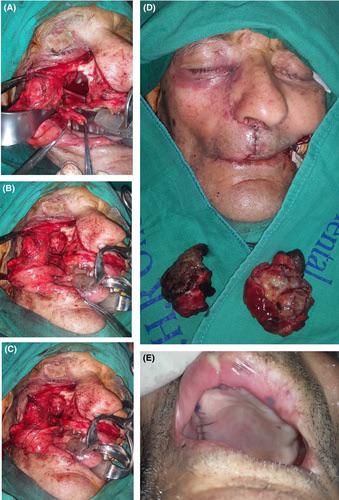 · 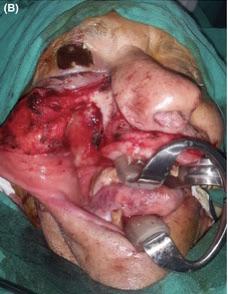 · 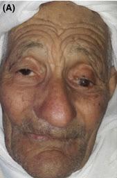 · 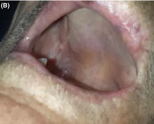 · |
|
#2
●
03-30-2023, 10:08 PM
| ||||||||
 ♚ Legacy Gold Member ♚ Poster Rank:206 Iceman Join Date: Aug 2017 Posts: 7,505 Mentioned: 19 Post(s) Quoted: 3630 Post(s)
| ||||||||
|
Re: Enormous Gossypiboma
Ill have what he's having in 2nd last photo.
|
|
#8
●
04-03-2023, 07:32 PM
| ||||||||
| These are the rooms Poster Rank:26 of ruin. Join Date: Sep 2014 Posts: 54,011 Mentioned: 145 Post(s) Quoted: 30403 Post(s)
| ||||||||
|
Re: Enormous Gossypiboma
He probably didn't realize the gauze was left in there and he's elderly, so probably worried about the cost of another procedure.
|