|
#1
●
12-01-2022, 04:26 PM
| ||||||||
| These are the rooms Poster Rank:25 of ruin. Join Date: Sep 2014 Posts: 54,011 Mentioned: 145 Post(s) Quoted: 30403 Post(s)
| ||||||||
|
Crush Injury to the Foot
The case in question involves a 44-year-old male who sustained a crush injury to both lower extremities after being run over by a large utility vehicle. In the trauma bay, he was noted to have a degloving injury of the distal right lower extremity in combination with an open fracture and non-pulsatile bleeding. Biphasic pedal signals were identified bilaterally during resuscitation. Plain films revealed open displaced fractures to right metatarsals 2-4 and displaced right medial malleolar and tibial metaphysis fractures with a spiral fracture of the proximal fibula as shown in Figure 1. His MESS score was 6, and Gustilo-Anderson score was 3b, which are not predictive of needing amputation but do predict an elevated risk of complication. The patient did undergo computed tomography angiography (CTA) of the extremities, which showed three-vessel runoff to the level of the malleolus with a transection of the dorsalis pedis. The orthopedic surgery team took the patient to the operating room where he underwent debridement of nonviable tissues with reduction of the fractures and placement of a negative pressure wound therapy system. Postoperatively the patient’s extremity was warm with palpable dorsalis pedis pulse. During rounds on hospital day three, the patient was noted to have a cool and pulseless foot distal to the malleolus, which is suggestive of ischemia. Therefore, the patient was taken urgently to the operating room by trauma surgery service. His preoperative wound is shown in Figure 2. In the operating room, the dorsalis pedis artery was exposed at the level of the ankle as identified by Doppler signals. This was exposed distally until a complete transection was identified 2 cm distal to the ankle. The distal portion was identified proximal to the webspace. Necrotic tissues were resected, and the vessel underwent embolectomy to restore brisk bleeding from the proximal segment before flushing and systemic heparinization. Distal greater saphenous vein was dissected, and an 8-cm segment was utilized to create a reversed interposition graft with spatulation of the ends. Fasciotomies were performed in the forefoot, and hematomas were evacuated from the compartments due to the presence of compartment syndrome. The wounds were packed with gauze, and, given the lack of nearby viable tissue for complete coverage, the exposed vessels and anastomosis were covered with IntegraTM (Integra LifeSciences, Princeton, NJ) before wrapping in bismuth-laden petroleum gauze and dry gauze. Postoperative images of the patient’s revascularized wound are shown in Figure 3. Postoperatively, the patient’s graft was maintained on a heparin infusion. His distal digits remained ischemic, but the patient had palpable pulses distally. Given the concern for continued ischemic insult with disruption of the microvasculature due to crush injury, the patient underwent hyperbaric therapy. Postoperative pictures of the wound are shown in Figure 4. Despite attempts to salvage the forefoot with continued hyperbaric therapy and serial debridement, the patient did require transmetatarsal amputation as shown in Figure 4 on the 19th hospital day before and after IntegraTM placement. The residual foot (and ankle) has remained viable and functional. 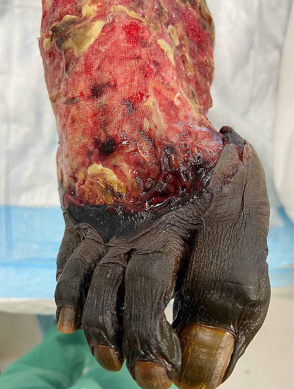 · 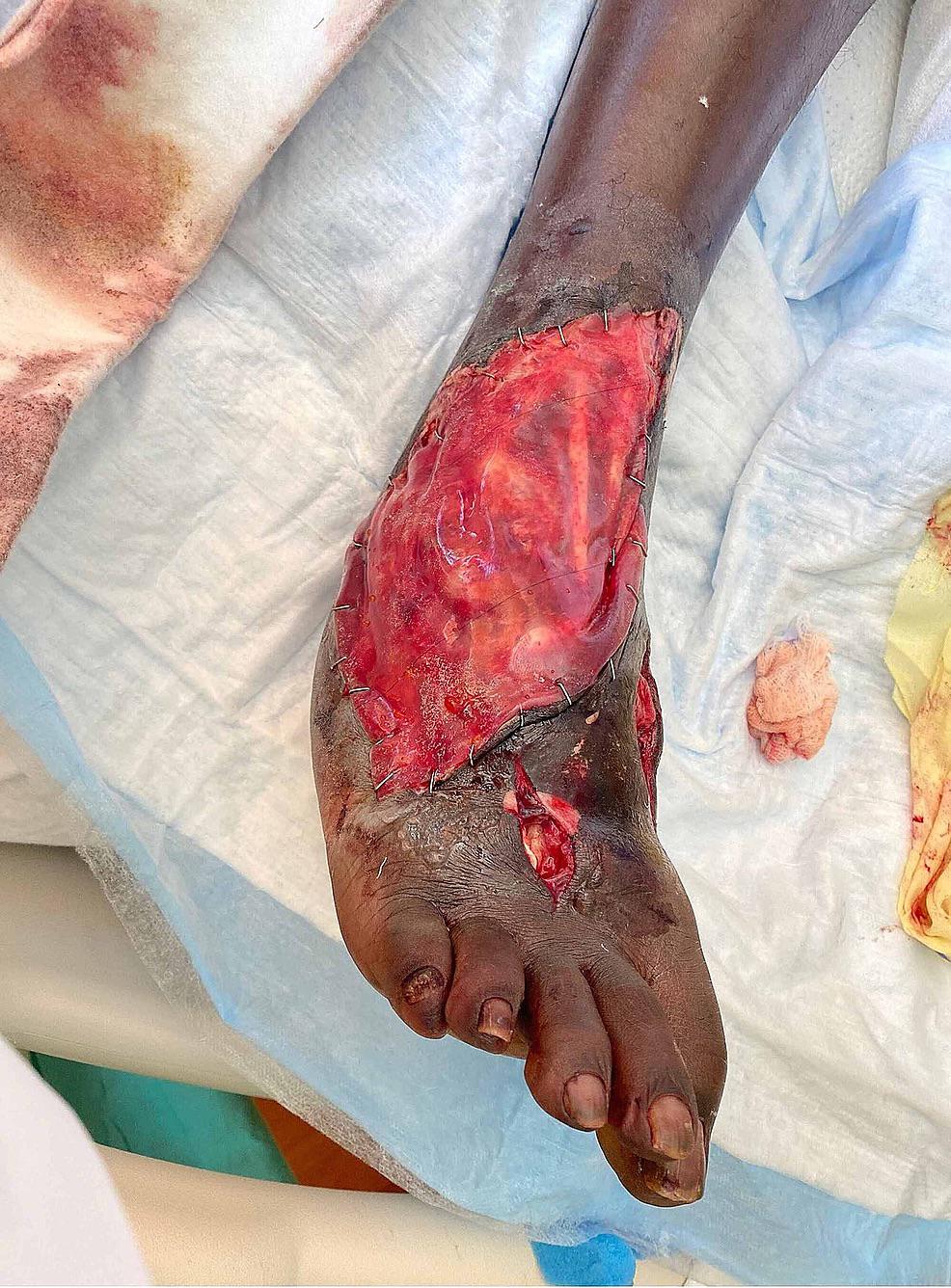 · 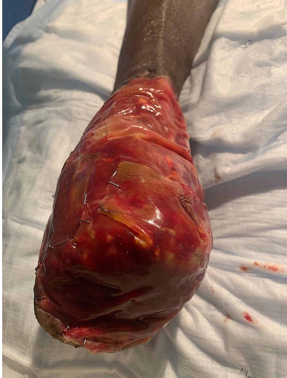 · |
|
#2
●
12-01-2022, 11:14 PM
| ||||||||
| ☾ Administrator ☽ Poster Rank:77 ஜᎻᎬᎩᎾᏦᎪஜ Join Date: Dec 2011 Posts: 19,441
Contributions: 32
Mentioned: 169 Post(s) Quoted: 6798 Post(s)
| ||||||||
|
Re: Crush Injury to the Foot
__________________ 💜🧿See Human | Be Human🧿💜 (War Section Hashtags) |
|
#3
●
12-02-2022, 12:20 AM
| ||||||||
| ★ Legacy Member ★ Poster Rank:247 So many choices now Join Date: Jul 2015 Posts: 5,548 Mentioned: 15 Post(s) Quoted: 2120 Post(s)
| ||||||||
|
Re: Crush Injury to the Foot
Why did they stick his foot into a canned ham?
|
|
#5
●
12-02-2022, 01:40 AM
| ||||||||
| ~So Much Blood~ Poster Rank:120 Gore Hag Join Date: Mar 2010 Posts: 13,161
Contributions: 1
Mentioned: 28 Post(s) Quoted: 822 Post(s)
| ||||||||
|
Re: Crush Injury to the Foot
That first pic looks like some kind of voodoo foot |
|
#6
●
12-02-2022, 02:54 AM
| ||||||||
| ✝Mudderator from Hell✝ Poster Rank:10 e-mail Join Date: Oct 2006 Posts: 94,975
Contributions: 817
Mentioned: 472 Post(s) Quoted: 10078 Post(s)
| ||||||||
|
Re: Crush Injury to the Foot
ring tone is a bit short, it looks like he has 2 pinky toes.
|
|
#7
●
12-02-2022, 06:32 AM
| ||||||||
| Addict Of Misery Poster Rank:69 Join Date: May 2012 Posts: 22,176 Mentioned: 18 Post(s) Quoted: 9547 Post(s)
| ||||||||
|
Re: Crush Injury to the Foot
Is that a mummy foot in the first pic?
__________________ A deep well of despair I found, the day my dreams came true... |