|
#1
●
07-09-2024, 11:07 PM
| ||||||||
| These are the rooms Poster Rank:25 of ruin. Join Date: Sep 2014 Posts: 54,011 Mentioned: 145 Post(s) Quoted: 30403 Post(s)
| ||||||||
|
Cervicothoracic Giant Cutaneous Squamous Cell Carcinoma
A 36-year-old male presented with a 3-year history of a growing mass on the anterior cervicothoracic wall. Biopsy of the lesion revealed moderately differentiated epithelial proliferation with focal keratinization consistent with cutaneous SCC. The mass was a protruding, ulcerated, multilobular, mostly necrotic, foul-smelling, cauliflower-like, firm tumor with hyperkeratotic zones, measuring 11 × 9.5 cm in size that had developed over a large region of erythematous skin (Fig. 1, Fig. 2). He had multiple adenomegalies in the left inframandibular region and right armpit. Complete blood count and biochemistry were normal, and total-body computed tomography (CT) showed that the tumor had not infiltrated deeply into the musculoskeletal layers of the thoracic wall. His medical history included a personality disorder for which he took medication intermittently because of poor compliance and social support. No lymph node or distant metastases were noted. He consented to surgical reconstruction, and the tumor was excised completely with a 2–3 cm clear margin around it. The tumor appeared to have infiltrated the subcutaneous tissue, and thus, a 4-cm margin of subcutaneous tissue was excised with the tumor. Following this, wide tumor excision of the surrounding skin could not be approximated, and the surrounding skin was left to heal slowly with re-epithelialization. Neck CT showed a lesion infiltrating the inferior margin of the sternocleidomastoid muscle and prethyroid muscles in the deep plane. The tumor was connected to the anterior margin of the isthmus and the upper margin of the left thyroid lobe, extending to the level of the thyroid cartilage. At its anterior margin, the tumor showed signs of ulceration and had a diameter of approximately 135 × 43 × 65 mm. Thoracic CT showed an irregular-appearing, ulcerated mass in the anterior margin of the base of the neck, without any apparent signs indicating compromise of the visceral space. The mass was in contact with the anterior margin of the sternal handle (Fig. 2). Needle aspiration biopsy of the axillary node was performed and was negative for malignancy. Based on the patient’s condition, oncologists considered her to be a candidate for surgical resection for improving overall survival. Multidisciplinary management was performed by surgery of the breast and soft tissues, head and neck, thorax, plastic and reconstructive microsurgery deparments; intraoperative findings showed an ulcerated, necrotic, and stinking tumor lesion measuring 15 × 12 cm in the cervical region with compromise of subcutaneous cellular tissues, prethyroid muscles, inner third of the left clavicle and anterior wall of sternal notch. Then, we decided to perform wide local resection of the neck tumor before margins marking (prethyroid muscles, sternocleidomastoid plus bilateral low parietal decompression plus dissection of blood vessels) neck plus bilateral cervical lymphatic drainage plus resection of the inner third of the left clavicle plus disinsertion of the sternal hairpin and resection of the anterior wall of the sternal notch (Fig. 3). Finally, microsurgery was performed through reconstruction with an antero-lateral thigh (ALT) flap from the left thigh (Fig. 4). 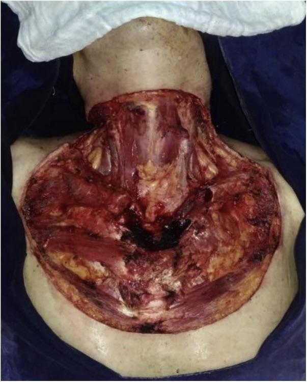 · 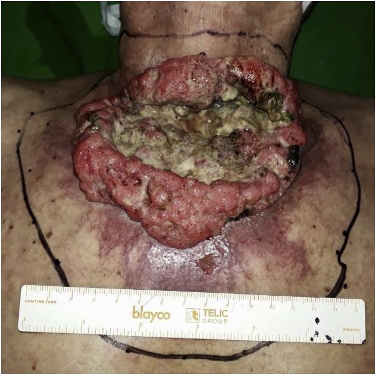 · 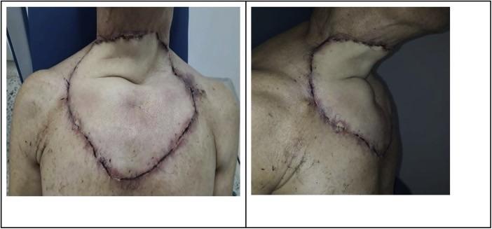 · |


