|
#1
●
12-18-2022, 11:41 PM
| ||||||||
 These are the rooms Poster Rank:26 of ruin. Join Date: Sep 2014 Posts: 54,011 Mentioned: 145 Post(s) Quoted: 30403 Post(s)
| ||||||||
|
Bilateral Ocular Necrotizing Fasciitis
She still has her eyes, they're just blacked out. A 58-year-old woman with a history of rheumatoid arthritis treated with tocilizumab, as well as depression treated with bupropion, presented to the emergency department with severe preseptal cellulitis and maxillary sinusitis. Her past ocular history was significant for uncomplicated cataract surgery in both eyes. She had been evaluated by her ophthalmologist one week prior to presentation, when she was prescribed brimonidine eye drops to treat persistently dilated pupils, thought to be a side effect of bupropion. Within one day she experienced rapidly progressive orbital swelling and pain, left greater than right. She also had increasing blurry vision, binocular diplopia, and upper respiratory symptoms, including sinonasal congestion and rhinorrhea. Her swelling was so significant that it caused obstruction of the visual axis secondary to eyelid edema. She presented to an outside hospital and was managed for a severe allergic reaction, where she received intravenous (IV) steroids and antihistamines. An outside CT scan was performed, which was concerning for extensive preseptal cellulitis and sinus disease. The patient was subsequently transferred to a tertiary care center for further evaluation and management. The differential diagnosis for painful, bilateral eye swelling is broad but commonly includes viral and bacterial conjunctivitis, allergic conjunctivitis, blepharitis, trauma, contact dermatitis, herpetic dermatitis, preseptal cellulitis, angioedema, eczema, and autoimmune diseases. On exam, the patient had erythema and swelling extending from the nose to forehead and laterally past the orbits bilaterally. Blackened, necrotic eyelids with skin breakdown and serosanginous discharge were present. There was tenderness of the malar prominence and neck bilaterally, without crepitus. The patient demonstrated significant lid edema, induration, ecchymosis, and tenderness; the left upper and lower lids were severely taught, and she had mild proptosis which was greater on the left. There was prominent conjunctival chemosis bilaterally. A 4 x 5 mm epithelial defect was present in the left inferior lid (Figure 1). Dilated fundus examination was within normal limits. Vision was measured with near card as 20/30 in the right eye and 20/400 in the left eye. The intraocular pressures were measured with tonopen at 19 mmHg in the right and 23 mmHg in the left. There was no afferent pupillary defect present, and extraocular motility was full without pain. The orbit was not noted to be tense or resistant to retropulsion. Nasal endoscopic exam was performed without anesthetic due to suspicion for invasive fungal sinusitis in the setting of medical immunosuppression (given the patient’s history of rheumatoid arthritis treated with tocilizumab). This showed significant leftward septal deviation and universally friable mucosa that was fully sensate with no signs of necrosis. She had mucopurulent discharge at the middle meatus bilaterally and the right sphenoethmoid recess. Relevant labs included a white blood cell count of 12.9 x 10³/mcL and C-reactive protein of 87.0 mg/L. Erythrocyte sedimentation rate (ESR) was normal. A maxillofacial CT scan with contrast demonstrated extensive periorbital soft tissue swelling with sinus inflammation and impaction of the bilateral maxillary and ethmoid sinuses and the left sphenoid sinus. She had bilateral conjunctival enhancement, with a rim-enhancing collection along the temporal right conjunctiva. This rim-enhancing fluid collection, indicative of an abscess, measured 5 mm x 13 mm with an intralesional focus of air (Figure 2). There was no postseptal stranding and no subperiosteal abscess identified. There was also bilateral cervical lymphadenopathy on neck CT. Magnetic resonance venography was obtained, which showed no involvement of the cavernous sinus. The patient was emergently taken to the operating room jointly by the ophthalmology and otolaryngology teams. Ophthalmology performed bilateral orbitotomy and washout. The orbital septum was noted to have a necrotic appearance, and there was “dishwater-appearing” darkened fluid in the preseptal space. Otolaryngology performed a functional endoscopic sinus surgery (FESS). Endonasal findings included diffuse inflammatory changes and purulence from bilateral maxillary and ethmoid sinuses; there was no evidence of devitalized tissue or necrosis, and frozen sections were negative for invasive fungal disease (Figure 3). The ophthalmology team placed penrose drains bilaterally for continued drainage (Figure 4). Intraoperative orbital pathology specimens were obtained, which showed coagulative necrosis and fibrinopurulent debris. Orbital cultures finalized with growth of Streptococcus pyogenes and methicillin-resistant Staphylococcus aureus (MRSA). Endonasal cultures grew only Streptococcus pyogenes. She was taken back to the operating room on hospital day 3 for a repeat orbital washout by ophthalmology and nasal debridement by otolaryngology, at which point the penrose drains were removed (Figure 5). She was also treated with four weeks of IV penicillin and vancomycin, IV steroids, and ophthalmic bacitracin ointment. Aggressive nasal hygiene was initiated, with saline irrigations and sprays, as well as fluticasone and oxymetazoline sprays. She also underwent temporary left tarsorrhaphy to protect the ocular surface. After completion of therapy, she was noted to have persistent left lower lid cicatricial ectropion, foreign body sensation, and lagophthalmos (Figure 6). Her best corrected visual acuity three months after presentation was 20/25 in the right eye and 20/60 in the left eye. She continued a nasal regimen and a follow-up nasal endoscopy demonstrated expected post-FESS changes with patent sinuses bilaterally and no evidence of sinonasal disease. 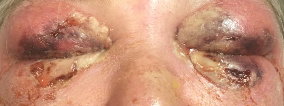 · 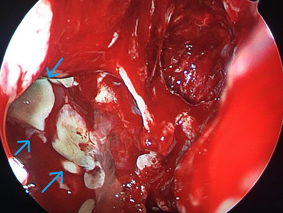 · 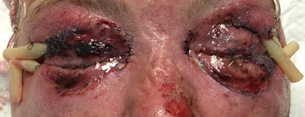 · 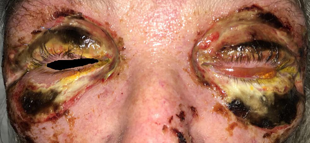 · 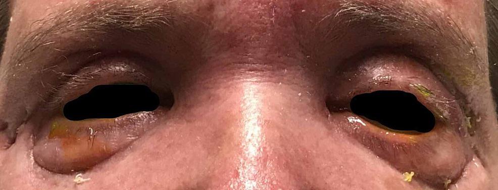 · |