|
#1
●
08-14-2024, 05:44 PM
|
 These are the rooms Poster Rank:26 of ruin. Join Date: Sep 2014 Posts: 54,008 54,008Posts 87,658Thanked 30,403Quoted 145Mentioned 0Contrib. |
|
Bike Accident
An 18-year-old man without previous comorbidities. He was presented to hospital after he was been involved in road traffic accident (RTA) while he rode a bike. After trauma, he was evacuated to a nearby primary health center, and examination showed a left foot wound without other obvious injuries. He underwent wound wash and dressing, then transferred to another city. It took 9 days to reach the hospital and regular wound dressing changes were ensured during this period. When he arrived at our hospital, he was evaluated through a comprehensive history followed by an examination alongside an x-ray. On examination, the skin of the left dorsum foot was missing, the wound bed shows exposed extensor commones tendons of the toes, and the wound extends from the level of the mid of the metatarsals to just distal ankle and from the medial aspect of the foot to the lateral side (Fig. 1). While the X-ray excluded the presence of bone fractures. Also, a swab was taken from the wound bed for culture and sensitivity; it grew later sapheloccocu auras and was found to be sensitive to meropenem, which was given to the patient 1 g every 8 h intravenous for 10 days. He underwent three sessions of debridement, the last one followed by a flap cover during the same surgery. Lateral supra malleolar fasciocutanous flap is considered to cover the defect. In the operating room, after spinal anesthesia administration and wound exposed, all dead tissue was excised and the resulting defect was 12*8 cm. Then bountiful irrigation with gentamicin diluted in saline was applied to the wound. Later, the tourniquet was inflated, and a template of defect was created using sterile paper followed by marking of the flap, while the lower margin of the flap was 5 cm proximal to the inter malleolar line. Anterior flap boarder was incised first down the subfascial plane, and the incision was extended distally to join the proximal wound edge and dissection was performed deeply on the subfascial plane. At this level, the anterior border of the flap was elevated to visualize septal perforators, then the posterior border was incised and dissection was carried out in a subfascial plane anterior to the inter-muscular septum and downward into the subperiosteal plane of the fibula. The superior flap incision was done and a superficial peroneal nerve was identified, then it was cut and buried between muscles, then the lateral intermuscular septum was then separated after ligation of the septal perforators leaving the lateral supramalleolar artery and one proximal perforator (Fig. 2, Fig. 3. At this point, the tourniquet was deflated to reassure vascularity of the flap and once this was confirmed, the flap was rotated to the defect (Fig. 4). Meshed split thickness graft was used to cover a raw area at the lateral foot beside the flap donor site. After the flap inset, multiple layers of dressing were used to close the wound and the posterior slab was applied to reduce sheering forces on the wound. The patient was discharged upon his request, but close follow-up was planned before discharge. Three days later, toe movement commenced, followed by an ankle joint on day 7 and finally weight bearing after three weeks post-surgery. He was followed twice per week at an outpatient clinic, and the wound healed uneventfully (Fig. 5). 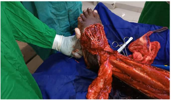 · 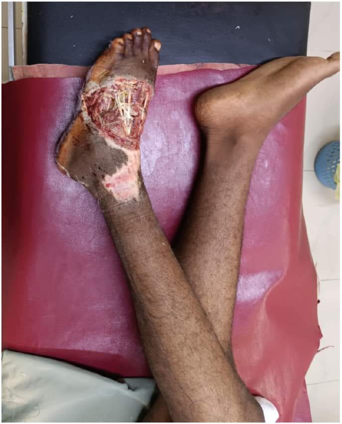 · 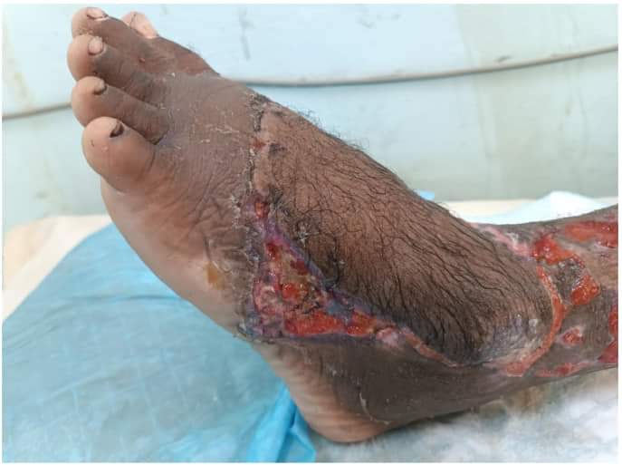 · 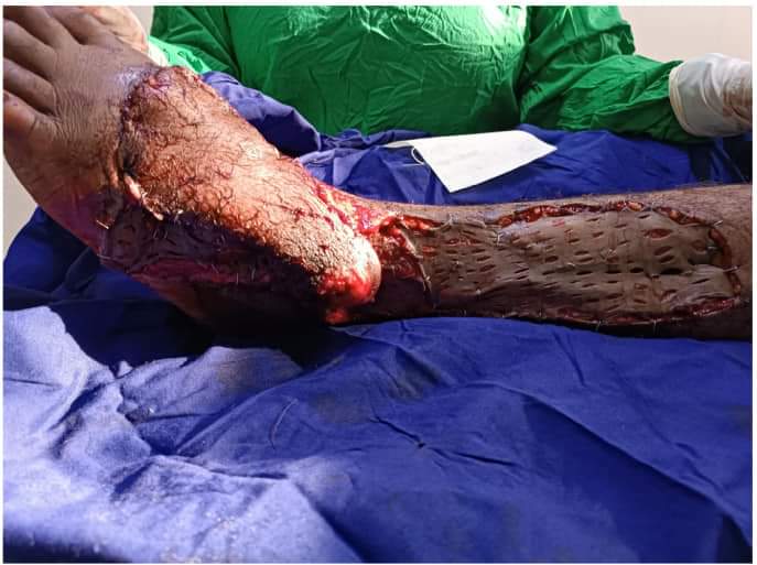 · 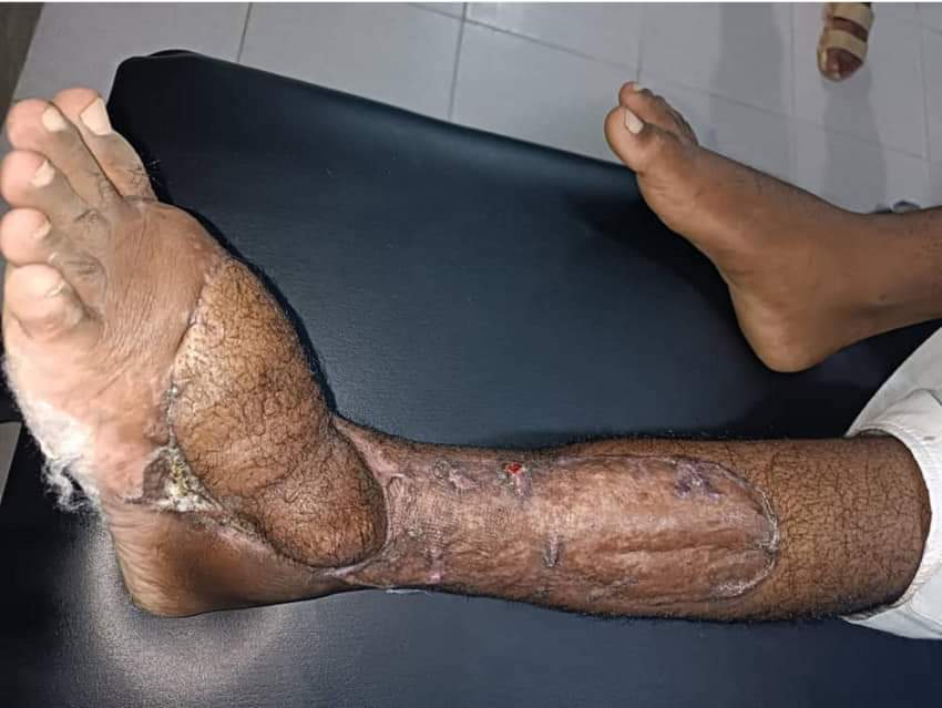 · |
|
#2
●
08-15-2024, 02:44 AM
|
 My Rank: FIRST SERGEANT Poster Rank:411 Male Join Date: Nov 2009 Posts: 2,964 2,964Posts 4,915Thanked 448Quoted 18Mentioned 0Contrib. |
|
Re: Bike Accident
"9 days to reach the hospital"  And a funky hairy foot to show for it. |
|
#3
●
08-15-2024, 03:29 AM
|
 ✝Mudderator from Hell✝ Poster Rank:10 e-mail Join Date: Oct 2006 Posts: 96,692
Contributions: 901
96,692Posts 543,647Thanked 10,372Quoted 479Mentioned 901Contrib. |
|
Re: Bike Accident
Hospital in Khartoum, Sudan. From the farest border place to Khartoum is 24 hour travel in a car and 13 days by foot. Maybe he went on his bike  It's official, they have Bigfoot in Sudan too. It's official, they have Bigfoot in Sudan too. |